


 medicine
Similar presentations:


Эндоскопический подход в хирургии гнойных отитов
1. Эндоскопический подход в хирургии гнойных отитов
Работу выполнил: Соков Ростислав ИгоревичНаучный руководитель: Ассистент кафедры оториноларингологии
Гергиев Владимир Феликсович
2.
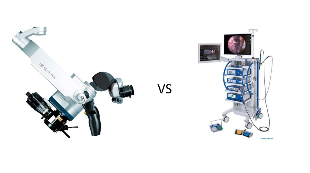
VS3.
4. ТЕОРИТИЧЕСКИЕ ПЛЮСЫ И МИНУСЫ
МИКРОСКОП• ДВЕ РАБОЧИЕ РУКИ
• ОГРАНИЧЕННАЯ ВИДИМОСТЬ
ЭНДОСКОП
• ОДНА РАБОЧАЯ РУКА
• ЯТРОГЕННЫЕ ТРАВМЫ
• ИНДУЦИРОВАННАЯ
ГИПЕРТЕРМИЯ
• ГИБКОСТЬ НАСТРОЙКИ
ВИЗУАЛИЗАЦИИ
5. Джентельменский набор
Figure 1. Instruments used. A: Traditional material for otological surgery added to curettes and speciallybuilt aspirator for ungluing. B: Endoscopes of 4 mm and 18 cm (0 and 45 degrees of angulation). Besides
these, traditional aspirators, tweezers and scissors are also required.
6.
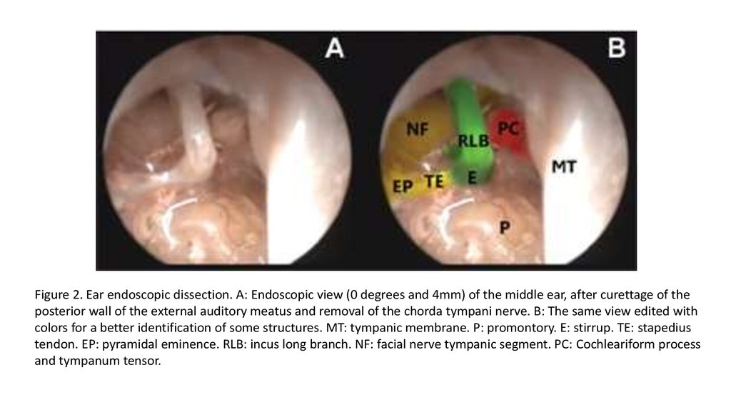
Figure 2. Ear endoscopic dissection. A: Endoscopic view (0 degrees and 4mm) of the middle ear, after curettage of theposterior wall of the external auditory meatus and removal of the chorda tympani nerve. B: The same view edited with
colors for a better identification of some structures. MT: tympanic membrane. P: promontory. E: stirrup. TE: stapedius
tendon. EP: pyramidal eminence. RLB: incus long branch. NF: facial nerve tympanic segment. PC: Cochleariform process
and tympanum tensor.
7.
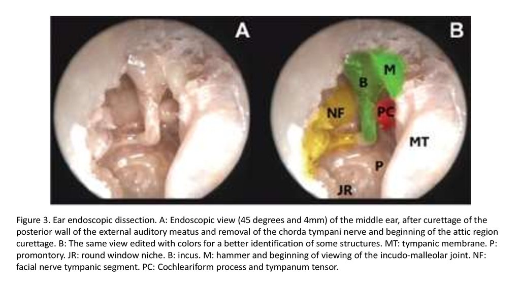
Figure 3. Ear endoscopic dissection. A: Endoscopic view (45 degrees and 4mm) of the middle ear, after curettage of theposterior wall of the external auditory meatus and removal of the chorda tympani nerve and beginning of the attic region
curettage. B: The same view edited with colors for a better identification of some structures. MT: tympanic membrane. P:
promontory. JR: round window niche. B: incus. M: hammer and beginning of viewing of the incudo-malleolar joint. NF:
facial nerve tympanic segment. PC: Cochleariform process and tympanum tensor.
8.
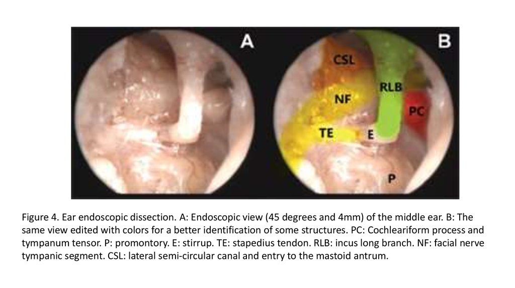
Figure 4. Ear endoscopic dissection. A: Endoscopic view (45 degrees and 4mm) of the middle ear. B: Thesame view edited with colors for a better identification of some structures. PC: Cochleariform process and
tympanum tensor. P: promontory. E: stirrup. TE: stapedius tendon. RLB: incus long branch. NF: facial nerve
tympanic segment. CSL: lateral semi-circular canal and entry to the mastoid antrum.
9.
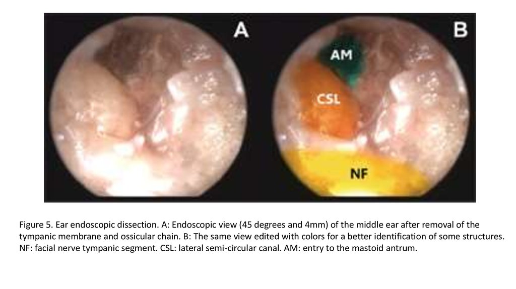
Figure 5. Ear endoscopic dissection. A: Endoscopic view (45 degrees and 4mm) of the middle ear after removal of thetympanic membrane and ossicular chain. B: The same view edited with colors for a better identification of some structures.
NF: facial nerve tympanic segment. CSL: lateral semi-circular canal. AM: entry to the mastoid antrum.
10.
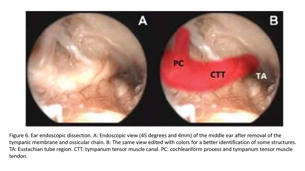
Figure 6. Ear endoscopic dissection. A: Endoscopic view (45 degrees and 4mm) of the middle ear after removal of thetympanic membrane and ossicular chain. B: The same view edited with colors for a better identification of some structures.
TA: Eustachian tube region. CTT: tympanum tensor muscle canal. PC: cochleariform process and tympanum tensor muscle
tendon.
11.
12.
13. ИССЛЕДОВАНИЕ
14. Объекты исследования
• 58 пациентов• Средний возраст 37 лет (15-63)
• 35 женщин, 23 мужчины
• Common complaints : Hearing loss, purulent discharge, dizziness
• Sensorineural hearing loss: 14.0 (+-5.8 dB)
• Conductive hearing loss: 31.9 (+-9.3 dB)
• + Rinne test in 16 patients
• + Weber test in 53 patients
15. Манипуляции
1)2)
3)
4)
5)
6)
Общая анестезия, заушный разрез
Lifted flap
!Examination!
Операция
!Examination!
=-=-=-=-
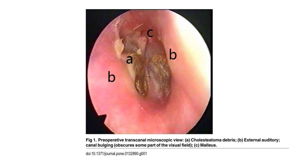
16.
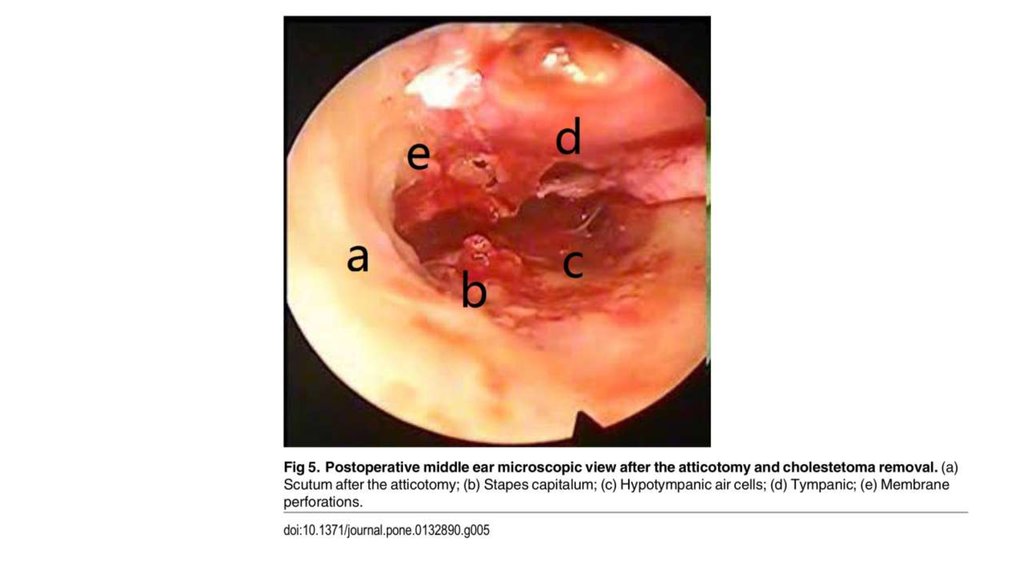
17.
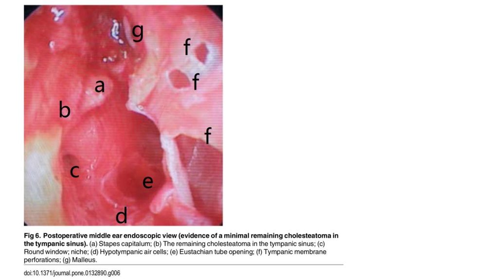
18.
19.
20.
21.
22.
23. А вот если бы…
• 4 из 13 холестеатом остались незамеченными• Остатки грануляционной ткани у 5
• Гипертрофированная слизистая у 23
• Тимпаносклеротические бляшки у 12
24. ВЫВОД
Один метод хорошо, но двалучше