


 medicine
Similar presentations:


")
Burns
1.
BurnsMedical Academy named
after S.I. Georgievsky
of Vernadsky CFU
General Surgery
Department
2.
Frequency• 67% occur in males
▫ Young adults (20-29 yr)
▫ Children < 9 years of age
▫ > 50 years of age fewest of serious burns
• Major causes of burns
▫ Flame (37%)
▫ Liquid (24%)
▫ Children < 2 years of age
Liquids/hot surfaces
• 5% die as a result of their
3.
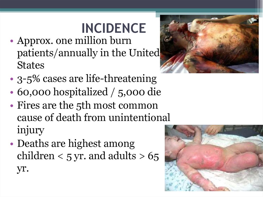
INCIDENCE• Approx. one million burn
patients/annually in the United
States
• 3-5% cases are life-threatening
• 60,000 hospitalized / 5,000 die
• Fires are the 5th most common
cause of death from unintentional
injury
• Deaths are highest among
children < 5 yr. and adults > 65
yr.
4.
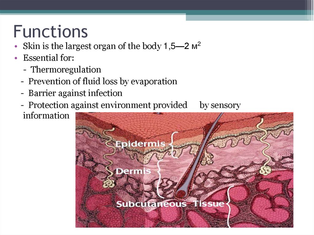
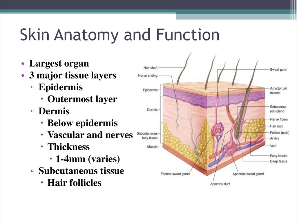
Functions• Skin is the largest organ of the body 1,5—2 м2
• Essential for:
- Thermoregulation
- Prevention of fluid loss by evaporation
- Barrier against infection
- Protection against environment provided by sensory
information
5.
Skin Anatomy and Function• Largest organ
• 3 major tissue layers
▫ Epidermis
Outermost layer
▫ Dermis
Below epidermis
Vascular and nerves
Thickness
1-4mm (varies)
▫ Subcutaneous tissue
Hair follicles
6.
Types of burn injuries• Thermal: direct contact with heat
(flame, scald, contact)
• Electrical
A.C. – alternating current (residential)
D.C. – direct current (industrial/lightening)
• Chemical
• Frostbite
7.
Classification• Burns are classified by depth, type and extent of
injury
• Every aspect of burn treatment depends on
assessment of the depth and extent
8.
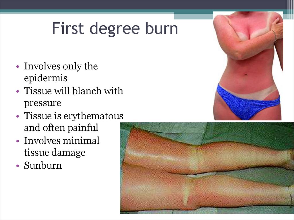
First degree burn• Involves only the
epidermis
• Tissue will blanch with
pressure
• Tissue is erythematous
and often painful
• Involves minimal
tissue damage
• Sunburn
9.
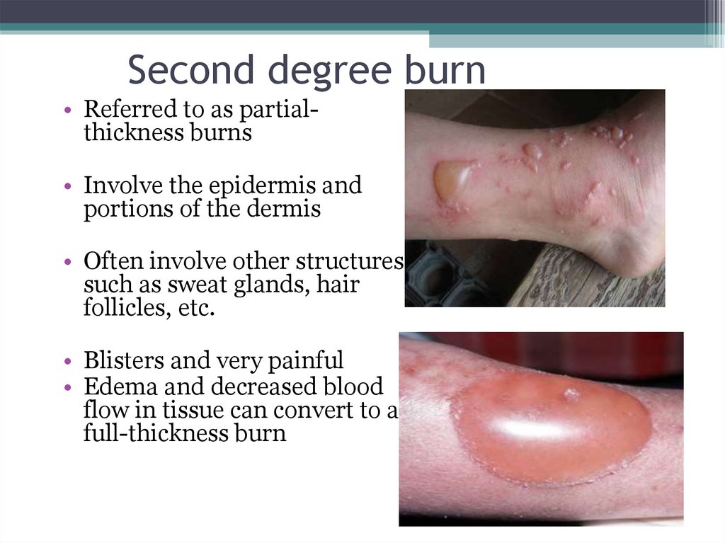
Second degree burn• Referred to as partialthickness burns
• Involve the epidermis and
portions of the dermis
• Often involve other structures
such as sweat glands, hair
follicles, etc.
• Blisters and very painful
• Edema and decreased blood
flow in tissue can convert to a
full-thickness burn
10.
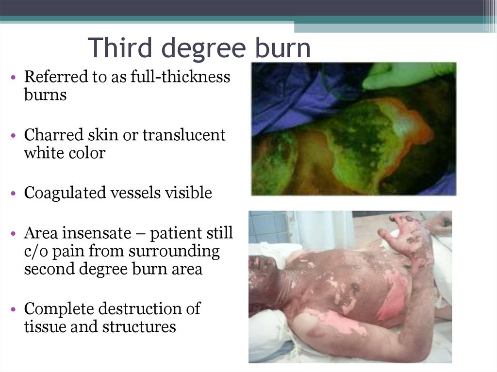
Third degree burn• Referred to as full-thickness
burns
• Charred skin or translucent
white color
• Coagulated vessels visible
• Area insensate – patient still
c/o pain from surrounding
second degree burn area
• Complete destruction of
tissue and structures
11.
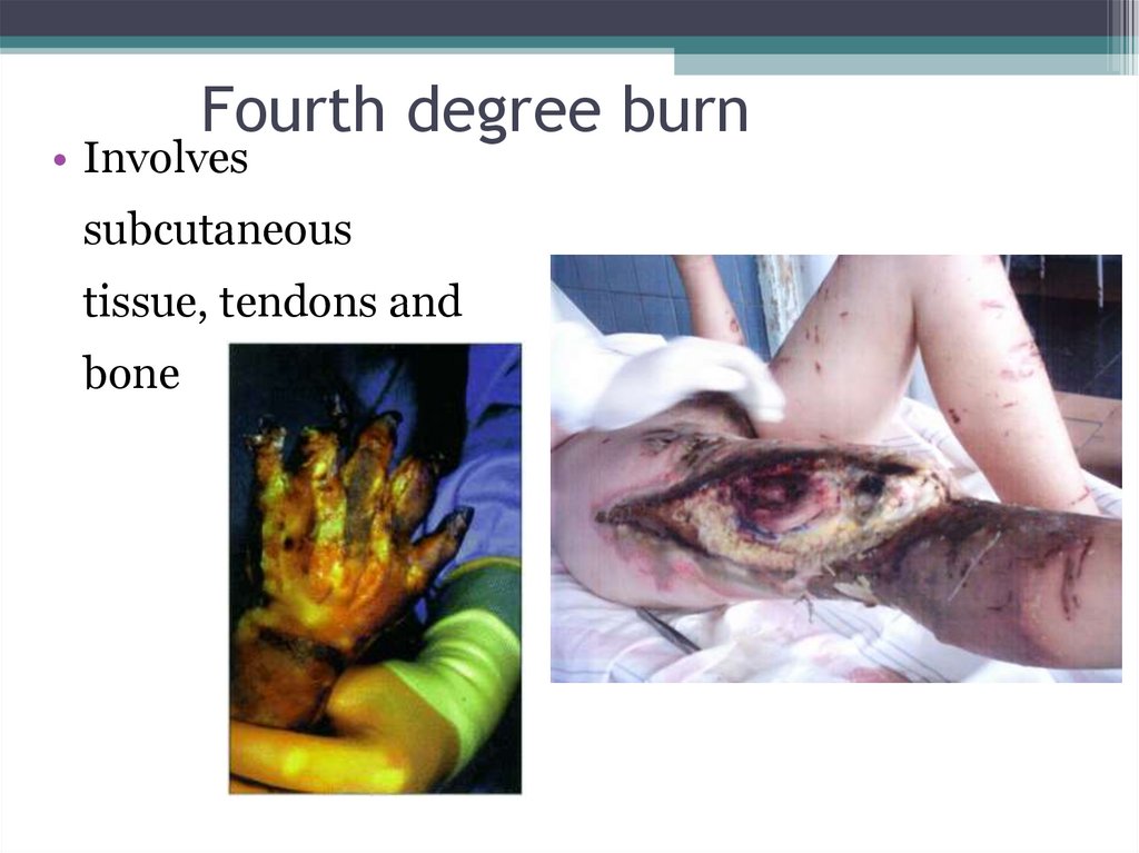
Fourth degree burn• Involves
subcutaneous
tissue, tendons and
bone
12.
Zones of Burn Wounds• Zone of Coagulation
▫
devitalized,
necrotic, white, no
circulation
• Zone of Stasis
‘circulation sluggish’
▫
may covert to full
thickness, mottled
red
• Zone of Hyperemia
▫
outer rim, good
blood flow, red
13.
Burn extent% BSA involved
morbidity
Burn extent is calculated only on individuals with
second and third degree burns
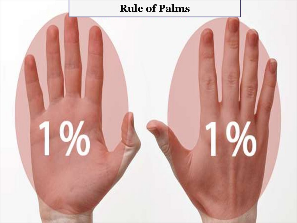
Palmar surface = 1% of the BSA
14.
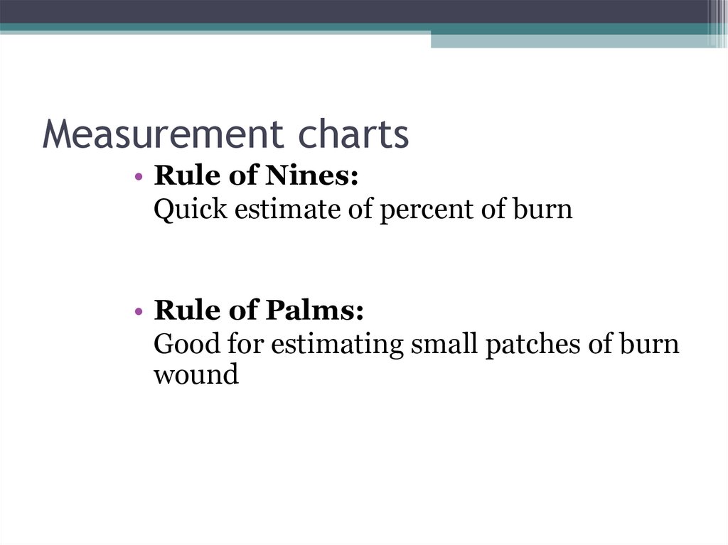
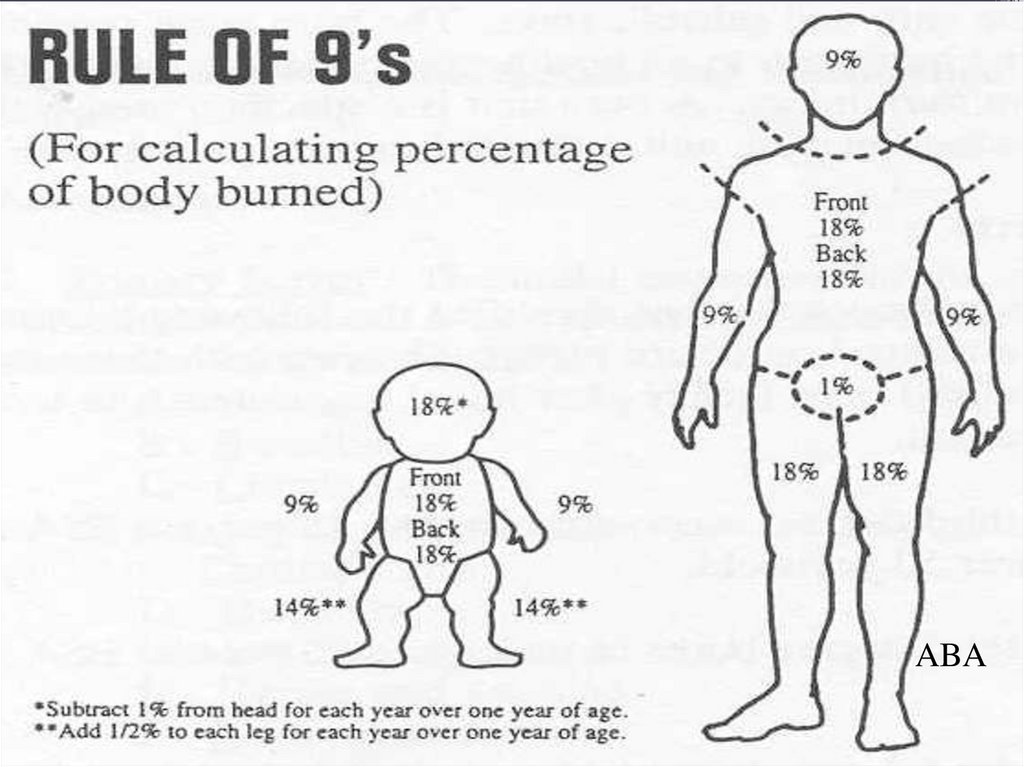
Measurement charts• Rule of Nines:
Quick estimate of percent of burn
• Rule of Palms:
Good for estimating small patches of burn
wound
15.
Rule of 9sABA
16.
Rule of Palms17.
Lab studiesSevere burns:
• CBC
• Chemistry profile
• Coagulation profile
• creatine phosphokinase and urine
myoglobin (with electrical injuries)
• 12 Lead EKG
18.
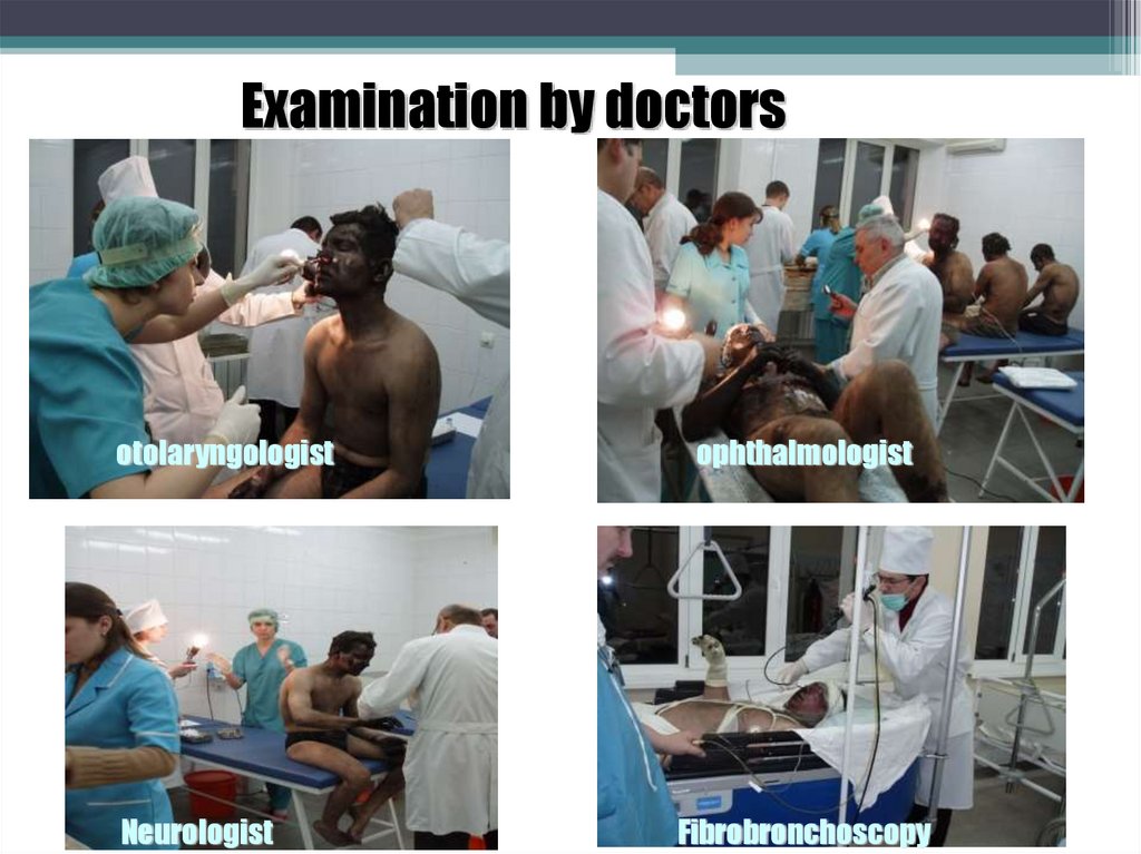
Examination by doctorsotolaryngologist
Neurologist
ophthalmologist
Fibrobronchoscopy
19.
Imaging studies• X-Ray
• Plain Films / CT scan: Dependent upon
history and physical findings
20.
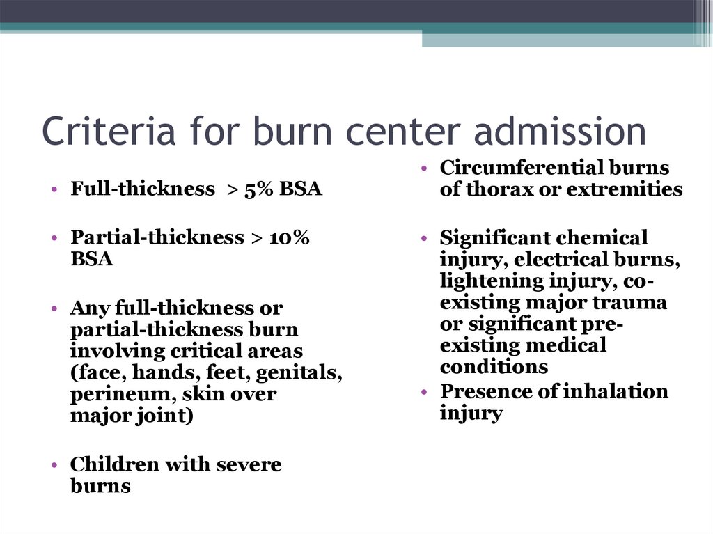
Criteria for burn center admission• Full-thickness > 5% BSA
• Partial-thickness > 10%
BSA
• Any full-thickness or
partial-thickness burn
involving critical areas
(face, hands, feet, genitals,
perineum, skin over
major joint)
• Children with severe
burns
• Circumferential burns
of thorax or extremities
• Significant chemical
injury, electrical burns,
lightening injury, coexisting major trauma
or significant preexisting medical
conditions
• Presence of inhalation
injury
21.
Initial patient treatment• Stop the burning process
• Consider burn patient as a multiple trauma
patient until determined otherwise
• Perform ABCDE assessment
• Avoid hypothermia!
Airway
Breathing
Circulation
Depth of Burn
Extent of Injury(s
• Remove constricting clothing and jewelry
22.
Details of the incident• Cause of the burn
• Time of injury
• Place of the occurrence (closed space, presence
of chemicals, noxious fumes)
• Likelihood of associated trauma (explosion,…)
• Pre-hospital interventions
23.
Care of small burnsWhat can YOU do?
24.
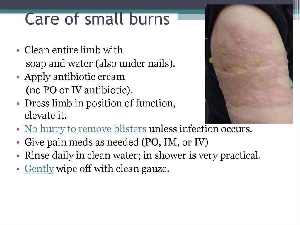
Care of small burns• Clean entire limb with
soap and water (also under nails).
• Apply antibiotic cream
(no PO or IV antibiotic).
• Dress limb in position of function,
and
elevate it.
• No hurry to remove blisters unless infection occurs.
• Give pain meds as needed (PO, IM, or IV)
• Rinse daily in clean water; in shower is very practical.
• Gently wipe off with clean gauze.
25.
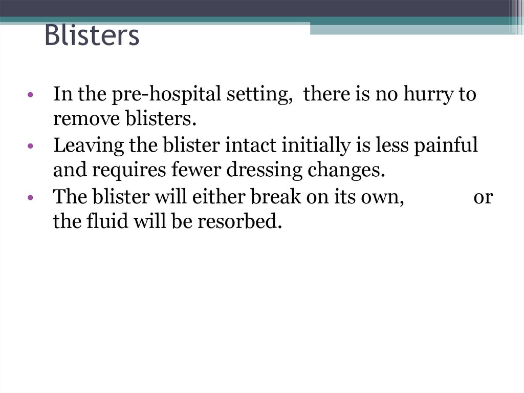
Blisters• In the pre-hospital setting, there is no hurry to
remove blisters.
• Leaving the blister intact initially is less painful
and requires fewer dressing changes.
• The blister will either break on its own,
or
the fluid will be resorbed.
26.
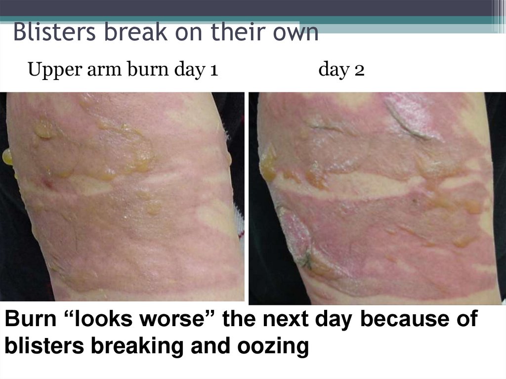
Blisters break on their ownUpper arm burn day 1
day 2
Burn “looks worse” the next day because of
blisters breaking and oozing
27.
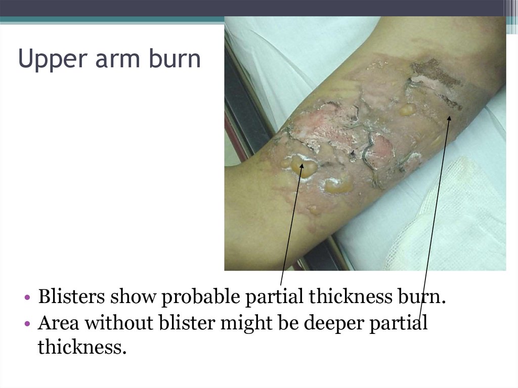
Upper arm burn121
• Blisters show probable partial thickness burn.
• Area without blister might be deeper partial
thickness.
28.
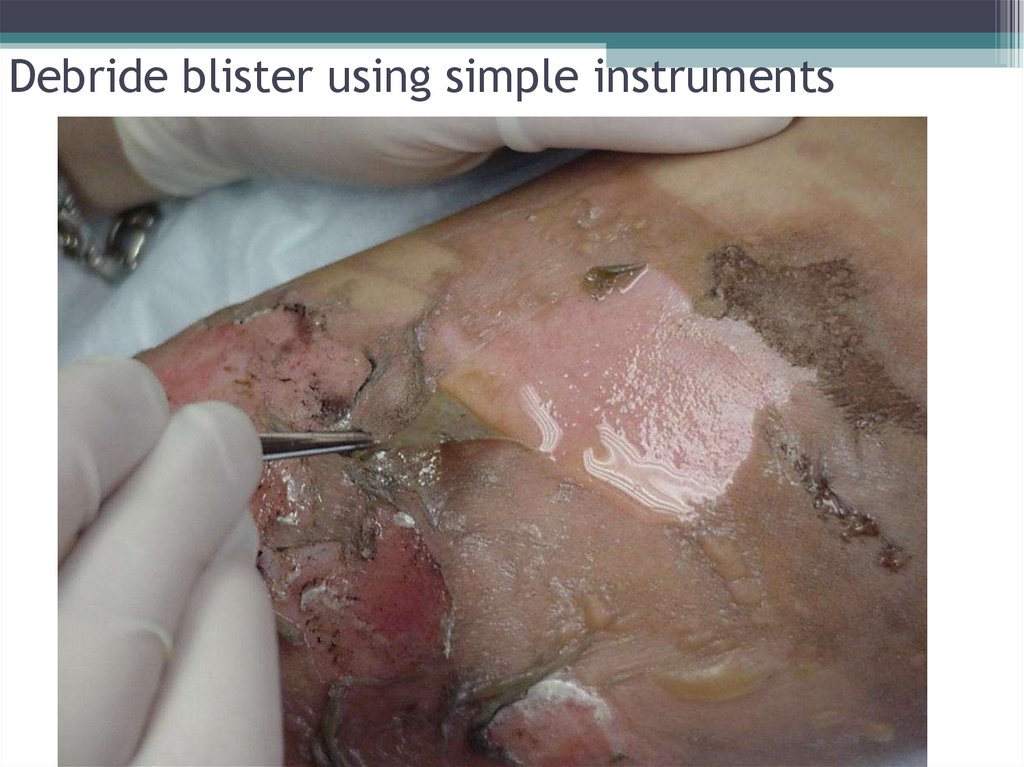
Debride blister using simple instruments29.
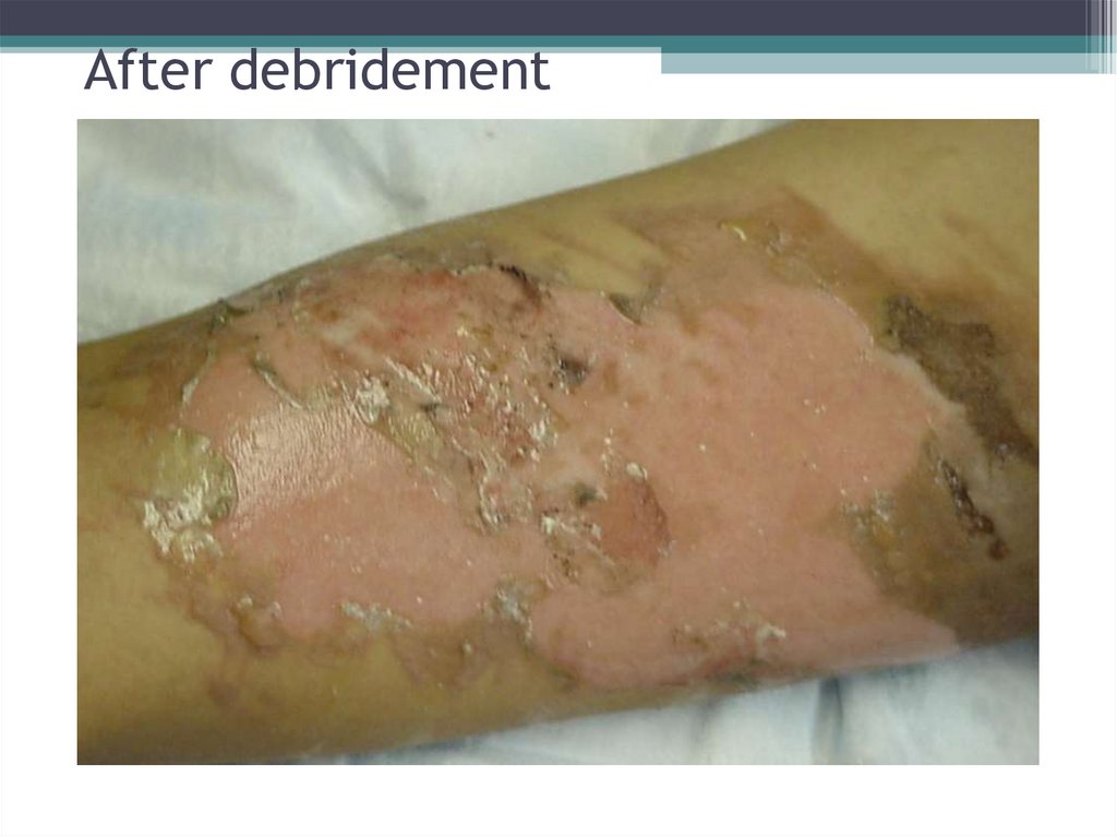
After debridement30.
Before and after debridement• Removing the blister leaves a weeping, very tender
wound, that requires much care.
31.
Silver sulfadiazene32.
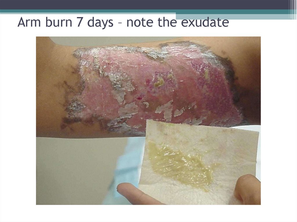
Arm burn 7 days – note the exudate33.
Burns of special areasof the body
Face
Mouth
Neck
Hands and feet
Genitalia
34.
FaceBe VERY concerned for the airway!!
Eyelids, lips and ears often swell.
In fact, they look even worse the next day.
But they will start to improve daily after that.
Cleanse eyes with warm water or saline.
Apply antibiotic ointment or liquid tears until
lids are no longer swollen shut.
• Bacitracin cream/ointment will serve
35.
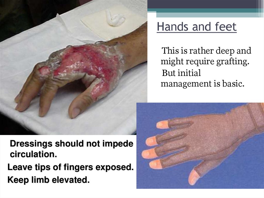
Hands and feetThis is rather deep and
might require grafting.
But initial
management is basic.
Dressings should not impede
circulation.
Leave tips of fingers exposed.
Keep limb elevated.
36.
Hands and feet• Allow use of the hands in dressings by day.
• Splint in functional position by night.
• Keep elevated to reduce swelling.
37.
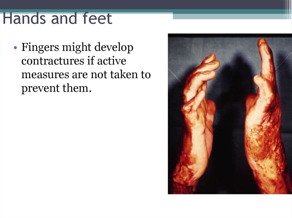
Hands and feet• Fingers might develop
contractures if active
measures are not taken to
prevent them.
38.
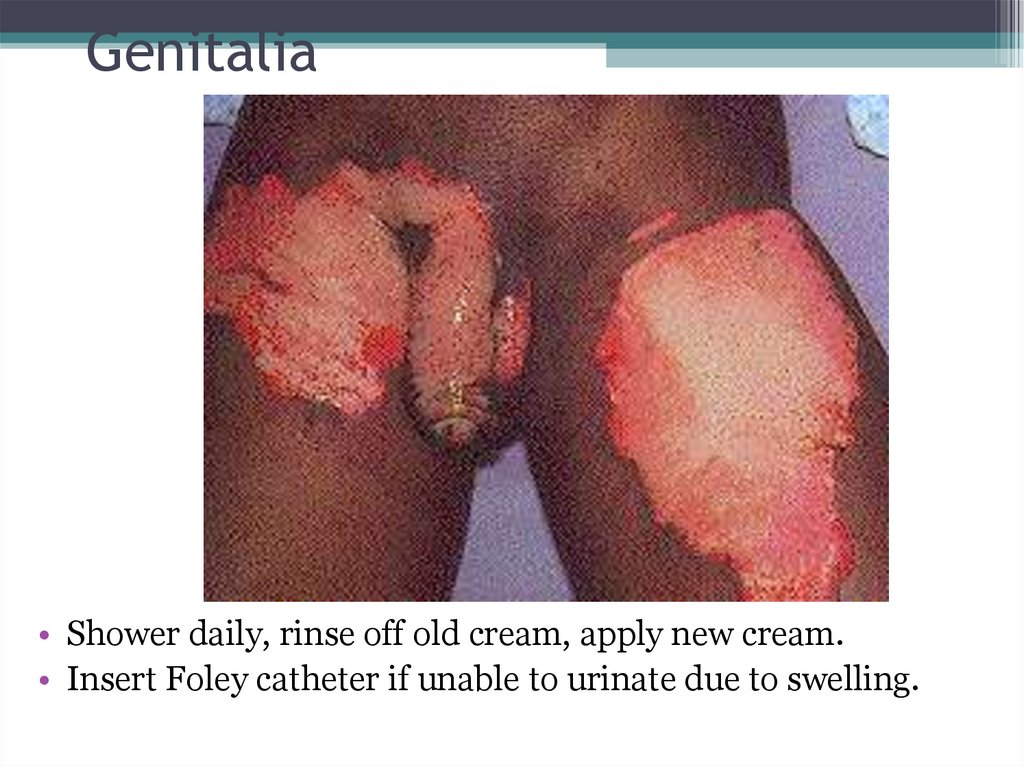
Genitalia• Shower daily, rinse off old cream, apply new cream.
• Insert Foley catheter if unable to urinate due to swelling.
39.
Large Burns40.
Causes of death in burn patients• Airway
▫ Facial edema, and/or airway edema
• Breathing
▫ Toxic inhalation (CO, +/- CN)
▫ Respiratory failure due to smoke injury or ARDS
41.
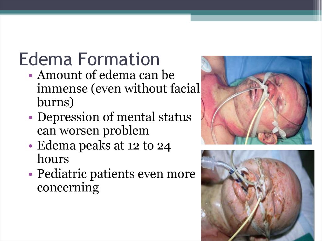
Edema Formation• Amount of edema can be
immense (even without facial
burns)
• Depression of mental status
can worsen problem
• Edema peaks at 12 to 24
hours
• Pediatric patients even more
concerning
42.
Causes of death in burn patients• Circulation: “failure of resuscitation”
▫ Cardiovascular collapse, or acute MI
▫ Acute renal failure
▫ Other end organ failure
• Missed non-thermal injury
43.
Patients with larger burnsFirst assess
• CBA’s
• “Disability” (brief neuro exam)
Later
• Examine rest of patient
• Calculate IV fluids
• Treat burn
44.
Airway considerations• Upper airway injury (above the glottis):
Area buffers the heat of smoke – thermal injury
is usually confined to the larynx and upper
trachea.
• Lower airway/alveolar injury (below the
glottis):
- Caused by the inhalation of steam or chemical
smoke.
- Presents as ARDS (Adult respiratory distress
syndrome) often after 24-72 hours
45.
Criteria for intubation• Changes in voice
• Wheezing / labored
respirations
• Excessive, continuous
coughing
• Altered mental status
• Carbonaceous sputum
• Singed facial or nasal
hairs
• Facial burns
• Oro-pharyngeal edema /
stridor
• Assume inhalation injury
in any patient confined in
a fire environment
• Extensive burns of the
face / neck
• Eyes swollen shut
• Burns of 50% TBSA or
greater
46.
Ventilatory therapiesRapid Sequence Intubation
Pain Management, Sedation and Paralysis
PEEP (positive end expiratory pressure)
High concentration oxygen
Avoid barotrauma
Hyperbaric oxygen
47.
Ventilatory therapies• Burn patients with
Acute respiratory distress syndrome
(ARDS) requiring
PEEP (positive end expiratory pressure) > 14
cm for adequate ventilation should receive
prophylactic tube thoracostomy.
48.
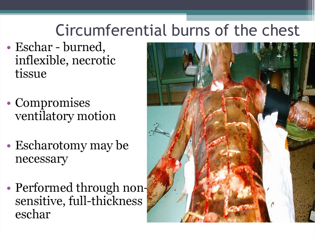
Circumferential burns of the chest• Eschar - burned,
inflexible, necrotic
tissue
• Compromises
ventilatory motion
• Escharotomy may be
necessary
• Performed through nonsensitive, full-thickness
eschar
49.
Carbon Monoxide IntoxicationCarbon monoxide has a binding affinity for
hemoglobin which is 210-240 times greater than
that of oxygen.
Results in decreased oxygen delivery to tissues,
leading to cerebral and myocardial hypoxia.
Cardiac arrhythmias are the most common fatal
occurrence.
50.
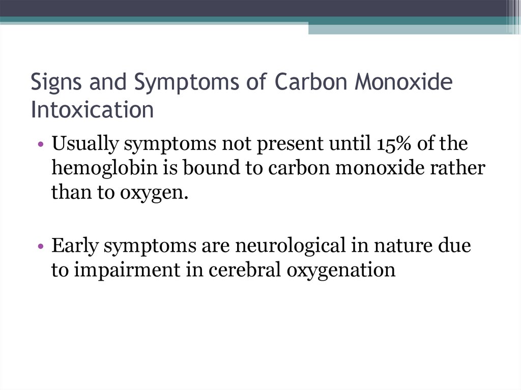
Signs and Symptoms of Carbon MonoxideIntoxication
• Usually symptoms not present until 15% of the
hemoglobin is bound to carbon monoxide rather
than to oxygen.
• Early symptoms are neurological in nature due
to impairment in cerebral oxygenation
51.
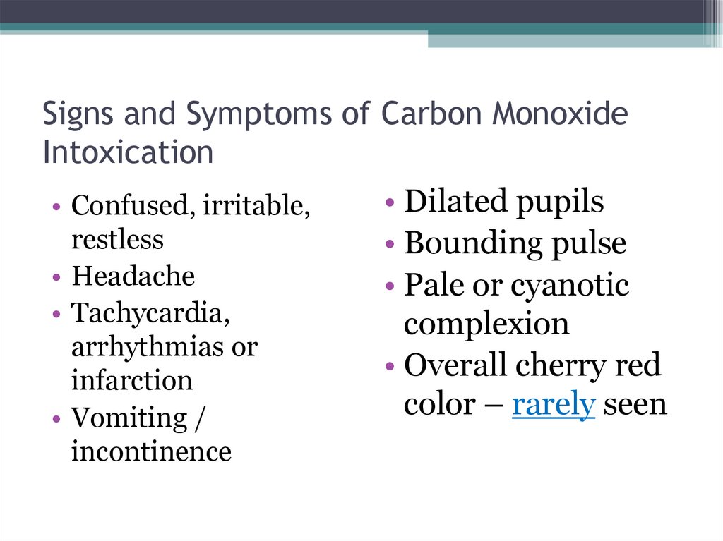
Signs and Symptoms of Carbon MonoxideIntoxication
• Confused, irritable,
restless
• Headache
• Tachycardia,
arrhythmias or
infarction
• Vomiting /
incontinence
• Dilated pupils
• Bounding pulse
• Pale or cyanotic
complexion
• Overall cherry red
color – rarely seen
52.
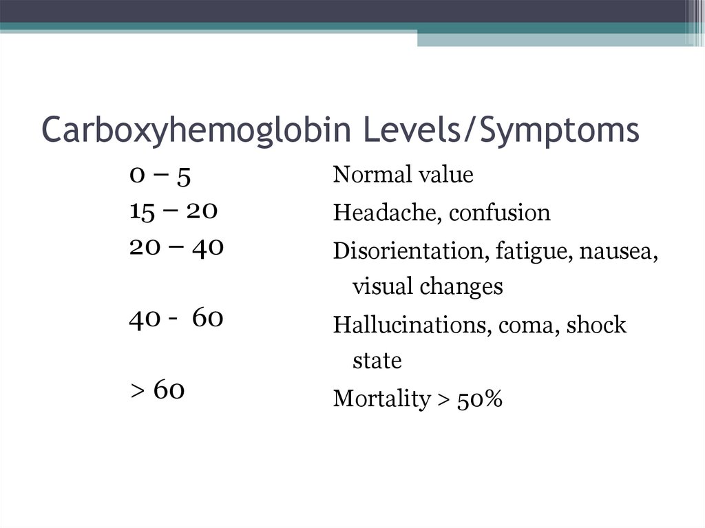
Carboxyhemoglobin Levels/Symptoms0–5
15 – 20
20 – 40
Normal value
40 - 60
Hallucinations, coma, shock
> 60
Headache, confusion
Disorientation, fatigue, nausea,
visual changes
state
Mortality > 50%
53.
Management of Carbon MonoxideIntoxication
• Remove patient from source of exposure.
• Administer 100% high flow oxygen
Half life of Carboxyhemoglobin in patients:
• Breathing room air 120-200 minutes
• Breathing 100% O2
30 minutes
54.
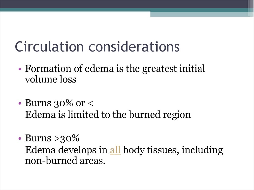
Circulation considerations• Formation of edema is the greatest initial
volume loss
• Burns 30% or <
Edema is limited to the burned region
• Burns >30%
Edema develops in all body tissues, including
non-burned areas.
55.
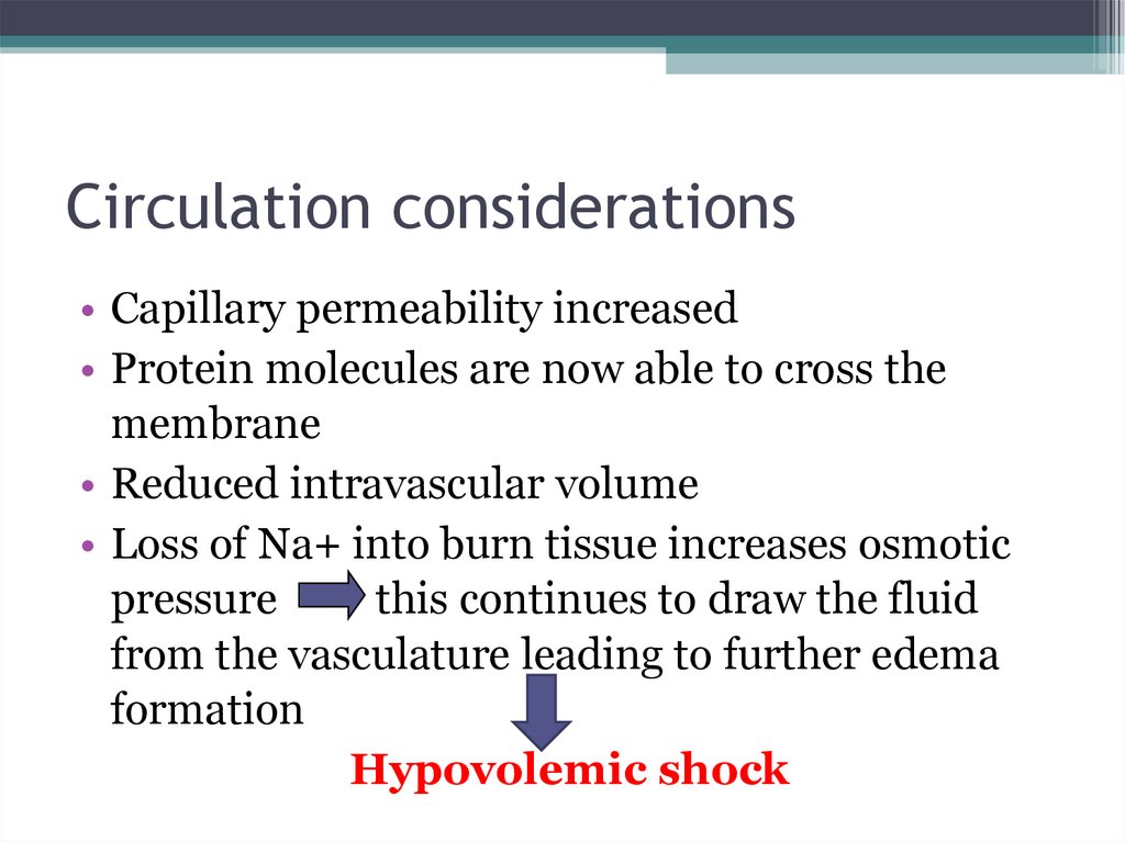
Circulation considerations• Capillary permeability increased
• Protein molecules are now able to cross the
membrane
• Reduced intravascular volume
• Loss of Na+ into burn tissue increases osmotic
pressure
this continues to draw the fluid
from the vasculature leading to further edema
formation
Hypovolemic shock
56.
Circulation considerations• Loss of plasma volume is greatest during the
first 4 – 6 hours, decreasing substantially in 8 –
24 hours if adequate perfusion is maintained.
57.
Fluid resuscitation• Goal: Maintain perfusion to vital organs
• Based on the TBSA, body weight and whether
patient is adult/child
• Fluid overload should be avoided – difficult to
retrieve settled fluid in tissues and may facilitate
organ hypoperfusion
58.
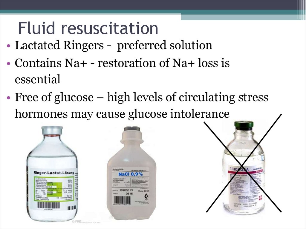
Fluid resuscitation• Lactated Ringers - preferred solution
• Contains Na+ - restoration of Na+ loss is
essential
• Free of glucose – high levels of circulating stress
hormones may cause glucose intolerance
59.
Fluid resuscitation• Burned patients have large insensible fluid
losses
• Fluid volumes may increase in patients with coexisting trauma
• Vascular access: Two large bore peripheral lines
(if possible) or central line.
60.
Fluid resuscitation• Fluid requirement calculations for infusion rates
are based on the time from injury, not from the
time fluid resuscitation is initiated.
61.
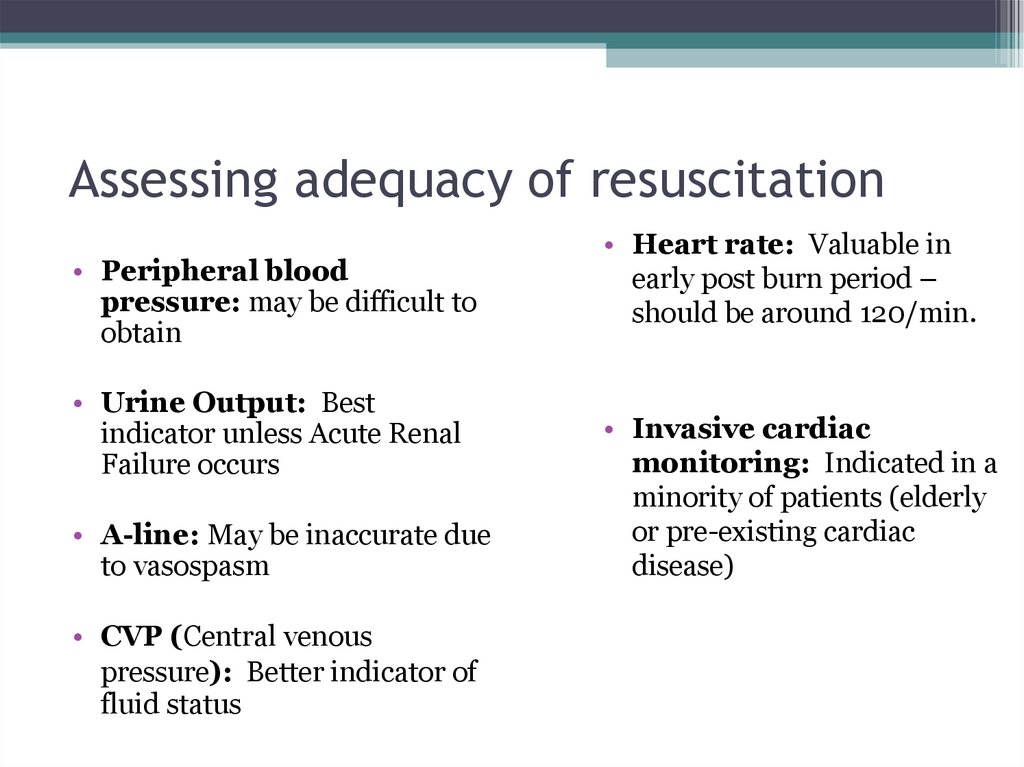
Assessing adequacy of resuscitation• Peripheral blood
pressure: may be difficult to
obtain
• Urine Output: Best
indicator unless Acute Renal
Failure occurs
• A-line: May be inaccurate due
to vasospasm
• CVP (Central venous
pressure): Better indicator of
fluid status
• Heart rate: Valuable in
early post burn period –
should be around 120/min.
• Invasive cardiac
monitoring: Indicated in a
minority of patients (elderly
or pre-existing cardiac
disease)
62.
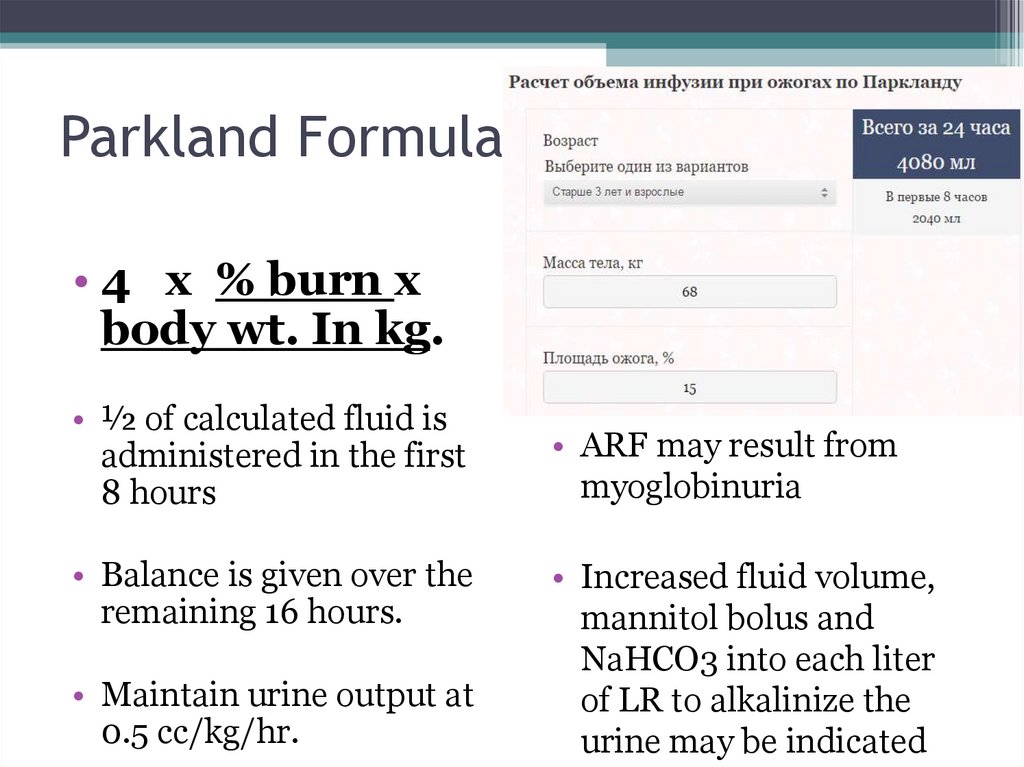
Parkland Formula• 4 x % burn x
body wt. In kg.
• ½ of calculated fluid is
administered in the first
8 hours
• Balance is given over the
remaining 16 hours.
• Maintain urine output at
0.5 cc/kg/hr.
• ARF may result from
myoglobinuria
• Increased fluid volume,
mannitol bolus and
NaHCO3 into each liter
of LR to alkalinize the
urine may be indicated
63.
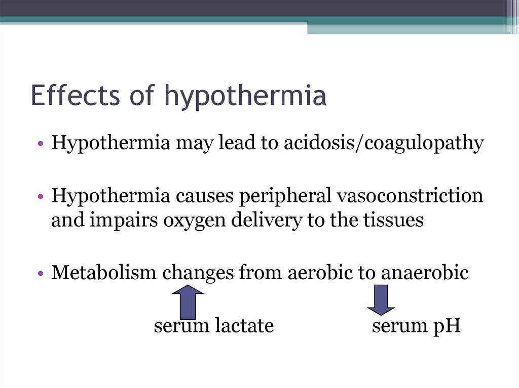
Effects of hypothermia• Hypothermia may lead to acidosis/coagulopathy
• Hypothermia causes peripheral vasoconstriction
and impairs oxygen delivery to the tissues
• Metabolism changes from aerobic to anaerobic
serum lactate
serum pH
64.
Prevention of hypothermia• Cover patients with a dry
sheet – keep head
covered
• Pre-warm trauma room
• Administer warmed IV
solutions
• Avoid application of
saline-soaked dressings
• Avoid prolonged
irrigation
• Remove wet / bloody
clothing and sheets
• Avoid application of
antimicrobial creams
• Continual monitoring of
core temperature via
foley or SCG temperature
probe
65.
Pain managementAdequate analgesia imperative!
DOC: Morphine Sulfate
Dose: Adults: 0.1 – 0.2 mg/kg IVP
Children: 0.1 – 0.2 mg/kg/dose IVP / IO
Other pain medications commonly used:
• Demerol
• Vicodin ES
• NSAIDs
66.
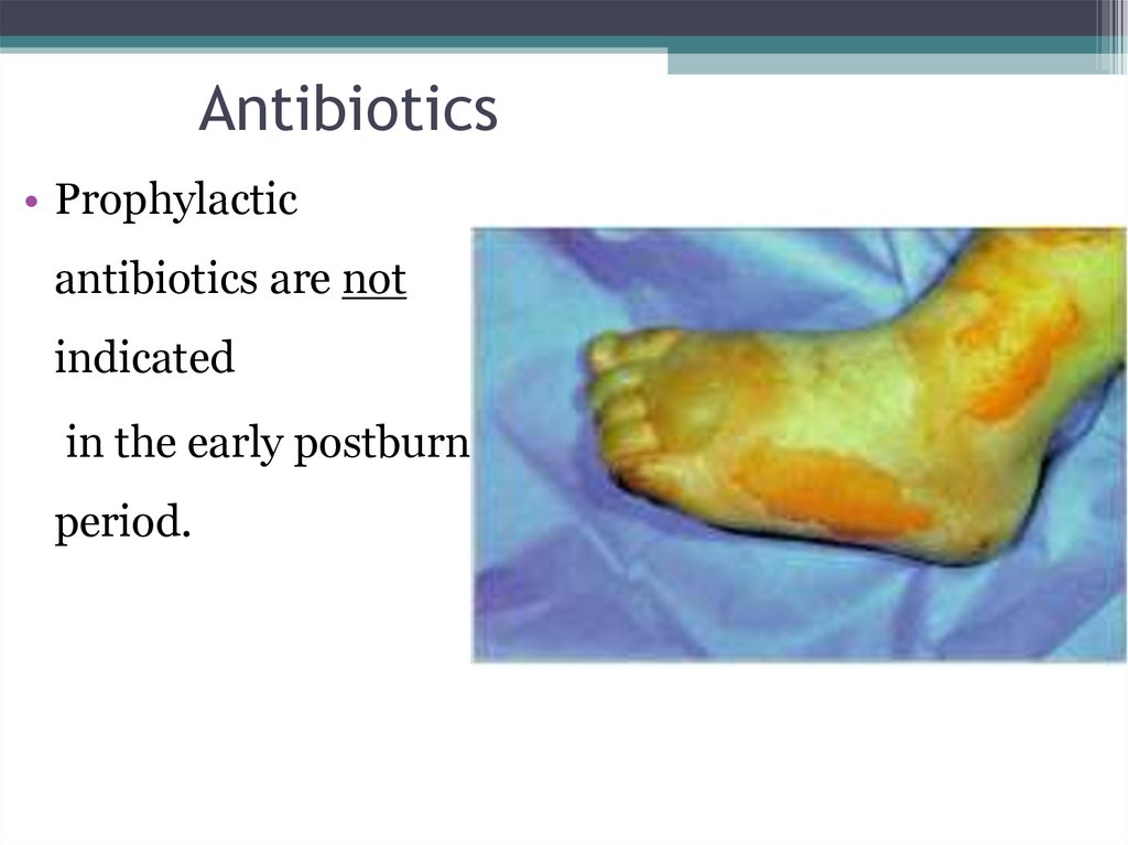
Antibiotics• Prophylactic
antibiotics are not
indicated
in the early postburn
period.
67.
Other considerations• Check tetanus status – administer Td as
appropriate
• Debride and treat open blisters or blisters
located in areas that are likely to rupture
• Debridement of intact blisters is controversial
68.
Electrical burns: are thermal injuries resultingfrom high intensity heat. The skin injury area may
appear small, but the underlying tissue damage
may be extensive.
Additionally, there may be brain or heart damage or
musculoskeletal injuries associated with the
electrical injuries.
Safely remove the person from the source of the
electricity.
69.
Chemical burns- Most often caused by strongacids or alkalis. Unlike thermal burns, they can
cause progressive injury until the agent is
inactivated.
a. Flush the injured area with a copious amount of
water while at the scene of the incident. Don’t
delay or waste time looking for or using a
neutralizing agent. These may in fact worsen the
injury by producing heat or causing direct injury
themselves.
70.
Burn Injury: Summary• Many risk factors age dependent
• Pediatricians primary role: prevention
• High risk of multiple organ system effects,
prolonged hospitalization
• Initial care: ABCs, then surgical issues
▫ special attention to airway, hemodynamics
• Chronic care issues: scarring, lean mass loss