


 medicine
Similar presentations:

")


")


Adult Nursing Care I
1.
1Adult Nursing Care I
NURS 241
2.
212/14/2022
Management of Patients
With Chest and Lower
Respiratory Tract Disorders
3.
12/14/20223
Learning outcomes
• 1. Identify patients at risk for atelectasis and nursing
interventions related to its prevention and management.
• 2. Compare the various pulmonary infections with regard
to causes, clinical manifestations, nursing management,
complications, and prevention.
• 3. Use the nursing process as a framework for care of the
patient with pneumonia.
• 4. Relate the therapeutic management techniques of
acute respiratory
4.
12/14/20224
Atelectasis
• Atelectasis refers to closure or collapse of alveoli and
often is described in relation to x-ray findings and clinical
signs and symptoms.
• Atelectasis may be acute or chronic and may cover a
broad range of pathophysiologic changes, from microatelectasis (which
is not detectable on chest x-ray) to
macro-atelectasis with loss of
segmental, lobar, or overall lung volume.
5.
12/14/2022Atelectasis
5
6.
12/14/20226
Causes
1.
2.
3.
4.
5.
6.
7.
Altered breathing patterns, retained secretions, alterations in
small airway function
Pain, prolonged supine positioning,
Reduced lung volumes due to musculoskeletal or neurologic
disorders,
Specific surgical procedures (eg, upper abdominal, thoracic,
or open heart surgery).
Postoperative patients are at risk for atelectasis.
Bronchial obstruction with impaired cough mechanism
Excessive pressure on the lung tissue, such as pressure
produced by fluid accumulating within the pleural space
(pleural effusion), air in the pleural space ( pneumothorax), or
blood in the pleural space (hemothorax), or tumor growth
within the thorax, or an elevated diaphragm.
7.
12/14/20227
Clinical manifestations
1. Increasing dyspnea
2. cough,
3. sputum production
4. In acute atelectasis involving a large amount of lung
tissue (lobar atelectasis), marked respiratory distress (
tachycardia, tachypnea , pleural pain, central cyanosis)
8.
12/14/20228
Assessment and diagnostic findings
1. Chest x-ray
2. Pulse oximetry demonstrate low saturation of
hemoglobin with O2 ( less than 90% )
9.
12/14/20229
Prevention
1. Change patient’s position frequently, especially from
supine to upright position, to promote ventilation and
prevent secretions from accumulating.
2. Encourage early mobilization from bed to chair followed
by early ambulation.
3. Encourage appropriate deep breathing and coughing to
mobilize secretions and prevent them from
accumulating.
4. Teach/reinforce appropriate technique for incentive
spirometry
10.
12/14/202210
Prevention
1. Administer prescribed opioids and sedatives to prevent
respiratory depression.
2.
Perform postural drainage and chest percussion, if
indicated.
3. Institute suctioning to remove tracheobroncial
secretions, if indicated.
11.
12/14/202211
Medical management
• The strategies to prevent atelectasis, which include
1. frequent turning,
2. early ambulation,
3. lung volume expansion maneuvers (eg, deep-breathing
exercises, incentive spirometry),
4. and coughing also serve as the first-line measures to
minimize or treat atelectasis by improving ventilation.
12.
12/14/202212
Medical management
• The secretions must be removed by coughing or
suctioning to permit air to re-enter that portion of the lung.
• Chest physical therapy (chest percussion and postural
drainage) may also be used to mobilize secretions.
• Nebulizer treatments with a bronchodilator medication
may be used to assist the patient
in the expectoration of secretions
13.
12/14/202213
Medical management
• A bronchoscopy is performed to remove secretions and
increase ventilation.
• Endotracheal intubation or mechanical ventilation may be
necessary.
• Thoracentesis may be indicated to remove the fluid by
needle aspiration.
14.
12/14/202214
Pneumonia
• Pneumonia is an inflammation of the lung parenchyma
that is caused by various microorganisms, including
bacteria, fungi, and viruses.
• Pneumonia and influenza are the most common causes
of death from infectious diseases in the United States.
15.
12/14/2022Pneumonia
15
16.
12/14/202216
Pneumonia
• Pneumonia is an inflammatory process, involving the
terminal airways and alveoli of the lung, caused by
infectious agents. It is classified according to its causative
agent.
17.
1712/14/2022
Pathophysiology and Etiology
• The organism gains access to the lungs through aspiration of
oropharyngeal contents, by inhalation of respiratory secretions
from infected individuals, by way of the bloodstream, or from
direct spread to the lungs as a result of surgery or trauma.
• Patients with bacterial pneumonia may have an underlying
disease that impairs host defense; pneumonia arises from
endogenous flora of the person whose resistance has been
altered, or from aspiration of oropharyngeal secretions.
A.
B.
Immunocompromised patients include those receiving
corticosteroids or immunosuppressants, those with cancer, those
being treated with chemotherapy or radiotherapy, those
undergoing organ transplantation, alcoholics, I.V. drug abusers,
and those with HIV disease and acquired immunodeficiency
syndrome.
These people have an increased risk of developing overwhelming
infection. Infectious agents include aerobic and anaerobic gramnegative bacilli; Staphylococcus; Nocardia; fungi; Candida;
viruses, such as cytomegalovirus; Pneumocystis carinii (also
known as P. jiroveci); reactivation of tuberculosis (TB); and others.
18.
12/14/202218
• When bacterial pneumonia occurs in a healthy person, there is
usually a history of preceding viral illness.
• Other predisposing factors include conditions interfering with normal
drainage of the lung, such as tumor, general anesthesia, and
postoperative immobility; depression of the central nervous system
(CNS) from drugs, neurologic disorders, or other conditions, such as
alcoholism, and intubation or respiratory instrumentation
• Pneumonia may be divided into three groups:
1. Community acquired, due to a number of organisms, including
Streptococcus pneumoniae
2. Hospital or nursing home acquired (nosocomial), due primarily
to gram-negative bacilli and staphylococci
3. Pneumonia in the immunocompromised person
19.
12/14/202219
• Clinical Manifestations
• For most common forms of bacterial pneumonia:
1. Sudden onset; shaking chill; rapidly rising fever of 101° F to
105° F (38.3° C to 40.5° C).
2. Cough productive of purulent sputum.
3. Pleuritic chest pain aggravated by respiration/coughing
4. Dyspnea, tachypnea accompanied by respiratory grunting,
nasal flaring, use of accessory muscles of respiration, fatigue
5. Rapid, bounding pulse
• Diagnostic Evaluation
1. Chest X-ray shows presence/extent of pulmonary disease,
typically consolidation.
2. Gram stain and culture and sensitivity tests of sputum—may
indicate offending organism.
3. Blood culture detects bacteremia (bloodstream invasion)
occurring with bacterial pneumonia.
4. Immunologic test detects microbial antigens in serum,
sputum, and urine.
20.
12/14/202220
• Management
1. Antimicrobial therapy ”depends on laboratory
identification of causative organism and sensitivity to
specific antimicrobials, or presumptive therapy with
broad spectrum agent in milder cases.
2. Oxygen therapy if patient has inadequate gas exchange
• Complications
1. Pleural effusion.
2. Sustained hypotension and shock, especially in gramnegative bacterial disease, particularly in elderly
patients.
3. Superinfection: pericarditis, bacteremia, and meningitis.
4. Delirium ”this is considered a medical emergency.
5. Atelectasis ”due to mucous plugs.
21.
12/14/202221
Nursing Diagnoses
• Impaired Gas Exchange related to decreased ventilation
secondary to inflammation and infection involving distal
airspaces
• Ineffective Airway Clearance related to excessive
tracheobronchial secretions
• Acute Pain related to inflammatory process and dyspnea
• Risk for Injury secondary to complications
22.
12/14/202222
Nursing Interventions
• Improving Gas Exchange
• Observe for cyanosis, dyspnea, hypoxia, and confusion,
indicating worsening condition.
• Follow ABG levels/Sao2 to determine oxygen need and
response to oxygen therapy.
• Administer oxygen at concentration to maintain Pao2 at
acceptable level. Hypoxemia may be encountered because of
abnormal ventilation-perfusion ratios in affected lung segments.
• Avoid high concentrations of oxygen in patients with COPD,
particularly with evidence of CO2 retention; use of high oxygen
concentrations may worsen alveolar ventilation by depressing
the patient's only remaining ventilatory drive. If high
concentrations of oxygen are given, monitor alertness and
Pao2 and Paco2 levels for signs of CO2 retention.
• Place patient in an upright position to obtain greater lung
expansion and improve aeration. Frequent turning and
increased activity (up in chair, ambulate as tolerated) should be
employed.
23.
12/14/202223
Nursing Interventions
• Enhancing Airway Clearance
• Obtain freshly expectorated sputum for gram stain and culture,
preferably early morning specimen as directed. Instruct the
patient as follows:
Rinse mouth with water to minimize contamination by normal flora.
Breathe deeply several times.
Cough deeply and expectorate raised sputum into sterile container.
• Encourage patient to cough; retained secretions interfere with
gas exchange. Suction as necessary.
• Encourage increased fluid intake, unless contraindicated, to
thin mucus and promote expectoration and replace fluid losses
caused by fever, diaphoresis, dehydration, and dyspnea.
• Humidify air or oxygen therapy to loosen secretions and
improve ventilation.
• Employ chest wall percussion and postural drainage when
appropriate to loosen and mobilize secretions
24.
12/14/202224
Nursing Interventions
• Relieving Pleuritic Pain
• Place in a comfortable position (semi-Fowler's) for resting
and breathing; encourage frequent change of position to
prevent pooling of secretions in lungs.
• Demonstrate how to splint the chest while coughing.
• Avoid suppressing a productive cough.
• Administer prescribed analgesic agent to relieve pain.
Avoid opioids in patients with a history of COPD.
• Apply heat and/or cold to chest as prescribed.
• Assist with intercostal nerve block for pain relief.
• Encourage modified bed rest during febrile period.
25.
12/14/2022PULMONARY TUBERCULOSIS
25
26.
12/14/202226
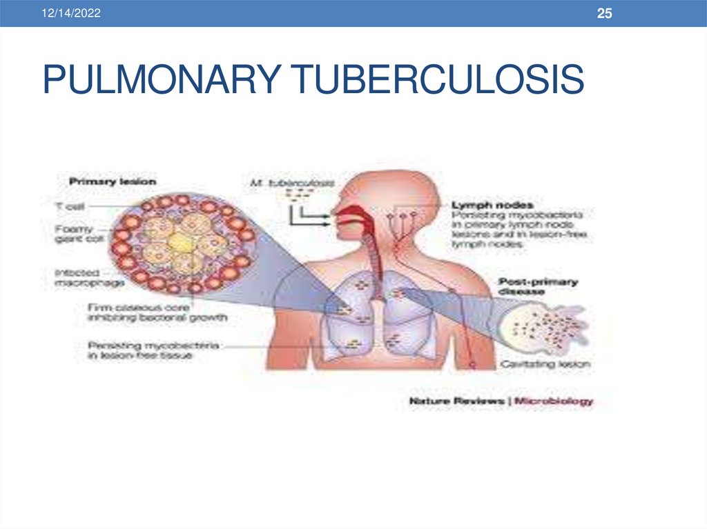
TUBERCULOSIS
• TB is an infectious disease caused by bacteria
(Mycobacterium tuberculosis) that are usually spread from
person to person through the air. It usually infects the lung
but can occur at virtually any site in the body. HIV-infected
patients are especially at risk. Drug-resistant TB is of
particular concern in certain parts of the United States.
27.
12/14/202227
Pathophysiology and Etiology
• Transmission
The term Mycobacterium is descriptive of the organism,
which is a bacterium that resembles a fungus. The
organisms multiply at varying rates and are characterized as
acid-fast aerobic organisms that can be killed by heat,
sunshine, drying, and ultraviolet light.
2. TB is an airborne disease transmitted by droplet nuclei,
usually from within the respiratory tract of an infected person
who exhales them during coughing, talking, sneezing, or
singing.
3. When an uninfected susceptible person inhales the dropletcontaining air, the organism is carried into the lung to the
pulmonary alveoli.
4. Most people who become infected do not develop clinical
illness, because the body's immune system brings the
infection under control.
1.
28.
12/14/202228
Clinical Manifestations
• Patient may be asymptomatic or may have insidious
symptoms that may be ignored.
• Constitutional symptoms
• Fatigue, anorexia, weight loss, low-grade fever, night sweats,
indigestion.
• Some patients have acute febrile illness, chills, and flu-like
symptoms.
• Pulmonary signs and symptoms
• Cough (insidious onset) progressing in frequency and producing
mucoid or mucopurulent sputum.
• Hemoptysis; chest pain; dyspnea (indicates extensive
involvement).
• Extrapulmonary TB: pain, inflammation, and dysfunction
in any of the tissues infected.
29.
12/14/202229
Diagnostic Evaluation
1. Sputum smear ”detection of acid-fast bacilli in stained
smears is the first bacteriologic clue of TB. Obtain first
morning sputum on 3 consecutive days.
2. Sputum culture ”a positive culture for M. tuberculosis
confirms a diagnosis of TB.
3. Chest X-ray to determine presence and extent of
disease.
4. Tuberculin skin test (purified protein derivative [PPD] or
Mantoux test) ”inoculation of tubercle bacillus extract
(tuberculin) into the intradermal layer of the inner aspect
of the forearm
30.
12/14/202230
Management
• Current recommended regimen of uncomplicated,
previously untreated pulmonary TB is an initial phase of 2
months of bactericidal drugs, including
1. isoniazid (INH),
2. rifampin (Rifadin),
3. pyrazinamide (PZA),
4. and ethambutol (EMB).
• This regimen should be followed until the results of drug
susceptibility studies are available, unless there is little
possibility of drug resistance.
31.
12/14/202231
Nursing Diagnoses
Ineffective Breathing Pattern related to pulmonary infection and
potential for long-term scarring with decreased lung capacity
Risk for Infection related to nature of the disease and patient's
symptoms
Imbalanced Nutrition: Less Than Body Requirements related to
poor appetite, fatigue, and productive cough
Noncompliance related to lack of motivation and long-term
treatment
32.
12/14/202232
Nursing Interventions
• Improving Breathing Pattern
• Administer and teach self-administration of medications as ordered.
• Encourage rest and avoidance of exertion.
• Monitor breath sounds, respiratory rate, sputum production, and
dyspnea.
• Provide supplemental oxygen as ordered.
• Preventing Transmission of Infection
• Be aware that TB is transmitted by respiratory droplets or secretions.
• Provide care for hospitalized patient in a negative-pressure room to
prevent respiratory droplets from escaping when door is opened.
• Enforce rule that all staff and visitors use well-fitted standard
dust/mist/fume masks (Class C) for contact with patient.
• Use high-efficiency particulate masks, such as HEPA filter masks, for
high-risk procedures, including suctioning, bronchoscopy,
• Use standard precautions for additional protection: gowns and gloves
for direct contact with patient, linens or articles in room, meticulous
hand washing.
• Educate the patient to control spread of infection through secretions.
33.
12/14/202233
Nursing Interventions
• Improving Nutritional Status
• Explain the importance of eating a nutritious diet to promote healing
and improve defense against infection.
• Provide small, frequent meals and liquid supplements during
symptomatic period.
• Monitor weight.
• Administer vitamin supplements, as ordered, particularly pyridoxine
(vitamin B6) to prevent peripheral neuropathy in patients taking
isoniazid.
• Improving Compliance
• Educate the patient about the etiology, transmission, and effects of
TB. Stress the importance of continuing to take medicine for the
prescribed time because bacilli multiply slowly and thus can only be
eradicated over a long period.
• Review adverse effects of the drug therapy (see Table 11-2). Question
the patient specifically about common toxicities of drugs being used,
and emphasize immediate reporting should these occur.
• Participate in observation of medication taking, weekly pill counts, or
other programs designed to increase compliance with treatment for
TB.
34.
12/14/202234
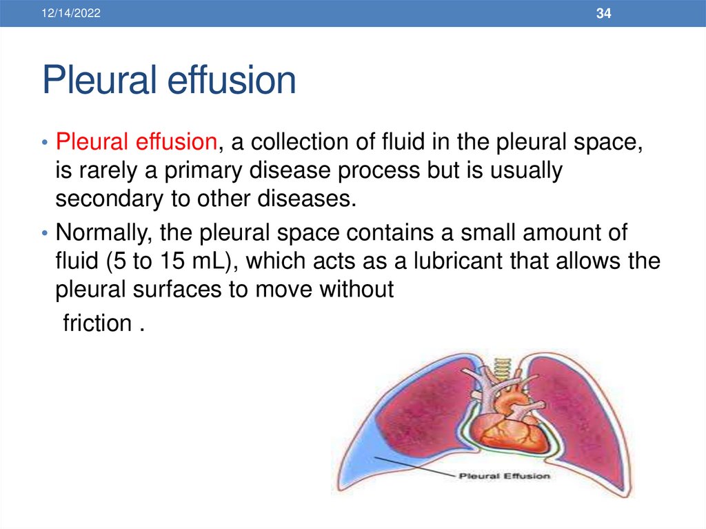
Pleural effusion
• Pleural effusion, a collection of fluid in the pleural space,
is rarely a primary disease process but is usually
secondary to other diseases.
• Normally, the pleural space contains a small amount of
fluid (5 to 15 mL), which acts as a lubricant that allows the
pleural surfaces to move without
friction .
35.
12/14/202235
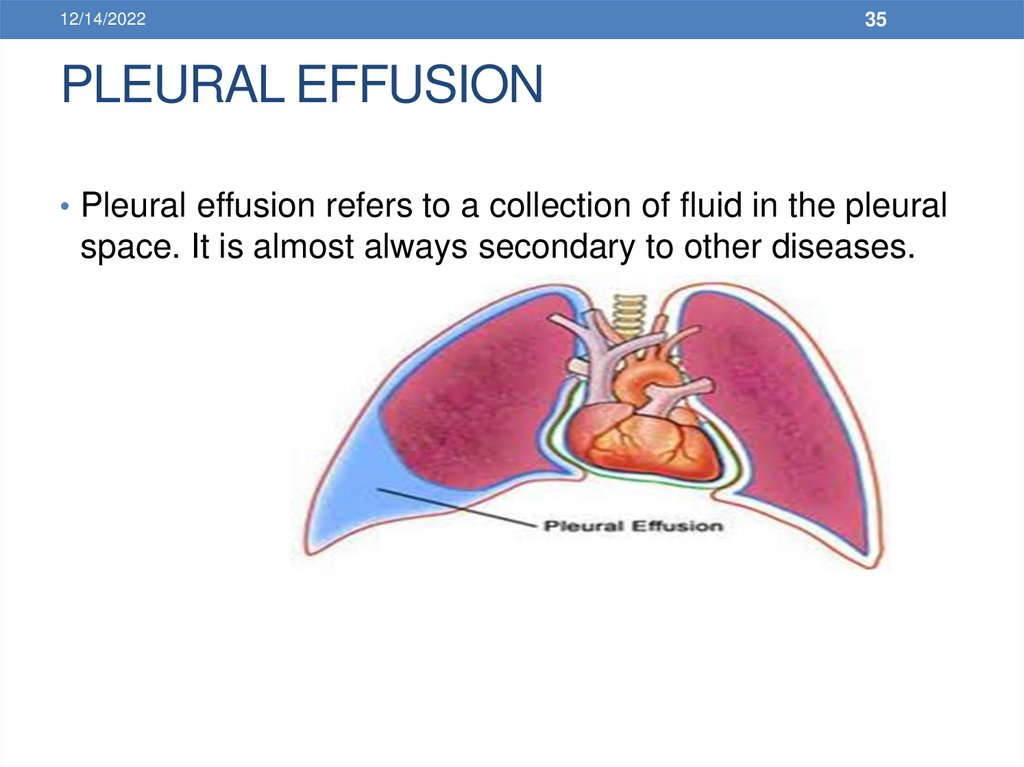
PLEURAL EFFUSION
• Pleural effusion refers to a collection of fluid in the pleural
space. It is almost always secondary to other diseases.
36.
12/14/202236
Pathophysiology and Etiology
• May be either transudative or exudative.
• Transudative effusions occur primarily in noninflammatory
conditions; is an accumulation of low-protein, low cell
count fluid.
• Exudative effusions occur in an area of inflammation; is
an accumulation of high-protein fluid.
• Occurs as a complication of:
• Disseminated cancer (particularly lung and breast), lymphoma.
• Pleuropulmonary infections (pneumonia).
• Heart failure, cirrhosis, nephrosis.
• Other conditions€”sarcoidosis, systemic lupus erythematosus
(SLE), peritoneal dialysis
37.
12/14/202237
• Clinical Manifestations
• Dyspnea, pleuritic chest pain, cough.
• Dullness or flatness to percussion (over areas of fluid)
with decreased or absent breath sounds
• Diagnostic Evaluation
• Chest X-ray or ultrasound detects presence of fluid.
• Thoracentesis €”biochemical, bacteriologic, and cytologic
studies of pleural fluid indicates cause.
• Management
• General
• Treatment is aimed at underlying cause (heart disease,
infection).
• Thoracentesis is done to remove fluid, collect a specimen,
and relieve dyspnea.
38.
12/14/202238
Nursing Diagnosis
• Ineffective Breathing Pattern related to collection of fluid in
pleural space
• Nursing Interventions
• Maintaining Normal Breathing Pattern
• Institute treatments to resolve the underlying cause as
ordered.
• Assist with thoracentesis if indicated
• Maintain chest drainage as needed .
39.
12/14/202239
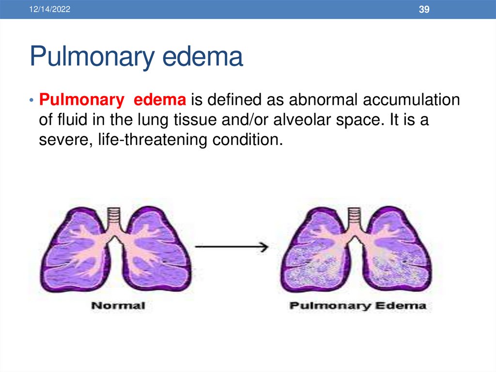
Pulmonary edema
• Pulmonary edema is defined as abnormal accumulation
of fluid in the lung tissue and/or alveolar space. It is a
severe, life-threatening condition.
40.
12/14/202240
Causes of pulmonary edema
• Inadequate left ventricular function
• Hypervolemia
• Sudden increase in the intravascular pressure in the lung.
41.
12/14/2022Clinical manifestations
• Respiratory distress, characterized by
dyspnea, and central cyanosis.
• The patient is very anxious and often agitated.
• The patient coughs up blood-tinged secretions.
41
42.
12/14/202242
Assessment and Diagnostic Findings
• Auscultation reveals crackles in the lung bases.
• Chest x-ray
• Pulse oximetry
• Arterial blood gas analysis
43.
12/14/2022Medical management
• Management focuses on correcting the underlying
disorder.
• Oxygen is administrated to correct hypoxemia
43
44.
12/14/202244
Nursing management
• Assisting with administration of oxygen and intubation and
mechanical ventilation if respiratory failure occurs.
• The nurse also administers medications as prescribed.
45.
12/14/202245
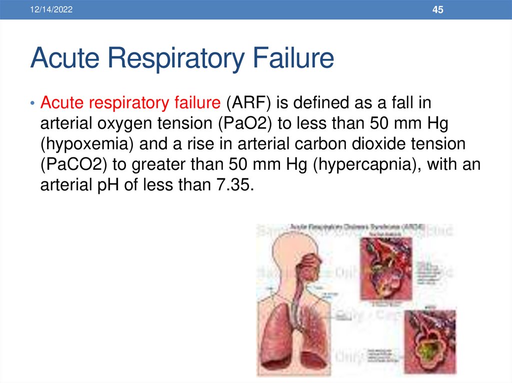
Acute Respiratory Failure
• Acute respiratory failure (ARF) is defined as a fall in
arterial oxygen tension (PaO2) to less than 50 mm Hg
(hypoxemia) and a rise in arterial carbon dioxide tension
(PaCO2) to greater than 50 mm Hg (hypercapnia), with an
arterial pH of less than 7.35.
46.
12/14/202246
RESPIRATORY FAILURE
• Respiratory failure is an alteration in the function of the
respiratory system that causes the partial pressure of
arterial oxygen (Pao2) to fall below 50 mm Hg
(hypoxemia) and/or the partial pressure of arterial carbon
dioxide (Paco2) to rise above 50 mm Hg (hypercapnia), as
determined by arterial blood gas (ABG) analysis.
Respiratory failure is classified as acute, chronic, or
combined acute and chronic.
47.
12/14/202247
Classification
• Acute Respiratory Failure
• Characterized by hypoxemia (Pao2 less than 50 mm Hg)
and/or hypercapnia (Paco2 greater than 50 mm Hg) and
acidemia (pH less than 7.35).
• Occurs rapidly, usually in minutes to hours or days.
• Chronic Respiratory Failure
• Characterized by hypoxemia (decreased Pao2) and/or
hypercapnia (increased Paco2) with a normal pH (7.35 to
7.45).
• Occurs over a period of months to years€”allows for
activation of compensatory mechanisms.
48.
12/14/202248
Acute and Chronic Respiratory Failure
• Characterized by an abrupt increase in the degree of
hypoxemia or hypercapnia in patients with preexisting
chronic respiratory failure.
• May occur after an acute upper respiratory infection or
pneumonia, or without obvious cause.
• Extent of deterioration is best assessed by comparing the
patient's present ABG levels with previous ABG levels.
49.
12/14/202249
Pathophysiology and Etiology
• Oxygenation Failure
• Characterized by a decrease in Pao2 and normal or
decreased Paco2.
• Primary problem is inability to adequately oxygenate the
blood, resulting in hypoxemia.
• Hypoxemia occurs because damage to the alveolarcapillary membrane causes leakage of fluid into the
interstitial space or into the alveoli and slows or prevents
movement of oxygen from the alveoli to the pulmonary
capillary blood.
• Hypocapnia results from hypoxemia and decreased
pulmonary compliance. Fluid within the lungs makes the
lung less compliant or stiffer.
50.
12/14/202250
Clinical Manifestations
1. Hypoxemia €”restlessness, agitation, dyspnea,
2.
3.
4.
5.
disorientation, confusion, delirium, loss of
consciousness.
Hypercapnia €”headache, somnolence, dizziness,
confusion.
Tachypnea initially; then when no longer able to
compensate, bradypnea
Accessory muscle use
Asynchronous respirations
51.
12/14/202251
NURSING ALERT
• Obtain ABG levels whenever the history or signs and
symptoms suggest the patient is at risk for developing
respiratory failure. Initial and subsequent values should
be recorded so comparisons can be made over time.
Need for ABG analysis can be decreased by using an
oximeter to continuously monitor oxygen saturation
(Sao2). Correlate oximeter values with ABG values and
then use oximeter for trending.
52.
12/14/202252
Diagnostic Evaluation
• ABG analysis €”show changes in Pao2, Paco2, and pH from patient's
normal; or Pao2 less than 50 mm Hg, Paco2 greater than 50 mm Hg,
pH less than 7.35.
• Pulse oximetry €”decreasing Sao2.
• End tidal CO2 monitoring €”elevated.
• Complete blood count, serum electrolytes, chest X-ray, urinalysis,
electrocardiogram (ECG), blood and sputum cultures €”to determine
underlying cause and patient's condition.
• Management
• Oxygen therapy to correct the hypoxemia.
• Chest physical therapy and hydration to mobilize secretions.
• Bronchodilators and possibly corticosteroids to reduce bronchospasm
and inflammation.
• Diuretics for pulmonary congestion.
• Mechanical ventilation as indicated. Noninvasive positive-pressure
ventilation using a face mask may be a successful option for shortterm support of ventilation
53.
12/14/202253
NURSING ALERT
• Avoid administration of oxygen at Fio2 of 100% for COPD
patients because you may depress the respiratory center
drive. For COPD patients, the drive to breathe may be
hypoxemia.
54.
12/14/202254
Nursing Diagnoses
• Impaired Gas Exchange related to inadequate respiratory
center activity or chest wall movement, airway
obstruction, and/or fluid in lungs
• Ineffective Airway Clearance related to increased or
tenacious secretions
55.
12/14/202255
• Nursing Interventions
• Improving Gas Exchange
• Administer antibiotics, cardiac medications, and diuretics as ordered
for underlying disorder.
• Administer oxygen to maintain Pao2 of 60 mm Hg or Sao2 > 90%
using devices that provide increased oxygen concentrations (aerosol
mask, partial rebreathing mask, nonrebreathing mask).
• Monitor fluid balance by intake and output measurement, urine
specific gravity, daily weight, and direct measurement of pulmonary
capillary wedge pressure to detect presence of hypovolemia or
hypervolemia.
• Provide measures to prevent atelectasis and promote chest
expansion and secretion clearance, as ordered (incentive spirometer,
nebulization, head of bed elevated 30 degrees, turn frequently, out of
bed).
• Monitor adequacy of alveolar ventilation by frequent measurement of
respiratory rate, VC, inspiratory force, and ABG levels.
• Compare monitored values with criteria indicating need for
mechanical ventilation (see section titled €Nursing Assessment €
).
Report and prepare to assist with noninvasive ventilation or intubation
and initiation of mechanical ventilation, if indicated.
56.
12/14/2022Pulmonary arterial hypertension
• Pulmonary hypertension exists when the systolic
pulmonary artery pressure exceeds 25 mm Hg.
• These pressures measured during right-sided heart
catheterization.
56
57.
12/14/202257
Pulmonary arterial hypertension
• In the absence of these measurements, clinical
recognition becomes the only indicator for the presence of
pulmonary hypertension.
• There are two forms of pulmonary hypertension: primary
(or idiopathic) and secondary.
58.
12/14/2022Causes of pulmonary arterial
hypertension
• Collagen vascular diseases
• Portal hypertension
• Altered immune mechanisms
• Chronic thrombotic or embolic disease
58
59.
12/14/2022Causes of pulmonary arterial
hypertension
• Pulmonary venous hypertension
• Pulmonary vasoconstriction due to hypoxemia
• Chronic obstructive pulmonary disease.
• Compression of pulmonary vessels
59
60.
12/14/202260
Clinical manifestations
• Dyspnea is the main symptom of pulmonary hypertension,
occurring at first with exertion and eventually at rest.
• Sub-sternal chest pain.
• Weakness, fatigue, syncope.
• Occasional hemoptysis, and signs of right-sided heart
failure (peripheral edema, ascites, distended neck veins,
liver engorgement)
61.
12/14/2022Assessment and diagnosis
• History
• Physical examination
• Chest x-ray
• Pulmonary function studies
• Electrocardiogram (ECG), echocardiogram
• cardiac catheterization.
61
62.
12/14/202262
Medical management
• Management of the underlying cardiac or pulmonary
condition:
• Anticoagulation
• Diuretics and oxygen
• Vasodilators as calcium channel blockers
63.
12/14/202263
Nursing management
• The nurse also must be alert for signs and symptoms of
pulmonary hypertension
• Administer oxygen therapy appropriately, and instruct
patients and their families about the use of home oxygen
supplementation.
64.
12/14/202264
Pulmonary embolism
• Pulmonary embolism (PE) refers to the obstruction of
the pulmonary artery or one of its branches by a thrombus
(or thrombi) that originates somewhere in the venous
system or in the right side of the heart.
65.
12/14/202265
PULMONARY EMBOLISM
• Pulmonary embolism refers to the obstruction of one or
more pulmonary arteries by a thrombus (or thrombi)
originating usually in the deep veins of the legs, the right
side of the heart or, rarely, an upper extremity, which
becomes dislodged and is carried to the pulmonary
vasculature.
• Pulmonary infarction refers to necrosis of lung tissue that
can result from interference with blood supply.
66.
12/14/202266
Pathophysiology and Etiology
• Obstruction, either partial or full, of pulmonary arteries, which
causes decrease or absent blood flow; therefore, there is
ventilation but no perfusion ([V with dot above]/[Q with dot
above] mismatch).
• Hemodynamic consequences:
Increased pulmonary vascular resistance
Increased pulmonary artery pressure (PAP)
Increased right ventricular workload to maintain pulmonary blood
flow
Right ventricular failure
Decreased cardiac output
Decreased blood pressure
Shock
• Pulmonary emboli can vary in size and seriousness of
consequences
67.
12/14/202267
Predisposing factors include:
• Stasis, prolonged immobilization.
• Concurrent phlebitis.
• Previous heart (heart failure, myocardial infarction [MI]) or lung
disease.
Injury to vessel wall.
Coagulation disorders.
Metabolic, endocrine, vascular, or collagen disorders.
Malignancy.
Advancing age, estrogen therapy.
68.
12/14/202268
NURSING ALERT
• Be aware of high-risk patients for pulmonary
embolism€”immobilization, trauma to pelvis (especially
surgical) and lower extremities (especially hip fracture),
obesity, history of thromboembolic disease, varicose
veins, pregnancy, heart failure, MI, malignant disease,
postoperative patients, elderly patients.
69.
12/14/202269
Clinical Manifestations
• Dyspnea, pleuritic pain, tachypnea, apprehension.
• Chest pain with apprehension and a sense of impending
doom occurs when most of the pulmonary artery is
obstructed.
• Cyanosis, tachyarrhythmia's, syncope, circulatory
collapse and, possibly, death encountered in patients with
massive pulmonary embolism
• Subtle deterioration in patient's condition with no
explainable cause
• Pleural friction rub
70.
12/14/202270
Diagnostic Evaluation
• ABG levels €”decreased Pao2 is usually found, due to
perfusion abnormality of the lung.
• Chest X-ray €”normal or possible wedge-shaped infiltrate.
• [V with dot above]/[Q with dot above] lung scans
€”perfusion scan investigates regional blood flow to
determine presence of perfusion defects; ventilation scan
may be done in patient with large perfusion defects.
• Pulmonary angiogram (most definitive)
71.
12/14/202271
Emergency Management
1.
2.
3.
4.
5.
6.
Oxygen is administered to relieve hypoxemia, respiratory
distress, and cyanosis.
An infusion is started to open an I.V. route for drugs and
fluids.
Vasopressors, inotropic agents such as dopamine (Intropin),
and antidysrhythmic agents may be indicated to support
circulation if the patient is unstable.
ECG is monitored continuously for right-sided heart failure,
which may have a rapid onset.
Small doses of I.V. morphine are given to relieve anxiety,
alleviate chest discomfort (which improves ventilation), and
ease adaptation to mechanical ventilator, if this is necessary.
Pulmonary angiography, hemodynamic measurements, ABG
analysis, and other studies are carried out.
72.
12/14/202272
• Subsequent Management €”Anticoagulation and
Thrombolysis
1. I.V. heparin €”stops further thrombus formation and
extends the clotting time of the blood; it is an
anticoagulant and antithrombotic.
2. Oral anticoagulation with warfarin (Coumadin) is usually
used for follow-up anticoagulant therapy after heparin
therapy has been established; interrupts the coagulation
mechanism by interfering with the vitamin K-dependent
synthesis of prothrombin and factors VII, IX, and X.
3. Thrombolytic agents, such as streptokinase (Streptase),
may be used in patients with massive pulmonary
embolism.
73.
12/14/202273
Nursing Diagnoses
Ineffective Breathing Pattern related to acute increase in alveolar dead
airspace and possible changes in lung mechanics from embolism
Ineffective Tissue Perfusion (Pulmonary) related to decreased blood
circulation
Acute Pain (pleuritic) related to congestion, possible pleural effusion,
possible lung infarction
Anxiety related to dyspnea, pain, and seriousness of condition
Risk for Injury related to altered hemodynamic factors and
anticoagulant therapy
74.
12/14/202274
Nursing Interventions
• Correcting Breathing Pattern
• Assess for hypoxia, headache, restlessness,
apprehension, pallor, cyanosis, behavioral changes.
• Monitor vital signs, ECG, oximetry, and ABG levels for
adequacy of oxygenation.
• Monitor patient's response to I.V. fluids/vasopressors.
• Monitor oxygen therapy€”used to relieve hypoxemia.
• Prepare patient for assisted ventilation when hypoxemia
is due to local areas of pneumoconstriction and
abnormalities of [V with dot above]/[Q with dot above]
ratios.
75.
12/14/202275
• Improving Tissue Perfusion
• Closely monitor for shock €”decreasing blood pressure,
tachycardia, cool, clammy skin.
• Monitor prescribed medications given to preserve right
ventricular filling pressure and increase blood pressure.
• Maintain patient on bed rest to reduce oxygen demands and
risk of bleeding.
• Monitor urinary output hourly, because there may be reduced
renal perfusion and decreased glomerular filtration.
• Relieving Pain
• Watch patient for signs of discomfort and pain.
• Ascertain if pain worsens with deep breathing and coughing;
auscultate for friction rub.
• Give prescribed morphine (Duramorph), and monitor for pain
relief and signs of respiratory depression.
• Position with head of bed slightly elevated (unless
contraindicated by shock) and with chest splinted for deep
breathing and coughing.
76.
12/14/202276
• Patient Education and Health Maintenance
• Advise patient of the possible need to continue taking
anticoagulant therapy for 6 weeks up to an indefinite period.
• Teach about signs of bleeding, especially of gums, nose,
bruising, blood in urine and stools.
• For patients on anticoagulants, instruct to use soft toothbrush,
avoid shaving with blade razor (use electric razor instead), and
avoid aspirin-containing products. Notify health care provider of
bleeding or increased bruising.
• Warn against taking medications unless approved by health
care provider, because many drugs interact with
anticoagulants.
• Instruct patient to tell dentist about taking an anticoagulant.
• Warn against inactivity for prolonged periods or sitting with legs
crossed to prevent recurrence.
• Warn against sports/activities that may cause injury to legs and
predispose to a thrombus.
• Encourage wearing a MedicAlert bracelet identifying patient as
anticoagulant user.
77.
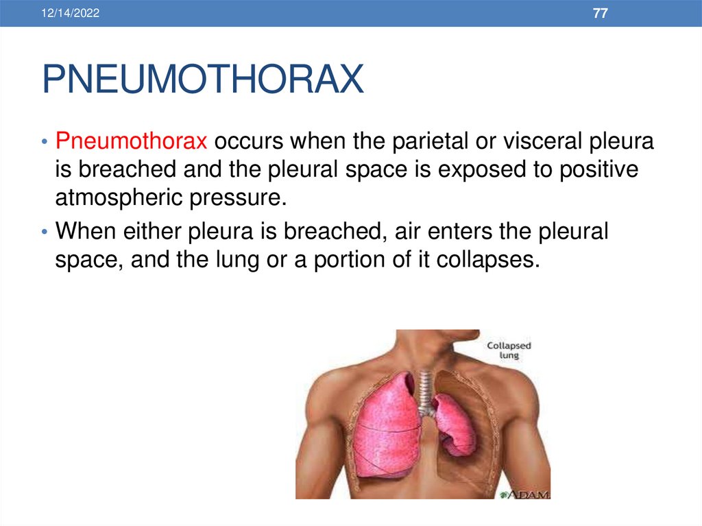
12/14/202277
PNEUMOTHORAX
• Pneumothorax occurs when the parietal or visceral pleura
is breached and the pleural space is exposed to positive
atmospheric pressure.
• When either pleura is breached, air enters the pleural
space, and the lung or a portion of it collapses.
78.
12/14/202278
TRAUMATIC DISORDERS
PNEUMOTHORAX
• Air in the pleural space occurring spontaneously or from
trauma (see Figure 11-4). In patients with chest trauma, it
is usually the result of a laceration to the lung
parenchyma, tracheobronchial tree, or esophagus. The
patient's clinical status depends on the rate of air leakage
and size of wound.
79.
12/14/202279
• Spontaneous pneumothorax €”sudden onset of air in
the pleural space with deflation of the affected lung in the
absence of trauma.
• Open pneumothorax (sucking wound of chest) €”implies
an opening in the chest wall large enough to allow air to
pass freely in and out of thoracic cavity with each
attempted respiration.
• Tension pneumothorax €”buildup of air under pressure
in the pleural space resulting in interference with filling of
both the heart and lungs.
80.
12/14/202280
Pathophysiology and Etiology
• When there is a large open hole in the chest wall.
• A portion of the tidal volume will move back and forth
through the hole in the chest wall, rather than the trachea
as it normally does.
81.
12/14/202281
Clinical Manifestations
1. Hyperresonance; diminished breath sounds.
2. Reduced mobility of affected half of thorax.
3. Tracheal deviation away from affected side in tension
pneumothorax
4. Clinical picture of open or tension pneumothorax is one
of air hunger, agitation, hypotension, and cyanosis
5. Mild to moderate dyspnea and chest discomfort may be
present with spontaneous pneumothorax
82.
12/14/202282
Diagnostic Evaluation
• Chest X-ray confirms presence of air in pleural space.
• Management
• Spontaneous Pneumothorax
• Treatment is generally nonoperative if pneumothorax is
not too extensive.
• Observe and allow for spontaneous resolution for less than 50%
pneumothorax in otherwise healthy person.
• Needle aspiration or chest tube drainage may be necessary to
achieve reexpansion of collapsed lung if greater than 50%
pneumothorax.
83.
12/14/202283
• Tension Pneumothorax
• Immediate decompression to prevent cardiovascular collapse
by thoracentesis or chest tube insertion to let air escape.
• Chest tube drainage with underwater-seal suction to allow for
full lung expansion and healing
• Open Pneumothorax
• Close the chest wound immediately to restore adequate
ventilation and respiration.
Patient is instructed to inhale and exhale gently against a closed
glottis (Valsalva maneuver) as a pressure dressing (petroleum
gauze secured with elastic adhesive) is applied. This maneuver
helps to expand collapsed lung.
• Chest tube is inserted and water-seal drainage set up to permit
evacuation of fluid/air and produce reexpansion of the lung.
• Surgical intervention may be necessary to repair trauma.
84.
12/14/202284
Nursing Diagnoses
• Ineffective Breathing Pattern related to air in the pleural space
• Impaired Gas Exchange related to atelectasis and collapse of
lung
• Nursing Interventions
• Achieving Effective Breathing Pattern
• Provide emergency care as indicated.
Apply petroleum gauze to sucking chest wound
• Assist with emergency thoracentesis or thoracotomy.
• Be prepared to perform cardiopulmonary resuscitation or administer
medications if cardiovascular collapse occurs.
• Maintain patent airway; suction as needed.
• Position patient upright if condition permits to allow greater
chest expansion.
85.
12/14/202285
Resolving Impaired Gas Exchange
• Encourage patient in the use of incentive spirometer.
• Monitor oximetry and ABG levels to determine
oxygenation.
• Provide oxygen as needed.
• Patient Education and Health Maintenance
• Instruct patient to continue use of the incentive spirometer
at home.
• For patients with spontaneous pneumothorax, there is an
increased risk for repeat occurrence; therefore,
encourage these patients to report sudden dyspnea
immediately.
86.
12/14/202286
• Hemothorax
• Blood in pleural space as a result of penetrating or blunt
chest trauma.
• Accompanies a high percentage of chest injuries.
• Can result in hidden blood loss
• Patient may be asymptomatic, dyspneic, apprehensive, or
in shock
87.
12/14/202287
Hemothorax
• Assist with thoracentesis to aspirate blood from pleural
space, if being done before a chest tube insertion.
• Assist with chest tube insertion and set up drainage
system for complete and continuous removal of blood and
air.
• Auscultate lungs and monitor for relief of dyspnea.
• Monitor amount of blood loss in drainage.
• Replace volume with I.V. fluids or blood products.