


 medicine
Similar presentations:


Development of the modern otorhinolaryngology, achievements of modern otorhinolaryngology
1. Development of the modern otorhinolaryngology, achievements of modern otorhinolaryngology. Clinical anatomy, physiology and methods of examination of the nose and paranasal sinuses. Acute and chronic rhinitis. Acute sinusitis..
Head of OtolaryngologyDepartment, professor
V.I. Troyan
1
2.
Because of great success of the modernotorhinolaryngology, of expansion and deepening of its
knowledge, it has developed so much that the following
parts have separated from it:
Phoniatria –the science of physiology and pathology
of the speech apparatus.
Surdology, that is the diagnostics and treatment of
hard hearing persons and deaf-and -dumbs.
Otoneurology – doctors- otorhinolaryngologists
working in neurosurgical departments and are engaged
in a diagnostics of the diseases of VIII pair of skullcerebral nerves.
ENT-prophpathology
Phthisiolaryngology.
ENT-diseases of childhood.
ENT-oncology.
2
3.
.In our clinic a wide volume of medical aid is given to the
patients with a various purulent pathology of ENT, ENToncology patients. The plastic and sanative operations on
middle ear, nasal cavity, paranasal sinuses, throat, and larynx
are performed.
Those students, who do not dream to be
otorhinolaryngologist, are to study the course of ear, throat
and nose diseases on the lectures and practical lessons.
Whoever you will be in the future - therapeustist, surgeon,
neuropathologist, dermatologist or other physician, you will
everyday meet ENT-diseases. Opportune revealing and
treatment of ENT-disease can avoid or improve the proceeding
of such disease as rheumatism, nephritis, kidney diseases,
diseases of liver, gall-bladder, lungs, central or peripheral
nervous system and so on.
3
4. Clinical anatomy, physiology and methods of investigation.
nose (nasusexternus). The skeleton of
the external nose is formed
by bones and cartilages.
The bony part of the nose is
formed by paired nasal
bones and by the frontal
processed of the maxilla.
The cartilaginous framework
of
the
nose
includes
triangular cartilage, paired
ala cartilage, and the
accessory cartilage.
External
4
5.
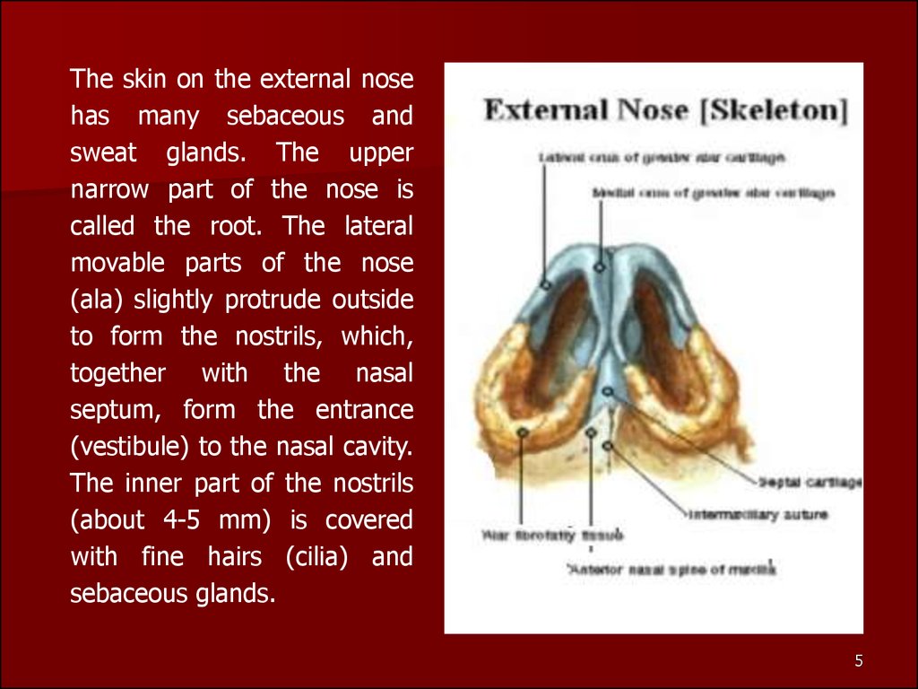
The skin on the external nosehas many sebaceous and
sweat glands. The upper
narrow part of the nose is
called the root. The lateral
movable parts of the nose
(ala) slightly protrude outside
to form the nostrils, which,
together with the nasal
septum, form the entrance
(vestibule) to the nasal cavity.
The inner part of the nostrils
(about 4-5 mm) is covered
with fine hairs (cilia) and
sebaceous glands.
5
6. External nose
The external nose is suppliedwith blood via branches of the
ophthalmic artery. The blood
outflows through the anterior facial
and angular veins into the superior
ophthalmic
vein
which
communicates with the cavernous
sinus. The external nose is
innervated by the fifth and seventh
pairs of the cranial nerves.
6
7.
Nasal cavity (cavum nasi). The nasal cavity is divided by theseptum into the right and left parts. The anterior part of the nasal
cavity opens with a piriform sinus (anteriorly) and choanae (posteriori).
The nasal cavity has four walls, namely, the superior, inferior, internal,
and external walls. The inferior wall (the floor) of the nasal cavity is the
hard (bony) palate. The superior wall (the roof) of the nasal cavity
includes the bones of the nose anteriorly, the cribriform plate of the
ethmoid bone in the middle (the greater part of the roof) and the
anterior wall of the sphenoidal sinus. The fibbers of the olfactory nerve
and the branches of the ethmoidal artery and the veins pass through
the perforations of the cribriform plate.
7
8.
The medial (internal) wall, or the septum, consists of the anteriorcartilaginous and posterior bony parts. The bony part of the septum is
formed by the perpendicular plate of the ethmoid and the vomer.
8
9.
The lateral (external) wall of the nasal cavity has a more complexstructure. Three nasal conch extend from the external wall toward the
nasal septum: the superior, middle and inferior conch. Three nasal
meatuses are distinguished accordingly: the superior, middle, and inferior
meatuses.
9
10.
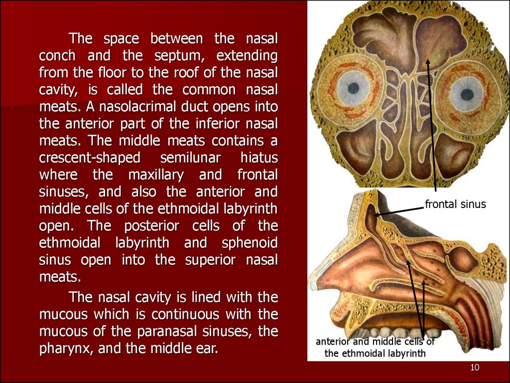
The space between the nasalconch and the septum, extending
from the floor to the roof of the nasal
cavity, is called the common nasal
meats. A nasolacrimal duct opens into
the anterior part of the inferior nasal
meats. The middle meats contains a
crescent-shaped semilunar hiatus
where the maxillary and frontal
sinuses, and also the anterior and
middle cells of the ethmoidal labyrinth
open. The posterior cells of the
ethmoidal labyrinth and sphenoid
sinus open into the superior nasal
meats.
The nasal cavity is lined with the
mucous which is continuous with the
mucous of the paranasal sinuses, the
pharynx, and the middle ear.
frontal sinus
anterior and middle cells of
the ethmoidal labyrinth
10
11.
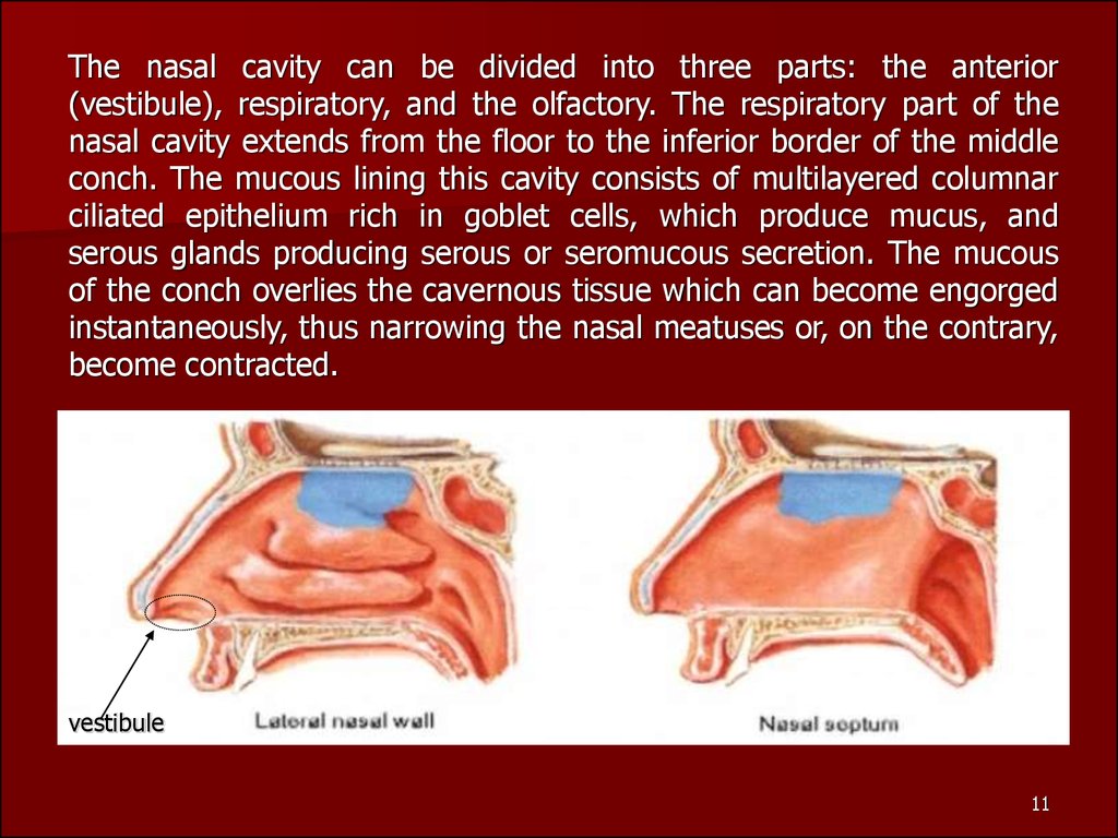
The nasal cavity can be divided into three parts: the anterior(vestibule), respiratory, and the olfactory. The respiratory part of the
nasal cavity extends from the floor to the inferior border of the middle
conch. The mucous lining this cavity consists of multilayered columnar
ciliated epithelium rich in goblet cells, which produce mucus, and
serous glands producing serous or seromucous secretion. The mucous
of the conch overlies the cavernous tissue which can become engorged
instantaneously, thus narrowing the nasal meatuses or, on the contrary,
become contracted.
vestibule
11
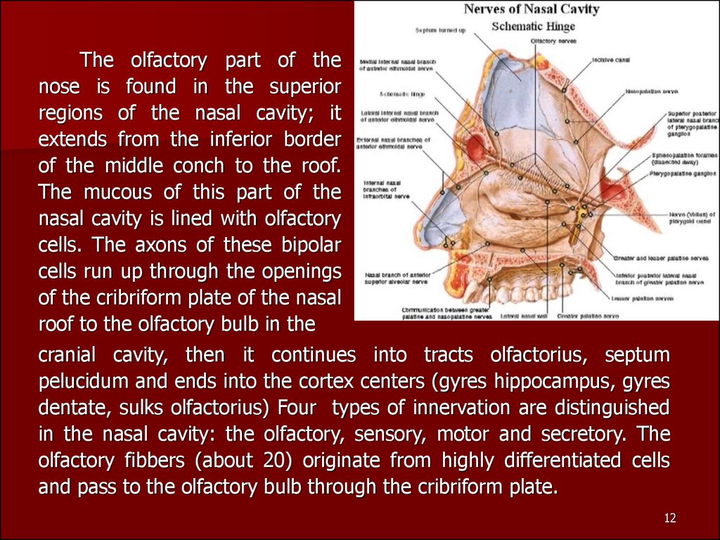
12.
The olfactory part of thenose is found in the superior
regions of the nasal cavity; it
extends from the inferior border
of the middle conch to the roof.
The mucous of this part of the
nasal cavity is lined with olfactory
cells. The axons of these bipolar
cells run up through the openings
of the cribriform plate of the nasal
roof to the olfactory bulb in the
cranial cavity, then it continues into tracts olfactorius, septum
pelucidum and ends into the cortex centers (gyres hippocampus, gyres
dentate, sulks olfactorius) Four types of innervation are distinguished
in the nasal cavity: the olfactory, sensory, motor and secretory. The
olfactory fibbers (about 20) originate from highly differentiated cells
and pass to the olfactory bulb through the cribriform plate.
12
13.
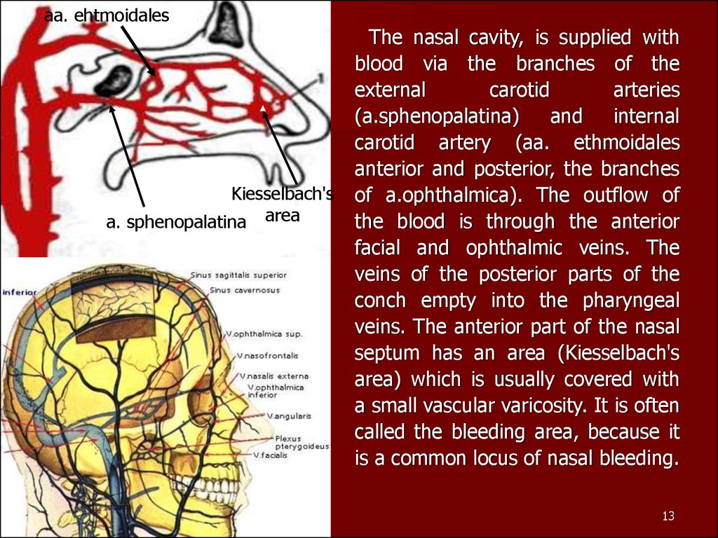
aa. ehtmoidalesKiesselbach's
a. sphenopalatina area
The nasal cavity, is supplied with
blood via the branches of the
external
carotid
arteries
(a.sphenopalatina)
and
internal
carotid artery (aa. ethmoidales
anterior and posterior, the branches
of a.ophthalmica). The outflow of
the blood is through the anterior
facial and ophthalmic veins. The
veins of the posterior parts of the
conch empty into the pharyngeal
veins. The anterior part of the nasal
septum has an area (Kiesselbach's
area) which is usually covered with
a small vascular varicosity. It is often
called the bleeding area, because it
is a common locus of nasal bleeding.
13
14.
The sensory innervation of the nasalcavity is accomplished by the first
and second branches of the
trigeminal
nerve.
The
motor
innervation of the external nose is
accomplished by facial nerve. The
secretory innervation of the nasal
cavity is represented by the
sympathetic nervous system. The
fibbers of the sympathetic nerve
pass from the pterygopalatine
ganglion.
They
serve
to
communicate with the sympathetic
nerves of the thoracic, abdominal,
and endocrine organs. All this
establishes
reflex
connection
between the nasal cavity and other
organs and systems.
14
15.
Paranasal sinuses.The paranasal sinuses are
located by sides of the nasal
cavity and communicate with it.
There are four paired air cavities,
namely, the maxillary, cells of the
ethmoidal labyrinth, frontal, and
sphenoid.
15
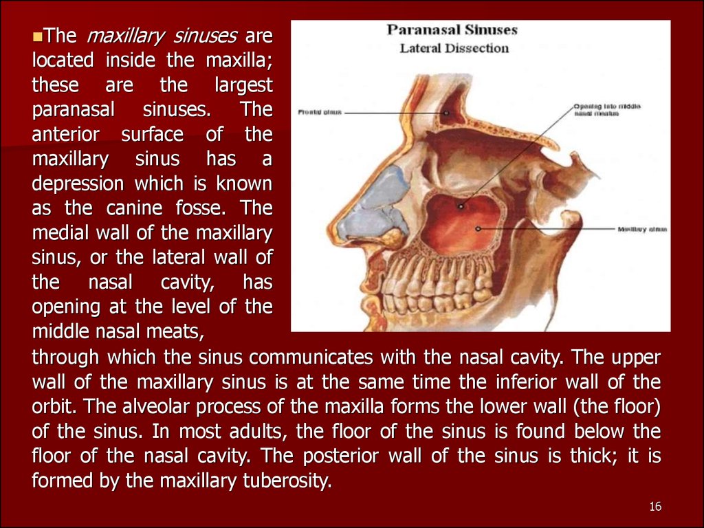
16.
Themaxillary sinuses are
located inside the maxilla;
these are the largest
paranasal sinuses. The
anterior surface of the
maxillary sinus has a
depression which is known
as the canine fosse. The
medial wall of the maxillary
sinus, or the lateral wall of
the nasal cavity, has
opening at the level of the
middle nasal meats,
through which the sinus communicates with the nasal cavity. The upper
wall of the maxillary sinus is at the same time the inferior wall of the
orbit. The alveolar process of the maxilla forms the lower wall (the floor)
of the sinus. In most adults, the floor of the sinus is found below the
floor of the nasal cavity. The posterior wall of the sinus is thick; it is
formed by the maxillary tuberosity.
16
17.
The ethmoidal sinuses (ethmoidal labyrinth) consist of air cells ofthe ethmoid which is located between the frontal and the sphenoid
sinuses. Anterior, middle, and posterior cells of the labyrinth are
distinguished (6-7 cells of each type on either side). In healthy man the
cells are filled with air.
17
18.
The frontal sinuses are found in the squama ofthe frontal bone. Each sinus has four walls: the
anterior (facial); the posterior, which borders with
the cranial fosse; the inferior, which in most
cases is the superior wall of the orbit and borders
with the cells of the ethmoid and the nasal cavity
over a small area; and the internal wall (the
septum).
The sphenoid sinuses are found in the body of
the sphenoid bone. The septum separating the
sinuses extends anteriorly to the nasal septum.
The roof is formed by the bone underlying the
optic chiasm, the clinoid processes, and the cella
turcica with the pituitary gland. The posterior
wall is formed by the solid bone of the
basissphenoid. The lateral wall is in relation to
the optic foramen and nerve, the cavernous sinus
and the internal carotid artery. The floor is the
roof of the nasopharynx. In the anterior wall is
the natural orifice which opens into superior
nasal meats.
18
19.
CLINICAL PHYSIOLOGYNasal respiration is very important because, in addition
to the respiratory function, the nose also performs the
protective, resonating, and olfactory functions.
The respiratory function of the nose is part of the
entire respiratory function in man. During inspiration, which
is due to creation of negative pressure in the chest, air
enters both parts of the nasal cavity mostly through the
respiratory part of the nose. The inspired air passes
upwards and then descends by the superior and middle
meatuses and passes posteriori to the choanae.
19
20.
The protective function of the nose consists in warmingthe inspired air, its moistening and filtering. Cold air
stimulates a rapid expansion of the cavernous sinuses and
their filling with blood. The inspired air is moistened by the
wet mucous. As the air passes through the vestibule of the
nose, large dust particles are retained by thick hairs. Fine
dust and airborne microbes, that pass first filter, are
precipitated on the nasal mucous moistened with mucous
secretion. Dust is also retained because the nasal passages
are narrow and curved. About 40-60 per cent of dust
particles and microbes inspired with air are retained in the
nose and then removed from it with mucus. This function is
performed by ciliated epithelium. Lysozyme, contained in
the nasal mucus and secretion of the lachrymal glands, has
a marked disinfecting property. The sneezing and lachrymal
reflexes are also important protective mechanisms.
20
21.
The olfactory function in man is provided by theolfactory mucous that contains the neuro-epithelial fusiform
olfactory cells, which are chemoreceptors. The molecules of
gases, vapor, mist, dust, or smoke stimulate the olfactory
receptors. It should be noted that man can also perceive
odor of some substances (e. g. spirit of ammonia that act
on the endings of the trigeminal nerve).
The resonating function of the nose accounts for the
special timbre of the human voice. Pathological changes in
the nasal cavity or in the nasopharynx (polyps, hypertrophy
of the conchae, inflammation of the nasal mucous, tumor,
adenoids, and other changes) cause rhinolalia clause (nasal
speech). If the nasal cavity has unusually large
communication with the nasopharynx (e.g. due to the
absence of the soft palate or its paralysis), the patient
develops rhinolalia aperta.
21
22.
METHODS OF EXAMINATIONThe external nose should be palpated. Palpation should
also be used to examine the anterior and inferior walls of
the frontal sinuses, the anterior walls of the maxillary
sinuses, and also the cervical regional lymph nodes.
The respiratory function of the nose should be
examined separately on each side. To that end, the wing of
the one nostril is pressed to the nasal and the patient is
asked to breathe air quietly in and out; a small piece of
cotton wool held close to that will show if the passage is
free. A special rhinopneumometer is used for a more
accurate assessment of the nasal breathing function.
The olfactory function of each side of the nose is tested
separately using odoriferous substances from a special
olfactometric set, or using a special instrument called
olfactometer. Olfaction can be normal (normosmia),
decreased (hyposmia), perverted (cacosmia), or it can be
absent (anosmia).
22
23.
Rhinoscopy can be anterior, middle,and posterior. Anterior rhinoscopy should
be carried out on both sides of the
nose.The normal color of the nasal
mucous is pink; its surface is smooth; the
normal position of the septum is central.
The other side of the nose should be
examined in a similar way.
Inspection of the posterior parts of
the nose is called posterior rhinoscopy
(epipharyngoscopy). The posterior parts
of the nasal cavity are inspected by
slightly turning the speculum to the
required side. The posterior ends of the
nasal conchae, the nasal meatuses, and
the vomer can thus be inspected. The
nasopharynx can be examined in a similar
way.
23
24.
Examination of the paranasal sinuses.Roentgenograph
y and clinical analyse of
rentgenological signs is
one
of
the
main
methods
of
investigation of PNS.
The
next special
projections are used for
the best observation of
sinuses:
naso-frontal,
naso-mental,
mento-parietal, lateral and semi-axial ones. Every type of pathology is
characterized by the certain structural shadings, changes of bone walls.
The typical signs of the inflammatory diseases
are: near-wall
thickening of mucous membrane, liquid level by the exudative forms,
"spotty" shading by polyposis. Osteo-destructive changes of
the
walls, dilation of sinuses, the presence of tissues of high intensity
with the clear contours are character for the volume formations
(tumours, cysts).
Layer investigation - tomography in the certain
depth, contrast investigation by jodolipol of the injuried sinus are used
to specify the pathological process.
24
25.
Compjuter X-ray tomography (CT).By CT the picture is got not in the X-ray
film but is synthetized with the help of
electronic compjuter (EC). X-rays, coming
from the tube in differents directions
(the set of irradiation is turned around the
patient), are perceived by semi-conducting
detectors, where the
quanta
make
flashes.. CT lets to see bones and soft
tissues of paranasal sinuses and nasal cavity
in the same time and measure their X-ray
density. So, with the help of CT we can
carry
out
differential
diagnose
of
inflammatory processes and tumours of
PNS, determine the presence
of osteodestructive changes.
25
26.
Nucleo-magneticresonance.
Diagnostic picture, got by magnetoresonance
tomography (MRT) (such
investigation is called MR-tomography).
MRT-investigation lets to carry
out
differentiation between
inflammatory
processes and tumours, determine their
localization, dimensions and spread,
contours, invasion of the neighbour
anatomical structures.
26
27.
Diseases of the noseAcute catarrhal rhinitis (common cold) is an acute non-specific
inflammation of the nasal mucosa. The aetiology of acute rhinitis is
determined by decreased local or general reactivity of the body and
activation of microflora of the nose. The disease usually occurs
following general or local chilling that interferes with the protective
nervous and reflex mechanisms.
The clinic of acute catarrhal rhinitis includes three stages, which
are continuous with one another: the first stage is dry irritation, the
second stage is characterized by increased mucous secretion, and the
third stage (resolution) is characterized by mucopurulent secretion.
Acute rhinitis begins with the feeling of dryness, tension, burning, and
itching in the nose and often in the pharynx and the larynx; sneezing is
annoying. The patient complains of indisposition, chill, discomfort and
headache (mostly pain in the forehead). The body temperature is
elevated. Nasal respiration becomes difficult-from insignificant
impediment to a complete obstruction due to obturation of the nasal
meatuses with swollen mucosa. Olfaction is impaired significantly.
27
28.
The sense of taste is also altered. The speech becomes nasal(rhinolalia clausa). Profuse watery discharge from the nose is
characteristic of the first day of acute rhinitis. The amount of mucus in
the discharge increases later. This can cause hyperemia and swelling of
the skin at the nose vestibule and of the upper lip. The nasal discharge
becomes seropurulent in 4 or 5 days. The amount of nasal discharge
decreases gradually during the next few days, swelling of the mucosa
subsides, respiration through the nose and olfaction are restored, and
the patient recovers in 8-14 days from the onset of acute catarrhal
rhinitis.
The treatment schem acute rhinitis of virus aetiology:
1. Laferon 100 000 ME
2. acetyisalicylic acid
3. Gelasoline
4. Ksisal,loratadin
5. aqua maris
28
29.
Chronic catarrhal rhinitis. Theonset of chronic rhinitis is connected
as a rule with frequently recurring
acute inflammation in the nasal cavity
(including inflammations associated
with various infections), irritating
environmental effects such as dust,
gas, dry or moist air, variations in
ambient temperature, etc.
The main symptoms of chronic
catarrhal
rhinitis
are
impeded
respiration through the nose and
rhinorrhoea;
both
signs
are
manifested moderately. Respiration
through the nose becomes periodically
difficult, mostly due to chilling. The
passageway through one side of the
nose
is
usually
obstructed
permanently. Nasal respiration is even
more difficult when the patient lies on
his side
29
30.
Chronichypertrophic
rhinitis. The main signs of
hypertrophic
rhinitis
are
impeded respiration through the
nose, mucous nasal discharge,
and thickened and swollen nasal
mucosa, mainly in the entire
inferior and middle concha. The
mucosa is usually red-blue, grayblue and covered with mucus. In
the presence of mucopurulent
discharge, inflammation of the
paranasal sinuses should be
excluded. The posterior ends of
the inferior conchae are usually
thickened;
application
of
vasoconstrictor
drops
don’t
causes the reduction of nasal
concha.
30
31.
Chronic atrophic rhinitis.Common chronic atrophic rhinitis
can be diffuse or circumscribed.
Mineral dust (silicates, cement) and
that of tobacco produce a strong
effect on the condition of the nose.
Common symptoms of the disease
are crusts in the nose. Meagre
tenacious mucus (or mucopurulent
discharge) adheres to the mucosa
and dries into crusts. The patient
complains of dryness in the nose
and the pharynx, and impairment
of olfaction. Separation of the
crusts often causes nosebleed,
usually from the Kiesselbach area.
31
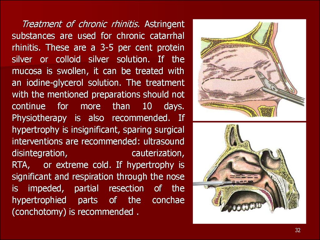
32.
Treatment of chronic rhinitis. Astringentsubstances are used for chronic catarrhal
rhinitis. These are a 3-5 per cent protein
silver or colloid silver solution. If the
mucosa is swollen, it can be treated with
an iodine-glycerol solution. The treatment
with the mentioned preparations should not
continue for more than 10 days.
Physiotherapy is also recommended. If
hypertrophy is insignificant, sparing surgical
interventions are recommended: ultrasound
disintegration,
cauterization,
RTA,
or extreme cold. If hypertrophy is
significant and respiration through the nose
is impeded, partial resection of the
hypertrophied parts of the conchae
(conchotomy) is recommended .
32
33.
Treatment of atrophic rhinitis. The patient should take care of hisnose so that crusts and nasal discharge should not accumulate in the
nasal cavity. The nose should be cleaned once or twice a day by
irrigating the nasal cavity with isotonic
solution (Аква
марис,ринофлуимуцил
containing
an
additive
биопарокс спрей). Irritants should periodically be used: а day in the
course of 10 days, this stimulates the secretion of the glands in the
nasal mucosa.
Ozaena is a pronounced atrophy of the nasal mucosa and the
nasal bones marked by formation of fetid crusts which produce a firm
layer on the nasal mucosa. Metaplasia of the columnar ciliated
epithelium into squamous epithelium associated with ozaena is
characteristic for the major part of the nasal mucosa. It mainly occurs
in women and begins in the young, its cause is unknown. The disease
persists during the whole life. Ozaena patients complain of marked
dryness in the nose, intensive crusting, and fetor. The respiration
through the nose is impeded. Olfaction is lost completely. Diagnosis is
established by the fetid odour from the nose, the presence of many
crusts and atrophy of the nasal mucosa and bony walls of the nose.
33
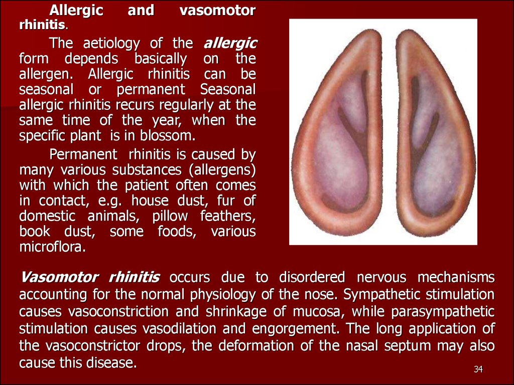
34.
Allergicrhinitis.
and
vasomotor
The aetiology of the allergic
form depends basically on the
allergen. Allergic rhinitis can be
seasonal or permanent Seasonal
allergic rhinitis recurs regularly at the
same time of the year, when the
specific plant is in blossom.
Permanent rhinitis is caused by
many various substances (allergens)
with which the patient often comes
in contact, e.g. house dust, fur of
domestic animals, pillow feathers,
book dust, some foods, various
microflora.
Vasomotor rhinitis occurs due to disordered nervous mechanisms
accounting for the normal physiology of the nose. Sympathetic stimulation
causes vasoconstriction and shrinkage of mucosa, while parasympathetic
stimulation causes vasodilation and engorgement. The long application of
the vasoconstrictor drops, the deformation of the nasal septum may also
cause this disease.
34
35.
The main symptom of both forms of rhinitis is paroxysmal sneezingattended by nasal hydrorrhoea and difficult nasal breathing. This triad
of symptoms is more or less pronounced in all cases. The rhinoscopic
signs of rhinitis are oedema and pallor of the mucosa, and cyanotic or
white spots on it.
The allergic form of the disease is characterized by increased eosinophil
counts and appearance of eosinophils in the nasal mucus.
Treatment depends on the findings of the allergological examination
and includes elimination from the patient's environment of allergens,
purulent foci or microbial allergy. Treatment includes specific and nonspecific hyposensitization of the patient, local procedures, including
surgery and action on the nervous system.
35
36.
Non-specific desensitization is used in both allergic and vasomotorforms of rhinitis. Antihistaminics (ериус, лоратидин, klaritin) and
hormones ( prednisolone, дексаметазон) are used for the purpose.
Topical steroids such as beclomethasone, назонекс are very effective
in the control of symptoms. Topical steroids have fewer systemic side
effects but their continuous use beyond 3 weeks is not recommended.
Аква марис
stabilises the mast cells and prevents them from
degranulation despite the formation of IgE antigen complex.
It is
useful both in seasonal and perennial allergic rhinitis.
36
37.
Endonasal electrophoresis with a 2 per centcalcium chloride solution is used most frequently.
Long-standing vasomotor rhinitis often increases
the volume of the conchae and imposes
permanent difficulties in nasal breathing. Surgical
treatment
(sparing
inferior
conchotomy,
submucous destruction of the inferior conchae
with ultrasound) is most rational in such cases.
37
38.
Inflammatory diseases of paranasal sinusesAcute and chronic inflammatory diseases of the paranasal sinuses
are frequent. They make 25-30 per cent of the hospitalized patients
with diseases of the ear, nose and throat. Maxillary sinusitis stands the
first in the list of incidence. Next comes ethmoiditis, then frontitis and
finally sphenoiditis_ Sometimes all paranasal sinuses are affected
(pansinusitis) or the sinuses of one side (hemisinusitis).
Acute inflammation of the sinuses is caused by acute respiratory
diseases, influenza, common cold, general microbial infections, and
injuries Chronic sinusitis can be secondary to protracted or frequently
recurring acute diseases in the presence of various local and general
harmful factors such as decreased reactivity and general weakening of
the body, impaired drainage of the sinuses in the presence of
hypertrophy or polyps of the mucosa in the region of the orifices,
deviated septum, and diseases of the teeth. The suppurative forms of
the disease are usually caused by streptococci and staphylococci or
other microorganisms.
38
39.
Classification of sinusitis:1.Acute sinusitis: a) catarrhal; b) suppurative.
2.Chronic sinusitis:
a) exudative (catarrhal, serous, suppurative, vasomotor, allergic)
b) polipous;
c) polipous-purulent;
d) hypertrophy;
e) atrophy (cholesteatomal, caseous, necrotic, ozaenous)
Acute maxillary sinusitis. Signs of acute inflammation of the
maxillary sinuses can be local and general. The local symptoms are
pain in the region of the involved sinus, forehead root of the nose, and
the cheek bone. Headache can be diffuse. Impeded respiration through
the involved side of the nose is a common symptom. Nasal discharge is
usually unilateral, and is first liquid serous, but then it becomes cloudy,
tenacious, and purulent. Olfaction is affected as a rule, but the severity
of other symptoms masks this disorder. The general symptoms are
elevated temperature of the body, indisposition. The temperature
reaction can begin with a chill and be intensive during the entire
disease.
39
40.
The objective symptom of acutemaxillary sinusitis is a narrow strip of
purulent discharge from the maxillary
sinus into the middle nasal meatus,
which is especially evident if the head
is inclined to the opposite side. Some
additional examinations should be
earned out: X-ray examination of the
paranasal sinuses, diagnostic antral
puncture and irrigation of the
maxillary sinus; contrast X-ray .
The
Kulikovsky
needle
is
commonly used for antral puncture.
The sinus wall is punctured by the
needle and the sinus contents are
aspirated; then, the sinus is irrigated
with
a
disinfectant
solution,
(флуимуцил-антибиотик,диоксидин,
ифиципро
40
41.
The liquid is passed intothe sinus through the
needle, while the sinus is
drained through the natural
orifice. The patient leans
downward so that the
washings are withdrawn
through the nose without
entering the nasopharynx.
.
antral puncture
41
42.
Treatmentincludes
local
use
of
vasoconstrictors
drops,
physiotherapy, and general antibacterial therapy in the presence of
high temperature and intoxication of the body. If these measures fail
to give the rapid effect, the sinus should be punctured and irrigated
and a mixture of antibiotics, steroid hormones, protheolitic enzyme
are instilled. The acute suppurative inflammation ends in 5-6 days.
42
43.
Chronic maxillary sinusitis. Chronic inflammation of the sinusis as a rule a sequel of acute sinusitis, which is recurrent in some
patients. Acute inflammation persisting for more than 3 weeks should
be considered as long-standing. If such inflammation does not
terminate by the end of the 6th week, the disease can be considered
chronic. Sometimes chronic maxillary sinusitis is associated with
spreading of pathology from a caries-affected tooth.
A common symptom and complaint of patients with the exudative
forms of chronic maxillary sinusitis is discharge from one side of the
nose, which can be copious during exacerbation and scarce in
remission. The purulent discharge in patients with maxillary sinusitis
can be thick or liquid and have a specific odour. The mucopurulent
discharge is tenacious and it dries in crusts. Catarrhal sinusitis is
marked by tenadous mucous discharge which is often retained in the
nasal cavity, and dries in crusts. The discharge in serous, or allergic
maxillary sinusitis accumulates in the sinus and drains in portions when
the patient assumes a certain position facilitating drainage of the sinus
through the nasal meatus.
43
44.
An unpleasant odour is sometimes the main complaint of thepatient who feels the smell himself. In bilateral chronic pathologies in
the maxillary sinuses patients always complain of decreased sense of
smell. Local or diffuse headache usually develops only during
exacerbations or in obstructed drainage of the sinus. During remission,
the general objective and subjective condition of the patient is
satisfactory. Exacerbation of a chronic process can be attended with
elevated temperature, worsening of the patient's condition, painful
swelling of the cheek, oedema of the eyelid and local or diffuse
headache.
Serous-catarrhal maxillary sinusitis facilitates formation of polyps
which usually grow from the middle nasal meatus.In rare cases, in the
presence of dental granuloma, cysts and fistulae in the sinus, a
cholesteatoma can form from the cells of the squamous epithelium.
44
45.
True (retention) cysts of the sinus form due to obstruction of themucous glands. Pseudocysts can also develop in the sinus, but they
differ from true cysts by the absence of the inner epithelial coat. The
main symptom of a cyst is headache arising due to compression of the
endings of the trigeminal nerve. Amber-coloured liquid can at times
issue from one side of the nose, after which the headache subsides.
This is a sign of spontaneous drainage of the cyst.
The pathological discharge from the nose and sinus (taken during
antral puncture) is examined in the laboratory for the presence of
microflora and for sensitivity to antibiotics.
Pathology of the maxillary sinus should be differentiated from
frontitis, ethmoiditis, and in rare cases from sphenoiditis. In adults it is
necessary to rule out the odontogenic nature of the disease, especially
in the presence of a suppurative process in the roots of the upper teeth
(4, 5, 6), whose apices are in the immediate vicinity of the floor of the
maxillary sinus.
45
46.
Conservative treatment. Treatment should begin with eliminationof causes of the disease. If maxillary sinusitis is odontogenic, the teeth
should first of all be treated. It should be noted that radical operations
on the sinus will be ineffective if the odontogenic cause remains
active.. As a rule, general antibacterial treatment is administered
during exacerbation.
Antral puncture and irrigation of the sinus with a disinfectant
solution (furacilin, флуимуцил-антибиотик,ифиципро,диоксидин) or
enzymes (chymopsin), and administration into the sinus of a solution of
the antibiotic to which the microflora is sensitive. In addition to the
irrigation of the sinus. If conservative treatment of chronic suppurative
maxillary sinusitis fails, a radical operation of the maxillary sinus is
indicated.
46
47.
Patients with the polypous and suppurative-polypous forms ofmaxillary sinusitis usually require radical surgical treatment which
should be followed by conservative treatment to prevent relapses of
polyposis. Postoperative conservative treatment includes regular
administration of astringent preparations, and if signs of allergy are
obvious, antiallergic treatment is indicated. (назонекс)
Surgical treatment. Operations on the maxillary sinus are
performed with endonasal and extranasal approach. The endonasal
technique can be used to open the medial wall of the sinus and to
perforate it for drainage and aeration of the sinus. The extranasal
approach operation ensures an easy access to all parts of the sinus and
the operation is therefore radical. This technique includes incision of
the soft tissues under the upper lip, separation of these tissues, and
approach to the anterior wall of the maxillary sinus. The sinus is then
opened, the pathological matter removed, and a communication with
the nasal cavity is made (through the inferior or middle nasal meatus).
47
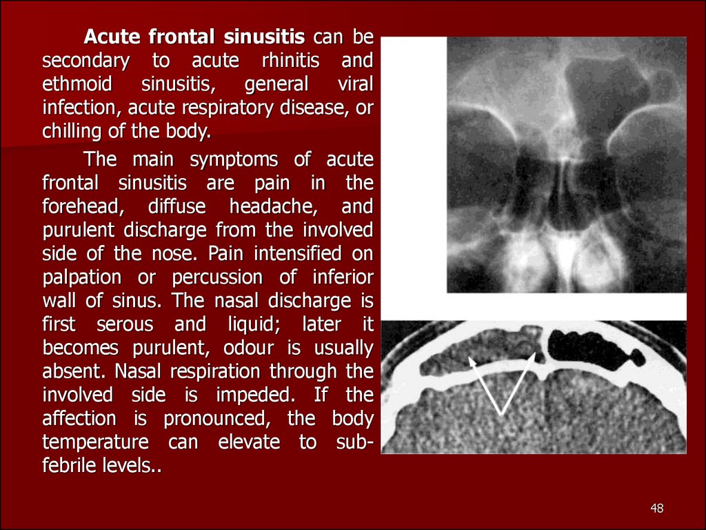
48.
Acute frontal sinusitis can besecondary to acute rhinitis and
ethmoid sinusitis, general viral
infection, acute respiratory disease, or
chilling of the body.
The main symptoms of acute
frontal sinusitis are pain in the
forehead, diffuse headache, and
purulent discharge from the involved
side of the nose. Pain intensified on
palpation or percussion of inferior
wall of sinus. The nasal discharge is
first serous and liquid; later it
becomes purulent, odour is usually
absent. Nasal respiration through the
involved side is impeded. If the
affection is pronounced, the body
temperature can elevate to subfebrile levels..
48
49.
X-ray examination and trepanation puncture of thefrontal sinus are used for diagnostic and therapeutic
purposes Treatment is usually conservative. But if the
disease is longstanding and complications develop in the
orbit, skull, or other organs, surgery should be performed
immediately to eliminate the purulent focus and to restore
patency of the frontonasal duct. Local treatment includes
application of preparations causing anaemization of the
nasal
mucosa:
vasoconstrictors
drops
(galasoline,
naphtiziine). Elevated temperature and headache can be
managed parenteral administration of antibacterial
preparations in the appropriate doses. The absence of the
desired effect is an indication for probing or puncture of the
sinus.
49
50.
Chronic frontal sinusitis. The most common cause ofconversion of acute frontal sinusitis into its chronic form is persistent
obstruction of the frontonasal duct and decreased reactivity of the
body, especially subsequent to general infectious diseases. This process
is promoted by hypertrophy of the middle concha, significant deformity
of the nasal septum, a narrow or tortuous frontonasal duct, or polyps
in the nasal cavity. There may be no complaints from the patient during
remissions. A small amount of the nasal discharge often escapes into
the nasopharynx to cause chronic pharyngitis, laryngitis, and tracheitis.
Palpation of the walls of the frontal sinus is often painful,
especially at the upper internal angle of the orbit, which can be
swollen. In the absence of microflora, obstruction of the frontonasal
duct sometimes stimulates the accumulation of discharge in the sinus
and the formation of mucocele consisting of secretions of the mucous
glands.
50
51.
Treatment. In the absence of local and general complications,conservative treatment is indicated. It is directed at providing
adequate drainage of the secretion from the sinus using
vasoconstrictors which are instilled into the nose, and administration
of antibacterial preparations (after preliminary testing of the microflora
for sensitivity to these preparations). Trephination puncture of the
frontal sinus with removal of its contents and subsequent irrigation
and administration of medicinal preparations are effective.
Long-standing and persistent chronic frontal sinusitis (despite
active treatment), and also symptoms of developing complications
(and complications themselves) are indications for surgical treatment
(operation of frontoethmoidotomy).
51
52.
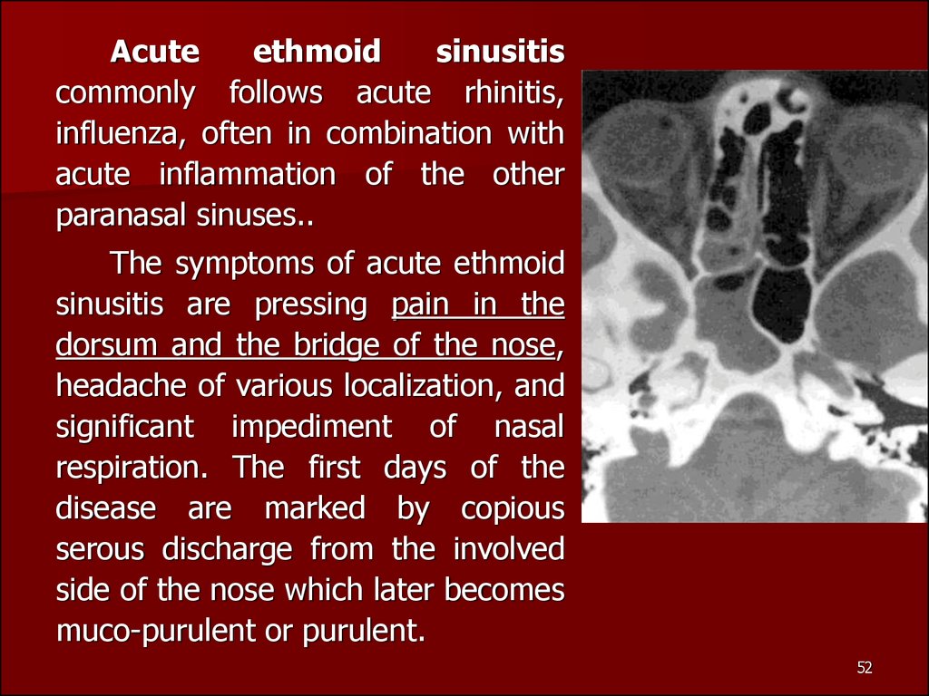
Acuteethmoid
sinusitis
commonly follows acute rhinitis,
influenza, often in combination with
acute inflammation of the other
paranasal sinuses..
The symptoms of acute ethmoid
sinusitis are pressing pain in the
dorsum and the bridge of the nose,
headache of various localization, and
significant impediment of nasal
respiration. The first days of the
disease are marked by copious
serous discharge from the involved
side of the nose which later becomes
muco-purulent or purulent.
52
53.
The discharge is usually odourless. Oedema and hyperaemia of theinternal angle of the orbit and the adjacent parts of the lower and upper
eyelids, and also conjunctivitis are frequent findings in children.
Hypoosmia are also frequent. The temperature is usually between 37.5
and 38 °C and persists for a week.
The diagnosis can be confirmed by X-ray examination. The nasal
discharge should be studied for microflora and its sensitivity to
antibiotics which will help assess the severity of the infection, prescribe
the appropriate antimicrobial therapy.
Treatment is conservative. If any complications develop, surgical
treatment is indicated. Vasoconstrictors are instilled into the nose. The
same preparations are applied under the middle concha. UHF or SHF on
the area of the ethmoidal sinus are indicated. If the body temperature is
elevated, antibacterial preparations are given. If a closed empyema or
ophthalmic complication develops, the cells of the ethmoidal labyrinth
should be opened to gain access to the purulent focus in the orbit.
53
54. Polypous and suppurative-polypous forms of sinusitis
Chronic ethmoid sinusitis. Thedisease is often secondary to the affection
of the other paranasal sinuses. Chronic
ethmoid sinusitis therefore often concurs
with frontal sinusitis, sphenoid sinusitis, and
more frequently, maxillary sinusitis. The
catarrhal-serous, catarrhal-suppurative and
polipous forms of chronic ethmoid sinusitis
prevail..
The symptoms depend on the activity
of the disease. During remission, the patient
complains of occasional headache, mostly in
the region of the nose root and bridge;
headache is sometimes diffuse. In serouscatarrhal ethmoid sinusitis, the nasal
discharge is clear and copious.
Polypous and suppurativepolypous forms of sinusitis
54
55.
The suppurative form is characterized by a meagre discharge thatdries to form crusts. Involvement of the posterior cells of the
ethmoidal labyrinth promotes accumulation of the discharge in the
nasopharynx, usually in the morning. Olfaction is impaired to some
degree.
Treatment
of
noncomplicated
forms
is
usually
conservative.
Sometimes it is combined with endonasal operations (polypotomy,
opening of cells of the ethmoidal labyrinth, partial resection of the
conchae, etc.). Opening of the cells of the ethmoidal labyrinth and
polypotomy with an endonasal approach are the most common
operations.
55
56.
Acute and chronic sphenoid sinusitis. Isolated affection of thesphenoidal sinuses is rare. The inflammation is usually combined with
lesion of the posterior cells of the ethmoidal labyrinth.
Acute sphenoid sinusitis is marked by severe oedema of the
mucosa. The most common subjective symptom of acute sphenoid
sinusitis is headache in the occipital region and inside the head;
the pain is sometimes felt in the orbit. Nasal discharge is often absent
because it passes from the superior nasal meatus into the nasopharynx
and further along the posterior wall of the pharynx, where it can easily
be seen during pharyngoscopy and posterior rhinoscopy. The body
temperature is usually subfebrile; the general condition is satisfactory;
the patient can complain of weakness, discomfort, and irritability.
X-ray examination is an important diagnostic tool. If the clinical
picture is obscure, the sphenoidal sinus can be punctured through its
anterior wall.
Treatment is usually conservative: local treatment with
vasoconstrictors and general antibacterial treatment. If the disease
lasts longer than 2 weeks, the sinus should be irrigated or opened
endonasally. Symptoms of complications (septic, intracranial,
ophthalmic) are indications for emergency operation on the sphenoidal
sinus.Chronic sphenoid sinusitis is provoked by the same conditions as
chronic affection of the other paranasal sinuses.
56
57.
RHINOGENIC ORBITALCOMPLICATIONS
(a) Inflammatory oedema of lids. This is
only reactionary. There is no erythema or
tenderness of the lids which characterises lid
abscess. Eyeball movements and vision are
normal. Generally, upper lid is swollen in
frontal, lower lid in maxillary, and both upper
and lower lids in ethmoid sinusitis.
(b) Subperiosteal abscess. Pus collects
outside the periosteum. A subperiosteal
abscess from ethmoids forms on the medial
wall of orbit and displaces the eyeball
forward, downward and laterally; from the
frontal sinus, abscess is situated just above
and behind the medial canthus and displaces
the eyeball downwards and laterally; from
the maxillary sinus, abscess forms in the
floor of the orbit and displaces the eyeball
upwards and forwards.
Inflammatory oedema of
lids
57
58.
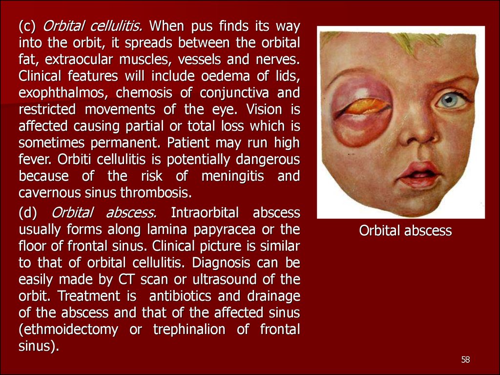
(c) Orbital cellulitis. When pus finds its wayinto the orbit, it spreads between the orbital
fat, extraocular muscles, vessels and nerves.
Clinical features will include oedema of lids,
exophthalmos, chemosis of conjunctiva and
restricted movements of the eye. Vision is
affected causing partial or total loss which is
sometimes permanent. Patient may run high
fever. Orbiti cellulitis is potentially dangerous
because of the risk of meningitis and
cavernous sinus thrombosis.
(d) Orbital abscess. Intraorbital abscess
usually forms along lamina papyracea or the
floor of frontal sinus. Clinical picture is similar
to that of orbital cellulitis. Diagnosis can be
easily made by CT scan or ultrasound of the
orbit. Treatment is antibiotics and drainage
of the abscess and that of the affected sinus
(ethmoidectomy or trephinalion of frontal
sinus).
Orbital abscess
58
59.
(e) Superior orbital fissure syndrome. Infection of sphenoid sinuscan rarely affect structures of superior orbital fissure. Symptoms
consist of deep orbital pain, frontal headache, and progressive paralysis
of CN VI, III and IV, in that order.
(f) Retrobulbar neuritis of CN I. Inflammation of the posterior cells
of the ethmoidal labyrinth and the sphenoidal sinus spreads to the orbit
impairing the visual acuity, narrowing the field of vision, and
intensifying scotoma.
Treatment is surgical with simultaneous general anti-inflammatory
treatment. In children, the paranasal sinuses, especially cells of the
ethmoidal labyrinth, should be opened by extranasal approach.
59