


 medicine
Similar presentations:


Clinical anatomy, physiology and methods of examination of the middle ear
1. Clinical anatomy, physiology and methods of examination of the middle ear. The contemporary methods of examination of the cochlear apparatus. Acute middle otitis. The peculiarities of acute otitis in children. Kinds of mastoiditis, clinical symptoms, diag
Clinical anatomy, physiology and methods ofexamination of the middle ear.
The contemporary methods of examination of the
cochlear apparatus. Acute middle otitis. The
peculiarities of acute otitis
in children. Kinds of mastoiditis, clinical symptoms,
diagnosis, treatment.
2. Actuality of the theme.
Acoustic analysator is of importance in process cognitionof surrounding world, is assist to forming speech function.
Diseases of the ear, the breach acoustic function are one
of the most frequent pathology; the fall of ear and deafness
are reflected on the capacity for work, on its condition.
Inflammatory diseases of the ear can be the reason of the
heavy lively dangerous intracranial complications.
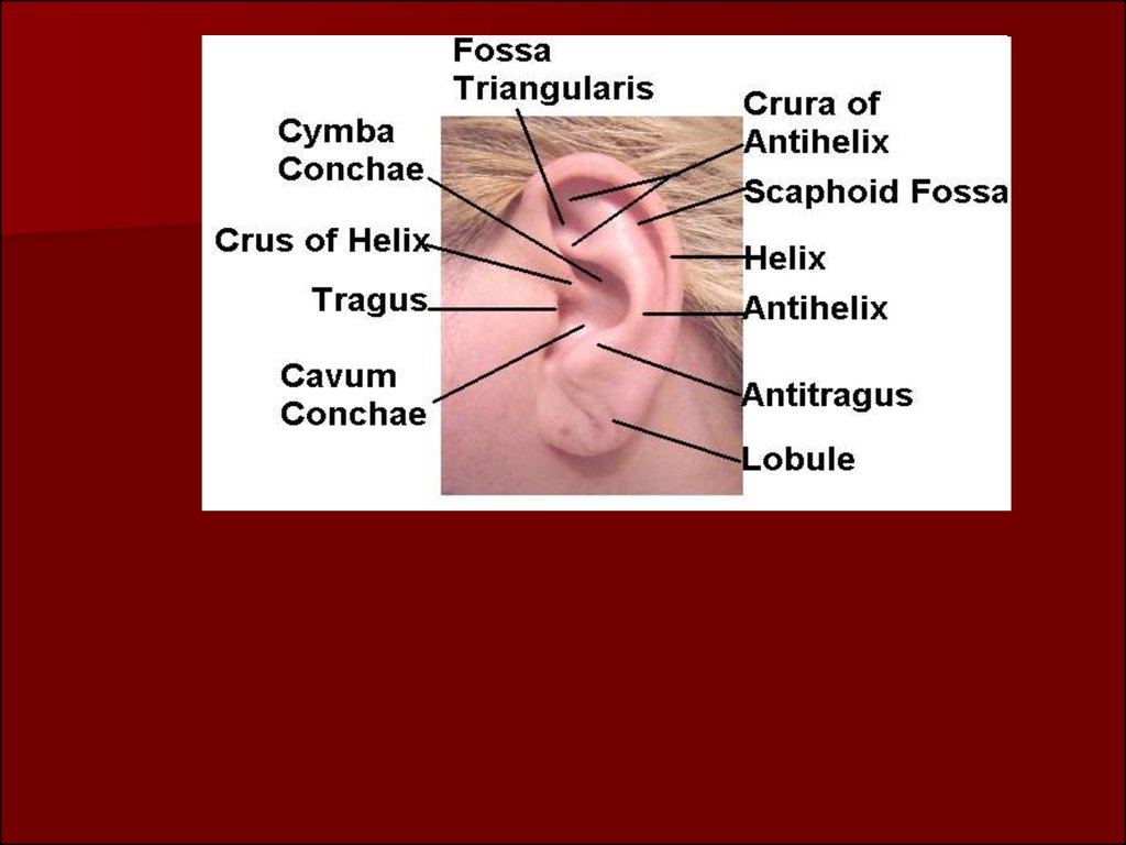
3. External and middle ear
Organ of hearing in anatomical relations isdivided into three parts: external, middle and
internal ear; functionally into - sound conducting
and sound apprehensive apparatus. The auricle,
external auditory tube passage, which gather
sound waves, tympanic membrane, chain of
ossicle bones and perilympha of internal ear
belong to the sound conduction apparatus.
4.
5.
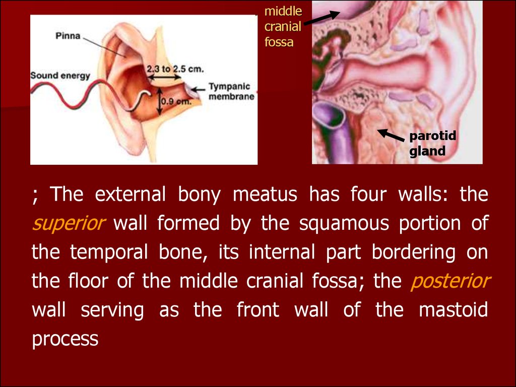
The external auditory meatus extends from the funnelshaped hollow on the outer surface of the pinna to thetympanic membrane end of the canal separates the
external and the middle ears. The outer third of the
auditory canal consists of cartilage and membranous tissue,
and both inner portions of bone.
Its narrowest part is the isthmus, where the cartilaginous
and bony portions form a junction and where foreign
bodies are most likely to lodge. The skin covering the
cartilaginous portion abounds in hair, sebaceous glands
and ceruminous glands which secrete the earwax, or
cerumen. The skin of the bony portion has neither hair, nor
glands.
6.
middlecranial
fossa
parotid
gland
; The external bony meatus has four walls: the
superior wall formed by the squamous portion of
the temporal bone, its internal part bordering on
the floor of the middle cranial fossa; the posterior
wall serving as the front wall of the mastoid
process
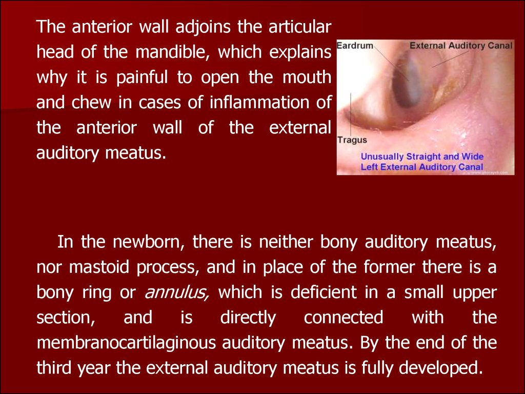
7.
The anterior wall adjoins the articularhead of the mandible, which explains
why it is painful to open the mouth
and chew in cases of inflammation of
the anterior wall of the external
auditory meatus.
In the newborn, there is neither bony auditory meatus,
nor mastoid process, and in place of the former there is a
bony ring or annulus, which is deficient in a small upper
section,
and
is
directly
connected
with
the
membranocartilaginous auditory meatus. By the end of the
third year the external auditory meatus is fully developed.
8.
The external ear is supplied with blood bybranches of the external carotid artery. It is
innervated, in addition to the trigeminal branches,
by the auricular nerve (ramus auricularis n. vagi) in
the posterior wall of the auditory meatus.
Mechanical irritation of the latter wall, as in wax
removal, often causes reflex cough. The lymph from
the walls of the auditory meatus drains into the
nearest lymph nodes located in front of the auricle,
on the mastoid process, and under the inferior wall
of the auditory meatus.
9. Tympanic Membrane
Thetympanic
membrane or drum
between the external
and middle ear. The
greater part of the
drum is called the pars
tensa; ,
smaller part of the pars flaccida or Shrapnell's membrane.
10.
The drum consists of three layers: an outer orepidermal layer continuous with that of the auditory
meatus, a middle layer of radiating and circular
connective tissue fibres, and an inner layer of
mucosa continuous with the mucous membrane of
the tympanic cavity. Shrapnell's membrane or pars
flaccida consists only of two layers and lacks the
middle stratum of fibrous tissue.
11.
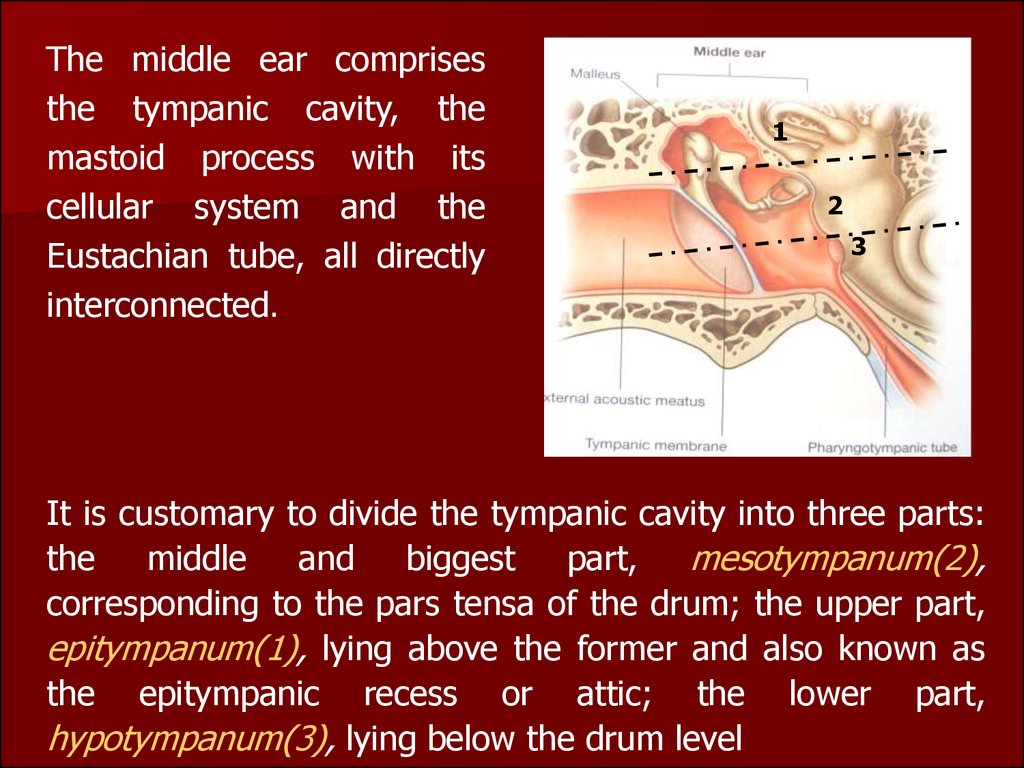
The middle ear comprisesthe tympanic cavity, the
mastoid process with its
cellular system and the
Eustachian tube, all directly
interconnected.
1
2
3
It is customary to divide the tympanic cavity into three parts:
the
middle
and
biggest
part,
mesotympanum(2),
corresponding to the pars tensa of the drum; the upper part,
epitympanum(1), lying above the former and also known as
the epitympanic recess or attic; the lower part,
hypotympanum(3), lying below the drum level
12.
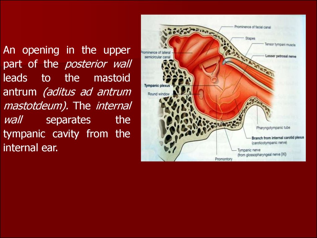
An opening in the upperpart of the posterior wall
leads to the mastoid
antrum (aditus ad antrum
mastotdeum). The internal
wall
separates
the
tympanic cavity from the
internal ear.
13.
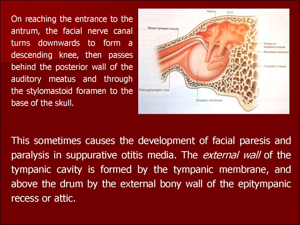
On reaching the entrance to theantrum, the facial nerve canal
turns downwards to form a
descending knee, then passes
behind the posterior wall of the
auditory meatus and through
the stylomastoid foramen to the
base of the skull.
This sometimes causes the development of facial paresis and
paralysis in suppurative otitis media. The external wall of the
tympanic cavity is formed by the tympanic membrane, and
above the drum by the external bony wall of the epitympanic
recess or attic.
14.
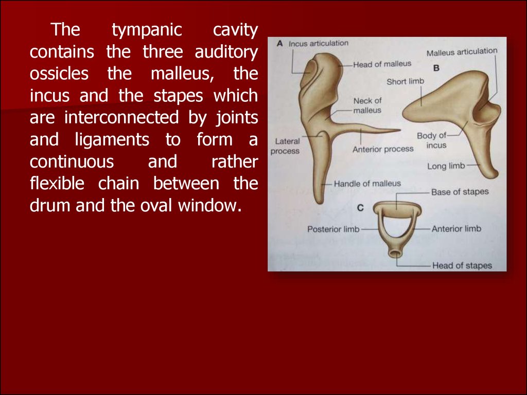
Thetympanic
cavity
contains the three auditory
ossicles the malleus, the
incus and the stapes which
are interconnected by joints
and ligaments to form a
continuous
and
rather
flexible chain between the
drum and the oval window.
15.
Thetympanic muscles.
There are two muscles in the
tympanic cavity: The tensor
tympani
muscle
which
stretches
the
tympanic
membrane.
The stapedius
muscle which arises from the
posterior wall of the tympanic
cavity and is attached to the
head of the stapes by a
slender tendon.
Eustachian
or auditory tube
which is about 3.5 cm in length
connects the tympanic cavity with
the nasopharynx.
16.
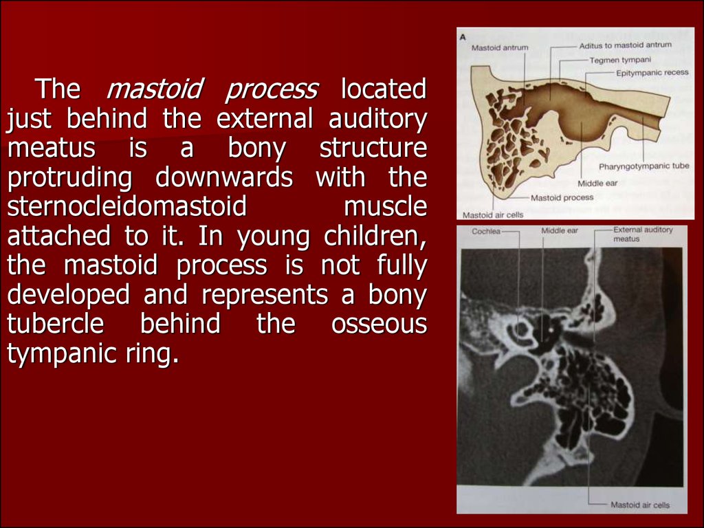
The mastoid process locatedjust behind the external auditory
meatus is a bony structure
protruding downwards with the
sternocleidomastoid
muscle
attached to it. In young children,
the mastoid process is not fully
developed and represents a bony
tubercle behind the osseous
tympanic ring.
17.
The anterior wall of the mastoid process is theposterior bony wall of the external auditory meatus.
The internal wall of the mastoid process abuts upon
the labyrinth, and more posteriorly is bordered by the
postcranial fossa. On the surface facing the postcranial fossa there is a rather wide S-shaped groove,
the sigmoid sulcus, containing part of the sigmoid
sinus of the dura mater. The central part of the
mastoid process is the antrum lying just behind the
epitympanic recess.
18.
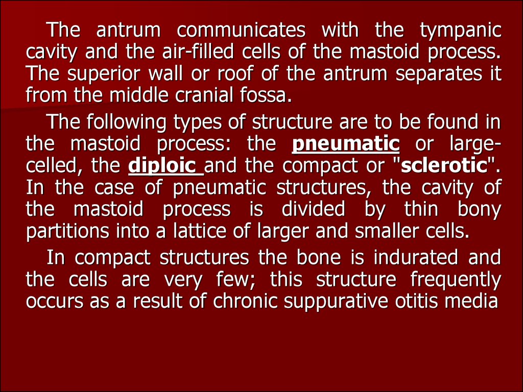
The antrum communicates with the tympaniccavity and the air-filled cells of the mastoid process.
The superior wall or roof of the antrum separates it
from the middle cranial fossa.
The following types of structure are to be found in
the mastoid process: the pneumatic or largecelled, the diploic and the compact or "sclerotic".
In the case of pneumatic structures, the cavity of
the mastoid process is divided by thin bony
partitions into a lattice of larger and smaller cells.
In compact structures the bone is indurated and
the cells are very few; this structure frequently
occurs as a result of chronic suppurative otitis media
19.
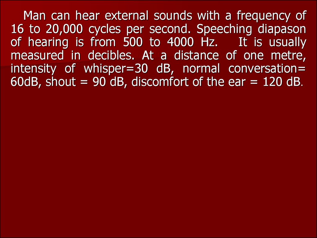
Man can hear external sounds with a frequency of16 to 20,000 cycles per second. Speeching diapason
of hearing is from 500 to 4000 Hz.
It is usually
measured in decibles. At a distance of one metre,
intensity of whisper=30 dB, normal conversation=
60dB, shout = 90 dB, discomfort of the ear = 120 dB.
20.
Methods of examination.Hearing test (whispered and spoken voice tests).
The patient is at a distance of 6 metres from the examiner,
with the examined ear toward the physician
2. The patient is asked to repeat loudly the words uttered by
the physician. In order to prevent visual hearing (lipreading),
the patient should not look at the physician.
3. The physician exhales normally, and then whispers words
with low vowels, e. g. "hawl, raw", etc., and then with high
vowels, such as "feet, cheese", etc.
4. If the patient cannot hear at a distance of 6 metres, the
physician should approach the patient to a distance of 5
metres, and examine the patient again.
1.
21.
The distance should thus be shortened by 1 metre eachtime until the patient repeats correctly all the words
pronounced by the physician.
5. The results of the test are expressed in metres at which the
examinee hears the whispered words.
6. The patient can be tested for hearing spoken voice using
the same technique as in the whispered voice testing.
Tuning-fork tests.
Test for air conduction. A set of tuning forks (Ci28, C512,
C2048) is used for the purpose. The test begins with the lower
frequency (C128).
22.
Weber's test. A vibrating fork (C128) is placed on the vertexof the patient's head so that the stem of the fork is in the
midline of the head.
Normally the patient hears the tuning fork in the middle of
the head, i. e. by both ears. If the sound is heard better by the
affected ear, the conduction system is probably damaged. If
the sound is better heard by the normal ear, this is probably
due to disease of the auditory apparatus.
Rinne's test A vibrating tuning fork (C128) is placed with
its stem on the mastoid. After the patient reports
discontinuation of sound perception, the fork (without
reactivation) is put to the external acoustic meatus. If the
patient hears the fork sound through air, the Rinne test is
considered positive (+). If the patient does not hear the fork
through the external acoustic meatus, the result is negative ().
23.
Federici's test. C128 tuning forks are used. Avibrating fork is applied to the mastoid process. As
the patient hears it no longer, the fork is placed on
the tragus.
Pure tone audiometry.
If the investigation by speech and turning forks not
The term “audiometry” means the methods of
investigation the ear with the help of electroacoustic
apparatus - audiometer.
24.
An audiometer is an electronic device whichproduces pure tones, the intensity of which can
be increased or decreased in 5 dB steps. Usually
air conduction thresholds are measured for
tones of 125,250,500,1000,2000,4000 and 8000
Hz and bone conduction thresholds for
250,500,1000and2000and 4000 Hz.
Maximum intensification of the sound by investigation ear conductivity 6080 DB. The investigation is accompanied in special soundsolate chamber. It
is charted in the form a graph called audiogram. On scale of audiometer the
level of normal (according international standart) ear correspond to line
0Db, that is loss of ear on this level is 0. The threshold of bone conduction
is a measure of cochlear function. The difference in the thresholds of air and
bone conduction is a measure of degree of conductive deafness.
25.
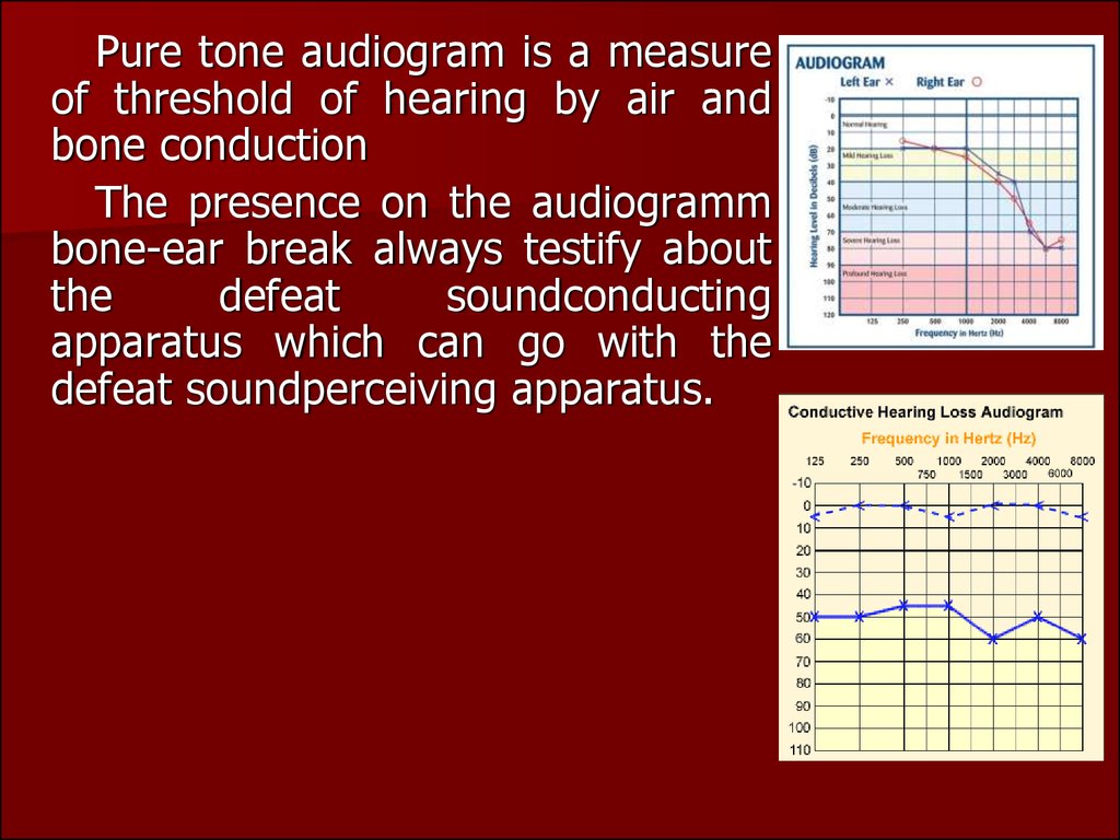
Pure tone audiogram is a measureof threshold of hearing by air and
bone conduction
The presence on the audiogramm
bone-ear break always testify about
the
defeat
soundconducting
apparatus which can go with the
defeat soundperceiving apparatus.
26.
ACUTE OTITIS.Acute purulent middle otitis is called inflammatory infectious
disease of mucous layer of air containing cavities of middle
ear. Today acute middle otitis occurs quite frequently within
the population of different age groups and particularly frequent
in early child age due to anatomic peculiarities of structure of
middle ear in this age, as well as tendency towards infectious
diseases, which are complicated by diseases of ear.
Suffered acute otitis may be the reason of stable hard
hearing, of development of chronic inflammation of middle ear,
threatening intracranial complications. Probability of the latter
is related with no diagnosis at right time, as well as with
mistakes in treatment tactics of acute purulent middle otitis
27.
The direct cause of acute otitis media is infection of themiddle ear with streptococci, staphylococci, pneumococci, and
less frequently other microbes; mixed flora is sometimes
responsible for the onset of the disease.
Acute otitis is often secondary. It can be a complication or
a manifestation of a systemic infection, for example, infection
of the upper airways and influenza; scarlet fever, measles,
and some other diseases provoke acute otitis media in
children. It can be due to acute and chronic inflammation of
the pharynx and the nose. The main pathological factor is
mechanical compression of the pharyngeal orifice of the
auditory tube and impairment of its ventilating and draining
functions. Among such diseases are adenoids, polyps of the
nose, tumors of the pharynx. Less frequently otitis is
secondary to injuries to the ear.
28.
Infection usually enters the middle ear through the auditorytube. Less frequently infection gets into the middle ear through
an injured tympanic membrane or through the damaged
mastoid process. In rare cases infection penetrates into the
middle ear by haematogenic routes (in infectious diseases).
Three periods are distinguished in a typical course of
acute suppurative otitis media.
The first period is characterized by the
onset and development of inflammation in
the middle ear, infiltration and exudation,
and development of minor symptoms, such
as hearing loss, noise, earache, hyperemia
of the tympanic membrane, protrusion of
the membrane due to the thrust of the
exudate, and some general symptoms such
as elevation of body temperature to 38-39
°C,
deranged
appetite
and
sleep,
indisposition.
29.
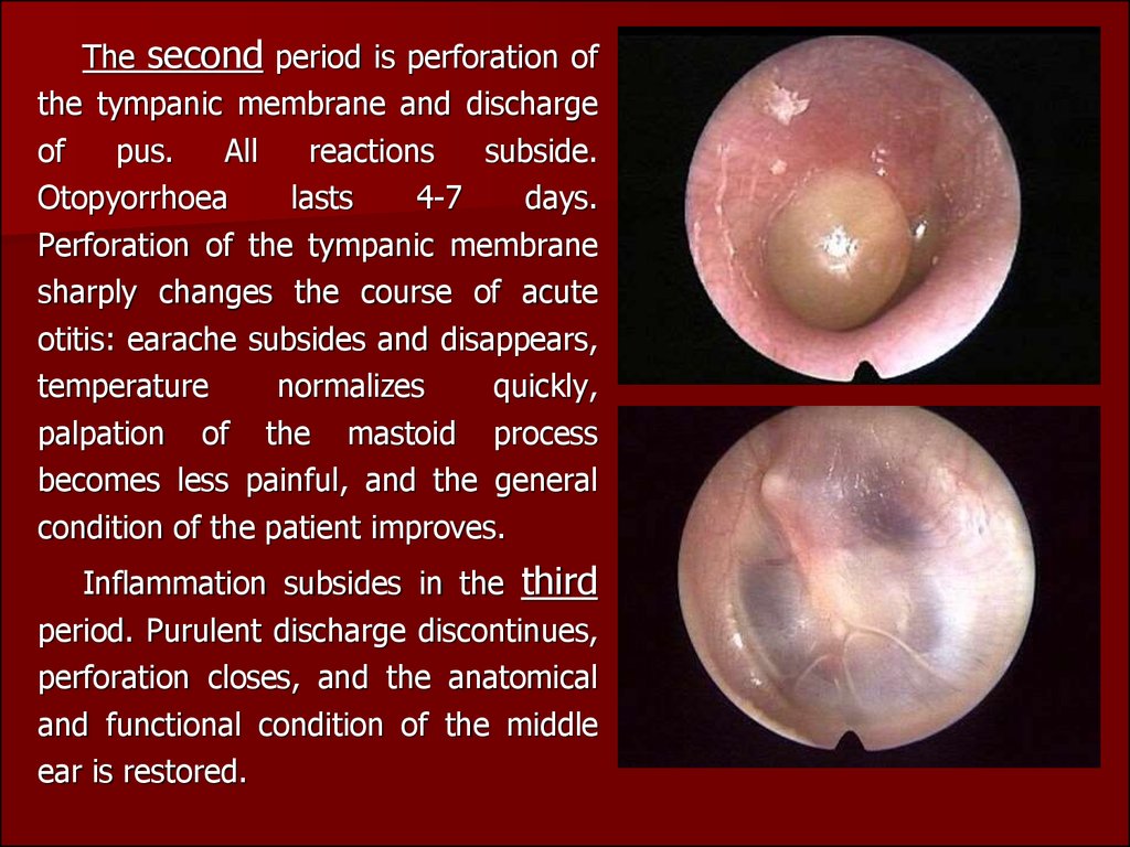
The second period is perforation ofthe tympanic membrane and discharge
of
pus.
All
reactions
subside.
Otopyorrhoea
lasts
4-7
days.
Perforation of the tympanic membrane
sharply changes the course of acute
otitis: earache subsides and disappears,
temperature
normalizes
quickly,
palpation of the mastoid process
becomes less painful, and the general
condition of the patient improves.
Inflammation subsides in the third
period. Purulent discharge discontinues,
perforation closes, and the anatomical
and functional condition of the middle
ear is restored.
30.
The first period of acute otitis media can sometimes be very grave andattended with hyperpyrexia, severe headache, vomiting, vertigo, and
drastic impairment of the general condition, painful palpation of the
mastoid process. Changes in the blood of patients with otitis during the first
days of the disease are characterized by high leukocyte count with a
considerable shift to the left. After perforation of the tympanic membrane
and discharge of pus, the blood picture gradually normalizes. If the disease
runs a typical benign course, the patient usually recovers with resolution of
the inflammation and complete restoration of the hearing function. If the
disease runs an atypical course, the outcomes can be different, with
adhesions and commissures between the tympanic membrane and the
medial wall of the middle ear and impairs hearing (adhesive otitis media);
persistent dry perforation (dry perforating otitis media); conversion of
acute disease into its chronic form with persistent perforation and periodic
otopyorrhoea; complications, such as mastoiditis, labyrinthitis, paresis of
the facial nerve, intracranial complications, etc.
31.
Dynamics of basic symptoms of AMO in 3 stages of development of processSymptoms
I stage
(before-perforate)
II stage
III stage
Pain in ear
sharp
(perforation or pus
flow)
insignificant
Noise in ear
moderate
less expressed
absent
Decrease in
hearing
Excretions
sharply
decreased
restores
no
serous-blood,
stops
Changes in
tympanic
membrane
infiltrated,
hyperemised,
protruded
Temperature of
body
high
mucous-purulent
perforation, pulsate
reflex
subfebril
(scaring or healing)
absent
tympanic membrane
becomes distinct, appear
recognising points (signs),
at the beginning short
process of malleus and at
the end - light cone; scars
of perforation of tympanic
membrane
normal
32. Differentiate symptoms of AMO from external otitis.
SymptomsAMO
External otitis
Pain in ear
Sharp, pulsate, irradiate;
Strong, sometimes irradiate,
accompanied with head ache, not accompanied by
heaviness and pressure in ear headache; increases during
chewing, movement of jaw
Decrease of hearing
Moderate
Hearing is not changed
Noise in ear
Of sharp intensity
Character of excretion in
acoustic meatus (auditory
passage)
Touching of acoustic meatus
and tragus
Change in tympanic
membrane
Mucous-purulent, serous;
blood.
Absent. May arise during
sharp infiltration of skin of
auditory passage and its
felling with pus
Purulent
Painless
Sharply painful
Depending upon stage of
process
Unchanged
33.
Treatment includes sparing conditions at home or athospital. The in vitamins to ensure the normal function diet
should be easily and rich of the gastrointestinal tract.
Vasoconstrictors or astringents should be instilled into the nose
for restoration or improvement of ventilation and drainage of
the auditory tube (naphtyzini, halasolini, sanorini) In cases of
shooting pains and marked redness of the drum,sp… drops
should be used. If acute otitis media runs a severe course with
marked general and local symptoms, antibiotic is injected
intramuscularly for at least 5-6 days. Analgesics and
antipyretics should be given for severe headache and pyrexia.
Warming compresses should be placed on the mastoid
process. Compresses should be prepared as follows: gauze
should be folded four or five times and soaked in alcohol
diluted with water (1:1). The compress should be changed at
4-5-hour intervals.
34.
In rare cases, when this treatment fails and severe pain in the earpersists, the body temperature remains high and the tympanic membrane
bulges outside, it is necessary to incise the tympanic membrane.
Paracentesis is positively indicated for irritation of the middle ear or
meningeal irritation which are manifested by vomiting, vertigo, severe
headache, and other signs. Paracentesis is more frequently indicated for
children because their tympanic membrane is thicker (especially in nursing
infants) and it resists rupture stronger than in adults, while the local and
general symptoms (pain, pyrexia) are more pronounced.
Paracentesis. The tympanic membrane is incised
using a special needle and observing the rules of
asepsis. When performing paracentesis in children,
not only the head but the whole body must be
immobilized. The incision is made on the drum bulge,
welllit, kept under direct observation and carried
downwards in the posterior-inferior quadrant of the
drum.
35.
Special conditions must be provided for unobstructeddrainage of pus from the ear after paracentesis. This can be
attained by inserting a special turunda. The external acoustic
meatus must be cleaned thoroughly using sterile hygroscopic
cotton with 3% hydrogen peroxide. The ear may be syringed
once or twice daily under low pressure along the posterior wall
of the auditory meatus. After them the medicinal preparations
can be administered into the middle ear through the external
acoustic meatus (transtympanic administration).To that end,
the mentioned mixture (1 ml) should be instilled into the
acoustic meatus and forced into the tympanic cavity by gently
pressing the tragus into the external orifice of the acoustic
meatus. The medicinal solution can pass the middle ear, the
auditory tube, and enter the mouth and nose. The blowing
with balloon of Politcer, catheterisation of the auditory tube
facilitates drainage of the middle ear and removes air
rarefaction which always attends acute otitis media.
36.
Moreover, this procedure normalizes the function of theauditory tube and has a favorable effect on the course of
inflammation. Blowing through a catheter is effective during
the third stages of acute otitis media. The procedure should be
performed once a day, during 3 or 4 days. A suspension of
hydrocortisone mixed with antibiotics should be administered
into the middle ear through a catheter.
Prevention includes a combination of measures such as
control of infectious diseases, timely treatment of acute and
chronic diseases of the nose, paranasal sinuses, and the
nasopharynx.
Acute otitis media in children. Acute otitis media in neonates and
infants occurs much more frequently than in adults. Its course is specific.
The special character of the symptoms is determined by the absence of
general and local immunity, the morphology of the mucous in the middle
ear and the structure of the temporal bone (residues of myxoid tissue, the
nutrient medium for infection growth, are present in the tympanic cavity).
37.
Inflammation of the middle ear in neonates often develops due topenetration of amniotic fluid into the middle ear through the auditory tube
during birth. The infection mechanism in nursing infants is the same, but in
addition to infection penetrating from the nose and nasopharynx, food can
also pass into the middle ear during regurgitation.
It is more difficult to establish the diagnosis of acute otitis media in a
nursing infant. But the behavior of a baby with a diseased ear differs
substantially from that of a healthy baby. The baby has bouts of
inconsolable crying, refuses the breast because of pain during swallowing,
rubs his diseased ear against the mother’s hand. The main symptoms of the
disease are painful palpation of the tragus (because of the absence of the
bony part of the acoustic meatus) and high body temperature (39.5-40°C).
A baby with otitis media is almost always depressed and sleeps a lot; his
gastrointestinal function is upset; vomiting develops and wasting ensues.
Meningeal symptoms with dimmed consciousness are possible.
38.
Influenzal otitis occurs usually during viral influenzaepidemics. The virus penetrates directly into the ear by the
haematogenic route or from the upper airways through the
auditory tube.
Specific influenzal otitis is characterized by
haemorrhagic inflammation which is manifested by a
pronounced dilatation of the vessels in the external acoustic
meatus and the middle ear with extravasation (haemorrhage)
under the epidermis in the bony part of the external acoustic
meatus and the tympanic membrane.
39.
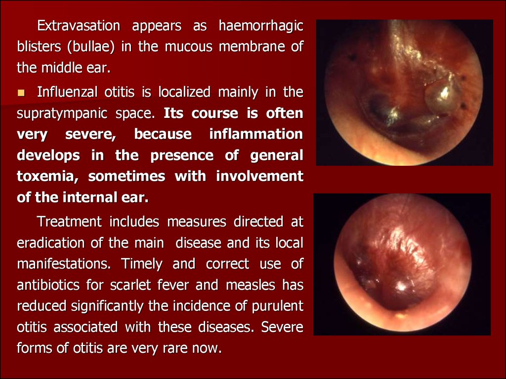
Extravasation appears as haemorrhagicblisters (bullae) in the mucous membrane of
the middle ear.
Influenzal otitis is localized mainly in the
supratympanic space. Its course is often
very severe, because inflammation
develops in the presence of general
toxemia, sometimes with involvement
of the internal ear.
Treatment includes measures directed at
eradication of the main disease and its local
manifestations. Timely and correct use of
antibiotics for scarlet fever and measles has
reduced significantly the incidence of purulent
otitis associated with these diseases. Severe
forms of otitis are very rare now.
40.
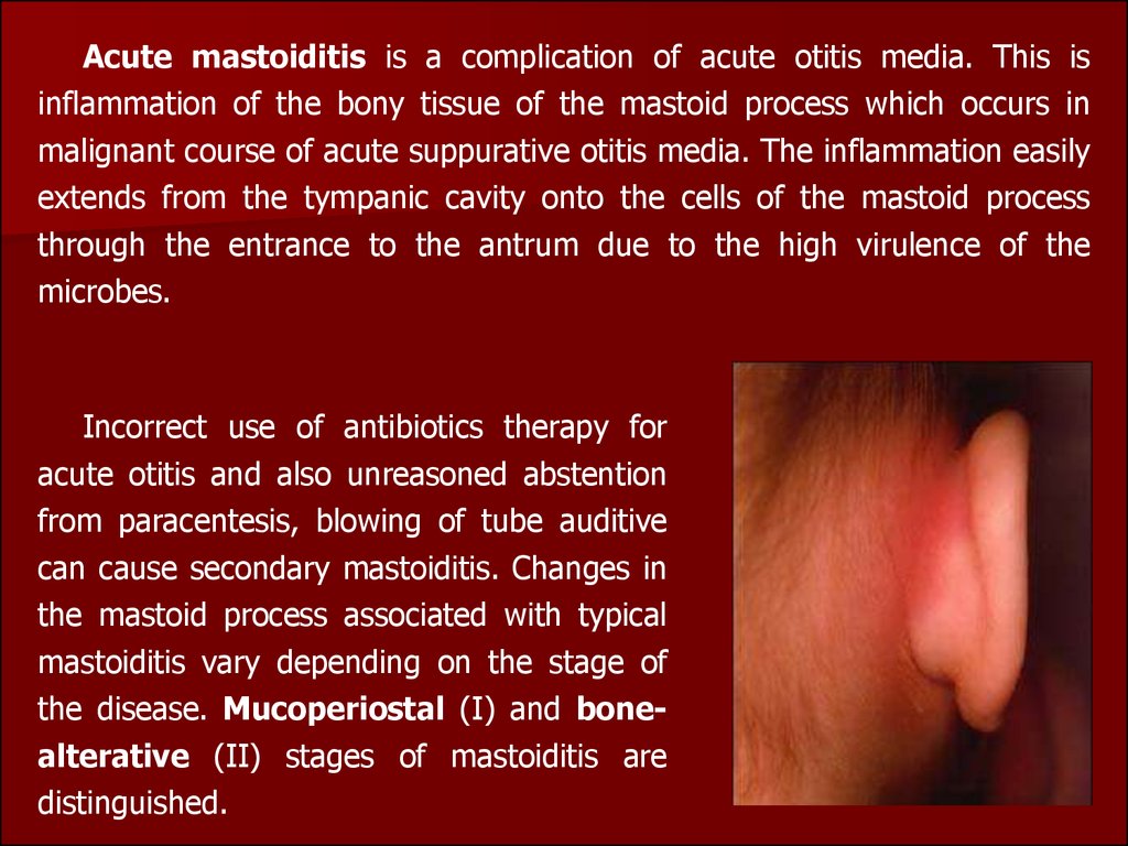
Acute mastoiditis is a complication of acute otitis media. This isinflammation of the bony tissue of the mastoid process which occurs in
malignant course of acute suppurative otitis media. The inflammation easily
extends from the tympanic cavity onto the cells of the mastoid process
through the entrance to the antrum due to the high virulence of the
microbes.
Incorrect use of antibiotics therapy for
acute otitis and also unreasoned abstention
from paracentesis, blowing of tube auditive
can cause secondary mastoiditis. Changes in
the mastoid process associated with typical
mastoiditis vary depending on the stage of
the disease. Mucoperiostal (I) and bonealterative (II) stages of mastoiditis are
distinguished.
41.
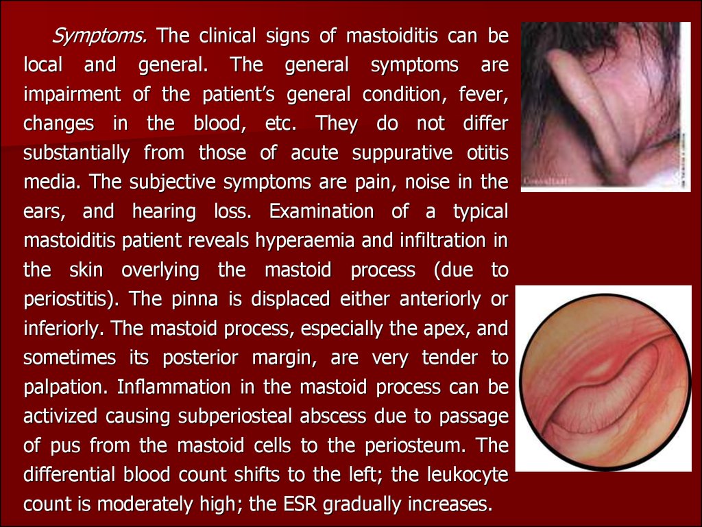
Symptoms. The clinical signs of mastoiditis can belocal and general. The general symptoms are
impairment of the patient’s general condition, fever,
changes in the blood, etc. They do not differ
substantially from those of acute suppurative otitis
media. The subjective symptoms are pain, noise in the
ears, and hearing loss. Examination of a typical
mastoiditis patient reveals hyperaemia and infiltration in
the skin overlying the mastoid process (due to
periostitis). The pinna is displaced either anteriorly or
inferiorly. The mastoid process, especially the apex, and
sometimes its posterior margin, are very tender to
palpation. Inflammation in the mastoid process can be
activized causing subperiosteal abscess due to passage
of pus from the mastoid cells to the periosteum. The
differential blood count shifts to the left; the leukocyte
count is moderately high; the ESR gradually increases.
42.
The specific otoscopic symptom of mastoiditis is sagging softtissue of the posterior-superior wall of the bony part of the
external acoustic meatus at the tympanic membrane (the
anterior wall of the antrum). Otopyorrhoea is often pulsating
and profuse. The consistency of pus is often creamy. Pus can
fill the acoustic meatus immediately after its cleaning.
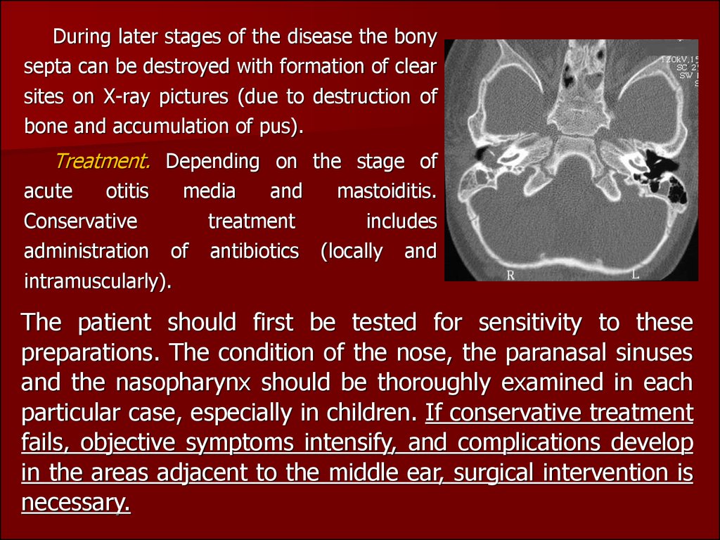
Diagnosis. Roentgenography of the temporal bone is very
important for diagnosis. An X-ray picture shows diffuse
reduction of pneumatization and shaded antrum and the cells.
43.
During later stages of the disease the bonysepta can be destroyed with formation of clear
sites on X-ray pictures (due to destruction of
bone and accumulation of pus).
Treatment. Depending on the stage of
acute
otitis
media
and
mastoiditis.
Conservative
treatment
includes
administration of antibiotics (locally and
intramuscularly).
The patient should first be tested for sensitivity to these
preparations. The condition of the nose, the paranasal sinuses
and the nasopharynx should be thoroughly examined in each
particular case, especially in children. If conservative treatment
fails, objective symptoms intensify, and complications develop
in the areas adjacent to the middle ear, surgical intervention is
necessary.
44.
Basic differential diagnostic symptoms of AMO and mastoiditisSymptoms
AMO
Mastoiditis
General (overall)
condition
Pain in ear
Improves
Inspite of treatment deteriorates
After perforation decreases
Inspite of perforation does not decrease
Noise in ear
Gradually decreases
In spite of treatment does not decrease
Hearing
Improves
Does not improve
Excretion from ear
Stands less, after then disappears. Purulent; purulent-blood in very big
From serous - blood and mucoid- quantities
purulent stands mucoid
Palpation of mastoid
process
Painless, may be painful during
Sharply painful
the first days of disease (mastoidal
reaction)
Unchanged
Infiltrated, swollen mastoid process,
smoothness of postauricular fold
Correlative to stages
Infiltrated, thickened (mastoidal type);
hanging of posterio-superior wall of
acoustic meatus
Painless
Painful
Skin of postauricular
region
Change in tympanic
membrane and external
acoustic meatus
Percussion of mastoid
process
45.
Differentiative symptoms of mastoiditis and furuncul of external acousticmeatus
Symptoms
Furuncul of external
acoustic meatus
Acute mastoiditis
Spontaneous pain
Increase during chewing
(mastication)
Does not increase while
chewing (mastication)
Pain caused by pressing
Maximum while pressing
on tragus
Maximum while pressing
on mastoid process
Pain cased by pulling the
auricle
Extremely painful
Painless
Condition of external
acoustic meatus
Swelling of skin of
cartilaginous part
Swelling of bony part
(hanging of posterior wall)
Tympanic membrane
Normal
Changed
Hearing
Normal
Decreased
Temperature
Normal or slightly
increased
Increased nearly always
46.
The operation on the mastoid process,known as mastoidectomy, is performed
under local and sometimes under general
anesthesia.The
operation
is
usually
concluded by filling the wound with
antibiotic powder and packing it lightly
with tampons. Sometimes mastoid cavity is
thoroughly irrigated with saline to remove
bone dust and the wound closed in two
layers. A rubber drain may be left at the
lower end of incision for 24-48 hours in
cases of infection or excessive bleeding.
Antibiotics started preoperatively are
continued postoperatively for at least one
week. Culture swab taken from the mastoid
during operation may dictate a change in
the antibiotic.