


 medicine
Similar presentations:


Methods of examination in gynecology
1. JSC “Astana Medical University” Obstetrics and gynecology department
Topic: “Methods of examination in gynecology”Performed by: Sakhi S.K.
Checked by: Gabdilashimova Z.T.
Astana 2018
2. Content
Anamnesis
-the main aspects
-the common cases
Gynecological examination
-examination of external genital organs
-bimanual vaginal-abdominal examination
-rectal and vaginal-rectal examination
Instrumental Research Methods
Tissue biopsy and cytology
Special examination methods
-laboratory diagnostics of pathogens
3. Anamnesis
• age;• complaints;
• family history;
• lifestyle, nutrition, bad habits, working and living conditions;
• past illnesses;
• menstrual and reproductive functions, the character of contraception;
• gynecological diseases and operations on the genitals;
• the history of the present disease.
• physical examination
Source: Gynecology, 4th edition, 2012, Savelieva G.M., Breusenko V.G., page 9
4. Anamnesis | the common cases
Amenorrhea - absence of menstruation;
Menorrhagia - bleeding associated with the menstrual cycle.
Menometrorrhagia - bleeding in the form of profuse menstruation,
continuing in the intermenstrual period.
Algodismenorea - painful menstruation.
Hypomencastral syndrome is expressed in reduction (hypomenorrhoea),
shortening (oligomenorrhea), and decreasing (opsonomena) of
menstruation.
Metrorrhagia is an acyclic uterine bleeding that is not associated with the
menstrual cycle and usually occurs with various ovarian disorders due to
disturbances in ovulation processes.
Source: Gynecology, 4th edition, 2012, Savelieva G.M., Breusenko V.G., page 10
5. Gynecological examination
*is carried out on the gynecological chair.In a healthy, mature, nonpregnant and unkempt woman, the vertical
position of the genitalia is considered normal (typical).
6. Gynecological examination
The normal position of female genital organs is provided by:• own tone of genital organs;
• the relationship between internal bodies and the activity of the diaphragm,
abdominal wall and pelvic floor;
• ligamentous apparatus of the uterus (suspension, fixation and holding).
Source: Pract Midwife. 2015 Sep;18(8):27-9. Introducing educational theory: vaginal examination. Killingley J.
7. Examination of external genitalia
Examination of external genitalia: the condition and magnitude of small andgreater the labia; condition of the mucous membranes; the size of the clitoris;
degree and nature of development of the hairline; a condition of a perineum;
pathological.
Pay attention also to the gaping of the genitals;
Source: Pract Midwife. 2015 Sep;18(8):27-9. Introducing educational theory: vaginal examination. Killingley J.
8. Bimanual vaginal-abdominal examination
The index and middle fingers of one hand (usually right), dressed in a glove,are inserted into the vagina. The other arm (usually the left one) is placed on
the anterior abdominal wall. The right hand palpates the walls of the vagina,
its arches and cervix, determine the volume of formation and anatomical
changes.
Source: Int J Gynaecol Obstet. 2017 Oct;139(1):114-119. doi: 10.1002/ijgo.12260. Epub 2017 Aug 14. Standardizing bimanual vaginal examination using cognitive task analysis.
Plumptre I1, Mulki O2, Granados A1, Gayle C1, Ahmed S2, Low-Beer N2, Higham J3, Bello F1.
9. Rectal and vaginal-rectal examination
is mandatory in postmenopause
to clarify the status of the uterine appendages
should be given to all women over 40
the tone of the sphincters of the anus and the state of the pelvic floor
muscles volumetric formations
Source: Pract Midwife. 2015 Sep;18(8):27-9. Introducing educational theory: vaginal examination. Killingley J.
10. Instrumental examination methods
!!!To women, leading sex life.Timely recognition of diseases of the cervix, erosions, polyps and other pathology is
possible by mirrors.
Source: Gynecology, 4th edition, 2012, Savelieva G.M., Breusenko V.G., page 14
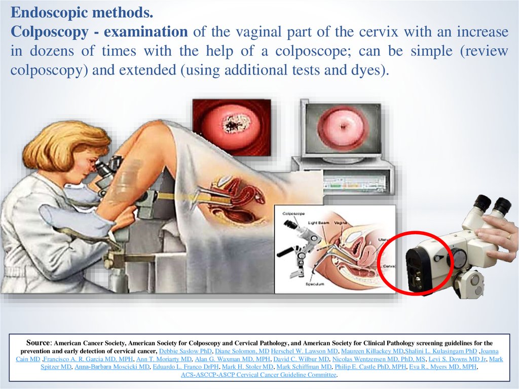
11.
Endoscopic methods.Colposcopy - examination of the vaginal part of the cervix with an increase
in dozens of times with the help of a colposcope; can be simple (review
colposcopy) and extended (using additional tests and dyes).
Source: American Cancer Society, American Society for Colposcopy and Cervical Pathology, and American Society for Clinical Pathology screening guidelines for the
prevention and early detection of cervical cancer, Debbie Saslow PhD, Diane Solomon, MD Herschel W. Lawson MD, Maureen Killackey MD,Shalini L. Kulasingam PhD ,Joanna
Cain MD ,Francisco A. R. Garcia MD, MPH, Ann T. Moriarty MD, Alan G. Waxman MD, MPH, David C. Wilbur MD, Nicolas Wentzensen MD, PhD, MS, Levi S. Downs MD Jr, Mark
Spitzer MD, Anna‐Barbara Moscicki MD, Eduardo L. Franco DrPH, Mark H. Stoler MD, Mark Schiffman MD, Philip E. Castle PhD, MPH, Eva R., Myers MD, MPH,
ACS‐ASCCP‐ASCP Cervical Cancer Guideline Committee.
12. Colposcopic microscopy
examination of the vaginal part of the cervix with an optical system (contrastluminescent colpomicroscope or colpomicroscope Hamo - type of
hysteroscope).
Source: Gynecology, 4th edition, 2012, Savelieva G.M., Breusenko V.G., page 22
13. Hysterocervical examination and Hysteroscopy
Hysterocervicaloscopy - examination with the help of optical systems of theinternal surface of the uterus and the cervical canal.
Hysteroscopy can be diagnostic and operating. Diagnostic hysteroscopy is
currently the optimal method of diagnosing all types of intrauterine
pathology.
2
1
Source: Gynecology, 4th edition, 2012, Savelieva G.M., Breusenko V.G., page 24
14. Probing the uterus
an invasive diagnostic method used to establishposition and direction of the uterine cavity, its length
Source: Gynecology, 4th edition, 2012, Savelieva G.M., Breusenko V.G., page 30
15. Puncture of abdominal cavity through posterior vaginal arch.
This puncture is performed when it is necessary to ascertain the presence orlack of free fluid (blood, pus) in the cavity of the small pelvis
Source: Gynecology, 4th edition, 2012, Savelieva G.M., Breusenko V.G., page 30
16. Ultrasound
The ultrasound technique assumes an assessment of the location of theuterus, its size, outer contour and internal structure. The size of the uterus is
subject to individual and is determined by a number of factors (age, number
of previous pregnancies, phase of the menstrual cycle). The size of the uterus
is determined by longitudinal scanning (length and thickness), the width is
measured with transverse scanning.
17. X-ray methods of research
Hysterosalpingography (at present - rarely)X-ray examination of the skull
Computed tomography (CT)
Magnetic resonance imaging (MRI)
18. Laparoscopy
- examination of the abdominal cavity organs by an endoscope insertedthrough the anterior abdominal wall while creating the pneumoperitoneum in
the background.
multiport
Uniport
Source: Laparoscopy in Gynecology SIEGLER ALVIN M. MD FACOG; BERENYI, KALMAN J. MD, Obstetrics & Gynecology: October 1969.
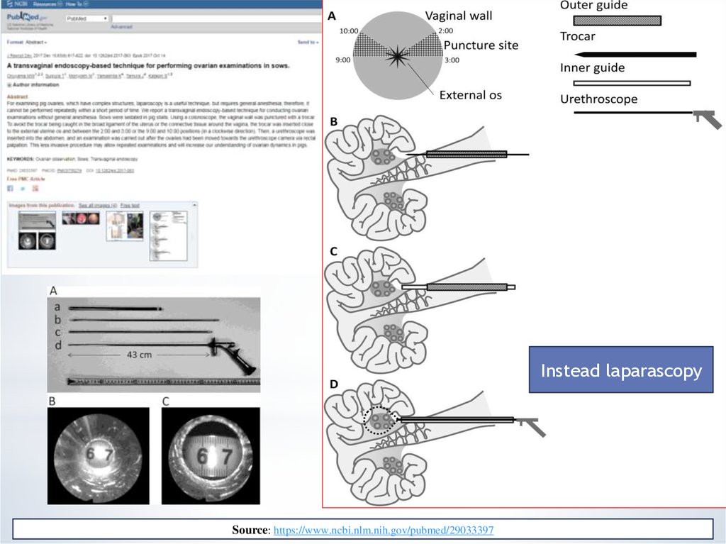
19.
Instead laparascopySource: https://www.ncbi.nlm.nih.gov/pubmed/29033397
20. Tissue biopsy and cytology
In gynecology used excisional biopsy (excision of a piece of tissue),targeted biopsy - under the visual control of an enlarged colposcopy or
hysteroscope and a puncture biopsy.
Cytological examination is a screening method for conducting mass
preventive examinations of women, in groups at increased risk for the
development of cancer.
Optic biopsy
Source: [Optical biopsy in gynecology]. Deligdisch L , Bulletin de L'Academie Nationale de Medecine, [01 Mar 2011, 195(3):605-11; discussion 611-2]
21. Cervical brush
The brush together with the dilator Cusco isinjected into the vagina. The brush is brought to
the neck, twisted, and evacuated from the
vagina.
Then
a
bacterioscopic
or
bacteriological study is performed.
Source: The efficiency of the Cytobrush versus the cotton swab in the collection of endocervical cells in cervical smears.
(PMID:3521175), Trimbos JB, Arentz NP, Acta Cytologica, [01 May 1986, 30(3):261-263]
22. Special examination methods
The symptom of “pupil”
The symptom of the extension of the
cervical mucus
KPI
Basal temperature
23. Bacteria
Bactrioscopic (microscopic)The cultural method
Molecular biological methods
Bacteriological diagnosis
Source: The vaginal microbiome: new information about genital tract flora using molecular based techniques, RF Lamont , JD Sobel , RA Akins, SS Hassan,
Chaiworapongsa, JP Kusanovic, R Romero.
24.
25.
26.
11
2
3
2
1
2
3
3
1
2
3