


 - inflammatory processes in the area of vaginal mucous membrane of the cervix and cervical canal of varying etiology. Subepithelial endometriosis – posttraumatic (after treatment or examination) implantation of endome")


 medicine
Similar presentations:


Background and precancerous diseases of female genital. Malignant neoplasms of female genital organs
1. .
Background andprecancerous diseases of
female genital.
Malignant neoplasms of
female genital organs
.
2.
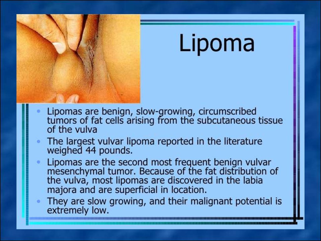
In benign tumors of the external genitalsinclude fibroma, leiomyoma, lipoma
(adipose tumor), myxoma (mucous
tumor), hemangioma, lymphangioma,
papilloma (papillary tumor),
hydroadenoma.
3. Fibroma – this tumor is mainly localized inside the labia majora or in a thin submucous layer of vagina and it’is formed by cells of the connective tissue and collagen fibers. Leiomyoma of the external genitals – a tumor that is formed from smooth m
Fibroma – this tumor is mainly localized inside the labia majora or in athin submucous layer of vagina and it’is formed by cells of the
connective tissue and collagen fibers.
Leiomyoma of the external genitals – a tumor that is formed from
smooth muscle fibers or transverse, mainly in the tissues of the labia
majora or vaginal wall.
4. Lipoma – a tumor that formed mature fatty tissue with connective tissue fibers in the pubic area and labia majora.
5.
6.
• Hemangioma– a tumor that arises due toatelectasis vessels of the skin (as node) and mucous
membranes of external genital organs.
Lymphangioma – a benign tumor that develops
from the lymphatic vessels of small nodules (blue
color and soft consistency).
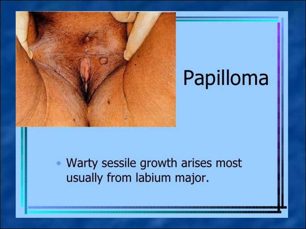
Papilloma – epithelial tumor with papillary
excrescence in the form of thin peduncle or wide
area in the labia majora .
Hydroadenoma – tumor, which is formed from
elements of the sweat glands in younger women in
the pubis and labia majora.
7.
8.
9.
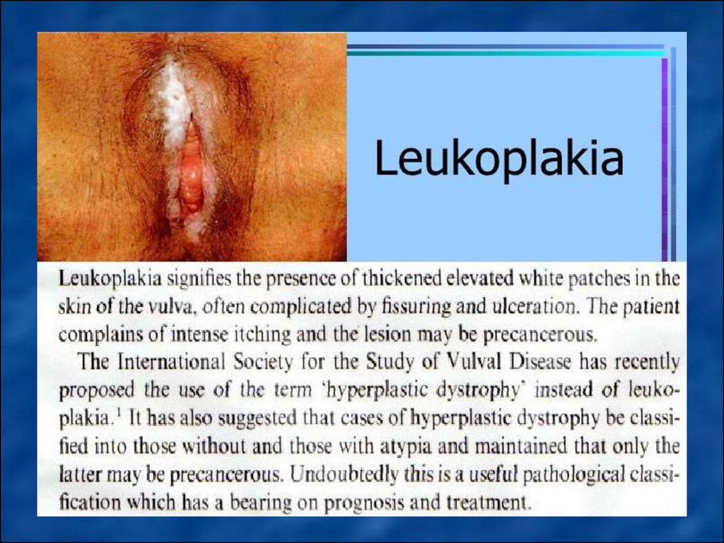
In precancerous diseases of externalgenitalia include leukoplakia, kraurosis
and Bowen’s and Paget disease.
10.
Vulvar leukoplakia developing mainly in theperimenopausal period (probably due to
hormonal disorders and immune status),
characterized by the proliferation of multilayered epithelium and violation of its
differentiation and maturation (para-and
hyperkeratosis, acanthosis without express
cellular and nuclear polymorphism, and no
violations of the basal membrane ) and shown
dry white or yellowish plaques of different size
with areas of sclerosis.
Treatment. Sedative therapy and also hormonal
therapy (androgens? Sometimes with small
doses of estrogens) are prescribed. Local
treatment is performed by corticosteroid
ointments. Good effect has magneto-laser
therapy.
11.
12.
Vulvar kraurosis develops mostly in theperimenopausal period is characterized by
papillary atrophy and mesh layers of skin,
loss of elastic fibers and connective tissue
and shown that skin and mucous
membrane of the external genitals
becomes atrophic, and fragile.
Treatment. Replacement therapy,
psychotherapy, sleeping-draughts,
sedative remedies are prescribed. But
treatment is not always effective. From
non-medicinous methods magneto-laser
therapy have been also used.
13. Vulvar kraurosis
14.
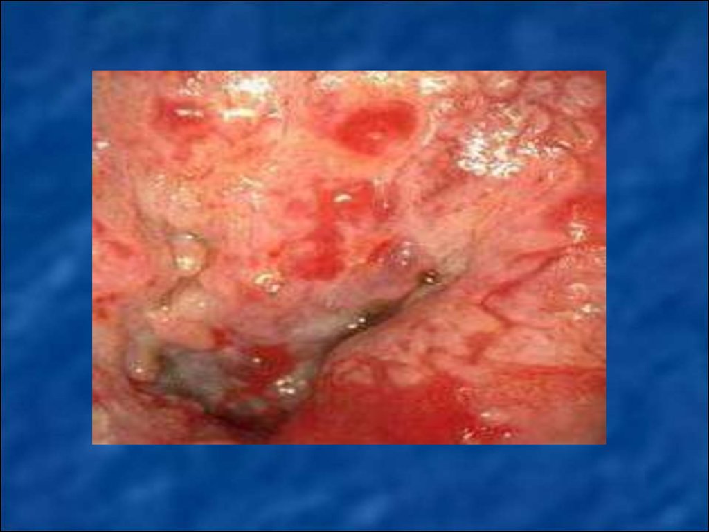
Bowen’s and Paget diseasecharacterized by hyperkeratosis and
acanthosis of the external genital
organs, shows bright-red, eczema-like
sharply limited spots with soft surface
and infiltration of surrounding tissues.
Treatment is surgical. Vulvectomy is
recommended.
15.
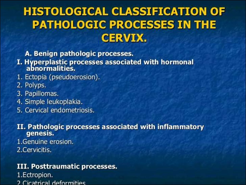
In benign (background) cervical diseasesinclude such pathological processes in which the
epithelium remains normoplaziya - ectopia of
columnar epithelium, benign transformation
zone (without atypia), cervicitis, subepithelial
endometriosis, true erosion, polyps of mucous
membrane.
16.
17.
18.
19.
20.
21.
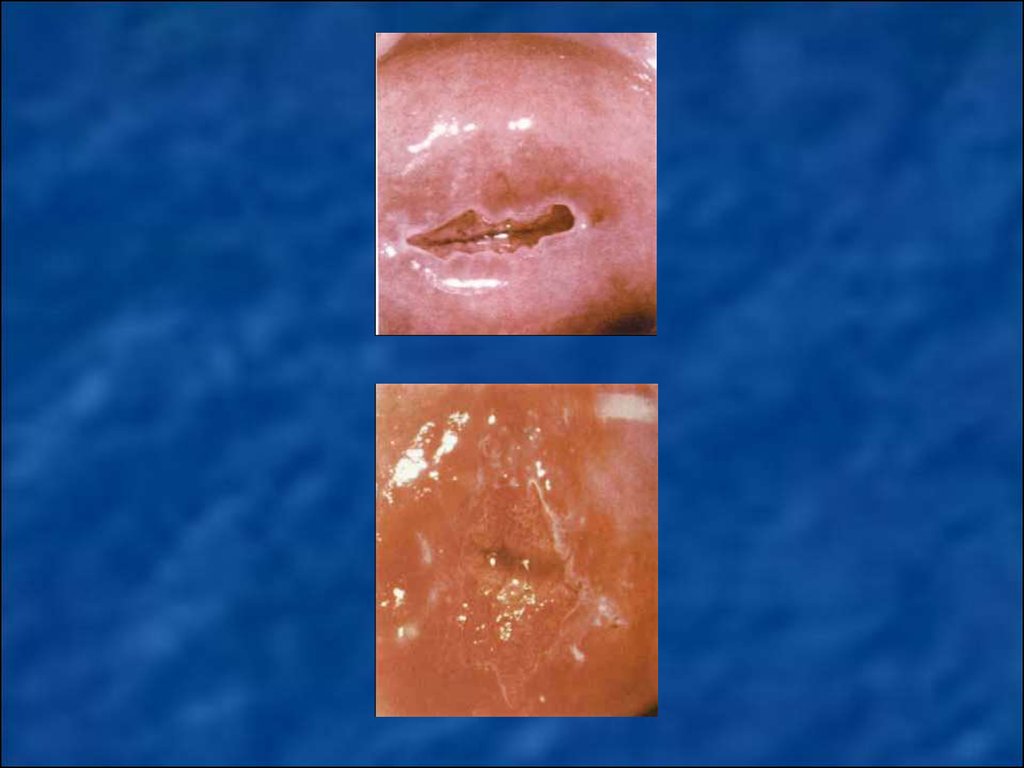
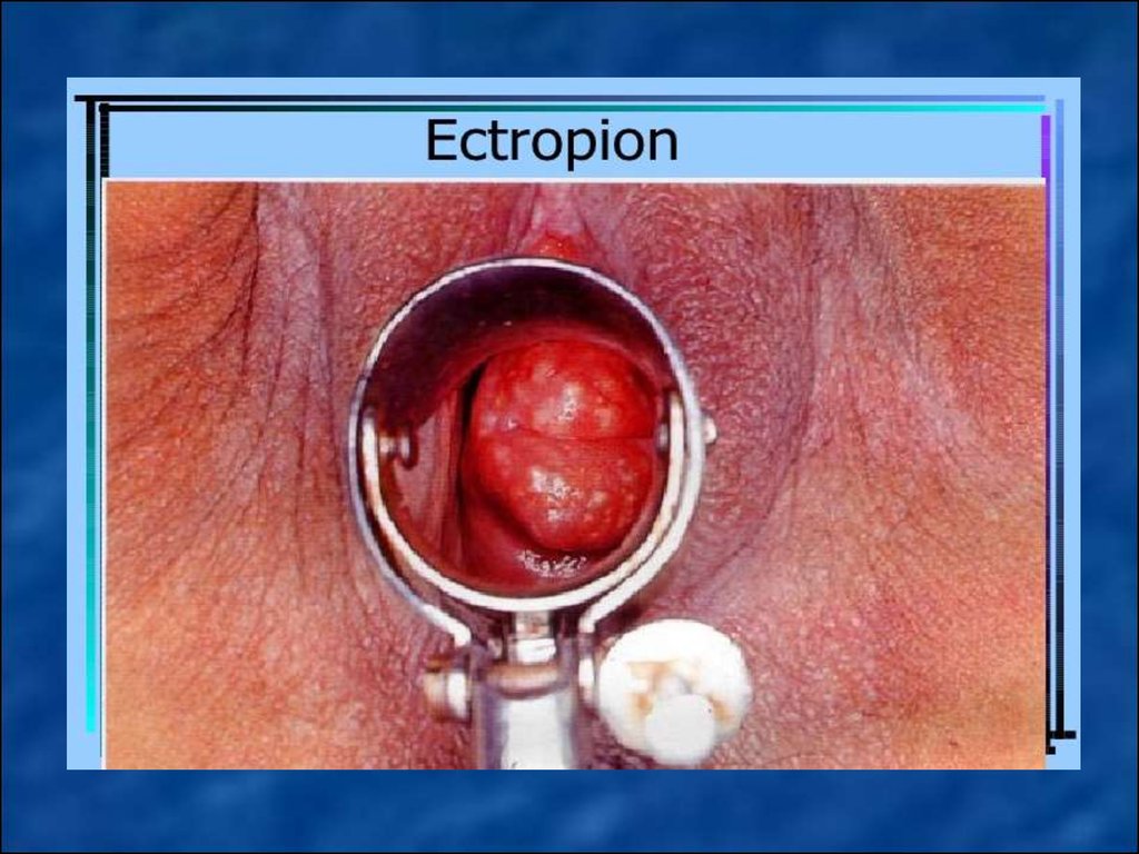
Ectopia of columnar epithelium(dishormonal, inflammatory,
posttraumatic - ektropion) - move the
cervical mucous membrane (columnar
epithelium) of the vaginal part of the
cervix.
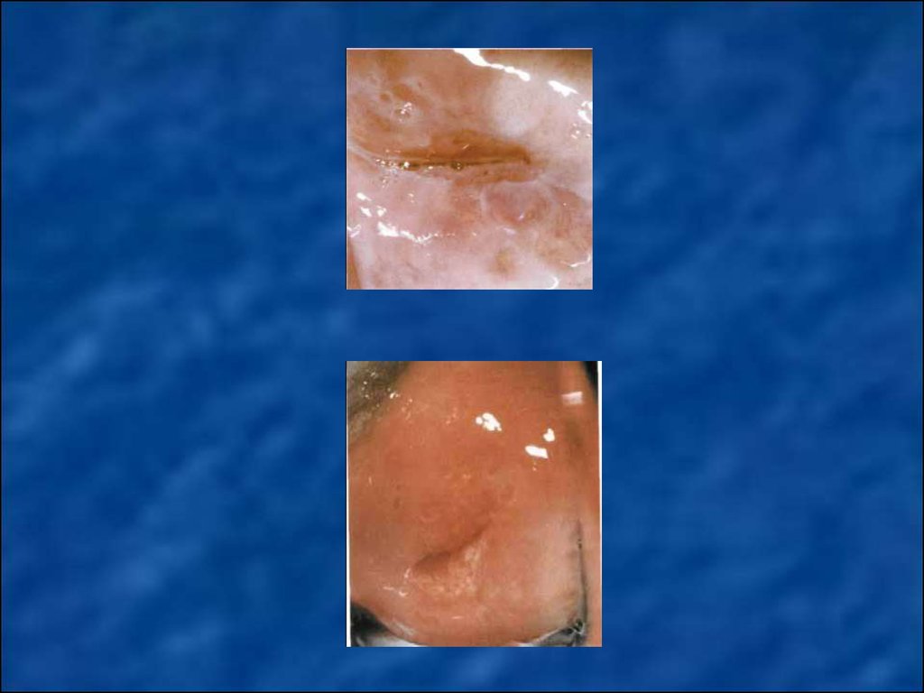
22. Cervicitis (endo-and exocervicitis) - inflammatory processes in the area of vaginal mucous membrane of the cervix and cervical canal of varying etiology. Subepithelial endometriosis – posttraumatic (after treatment or examination) implantation of endome
Cervicitis (endo-and exocervicitis) - inflammatory processes in thearea of vaginal mucous membrane of the cervix and cervical canal of
varying etiology.
Subepithelial endometriosis – posttraumatic (after treatment or
examination) implantation of endometrial cells and their proliferation,
depending on the phase of menstrual cycle, and shown from red to
brown plaques on the exocervix surface.
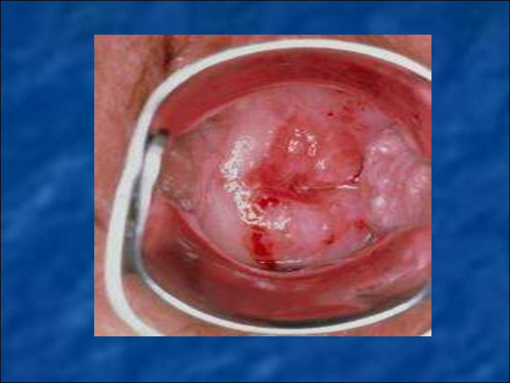
True cervical erosion is a pathological process, which is a result of
damage and following exfoliation of original squamous epithelium on
cervical vaginal part if it undergoes harmful influences, especially
mechanical traumatization or infection.
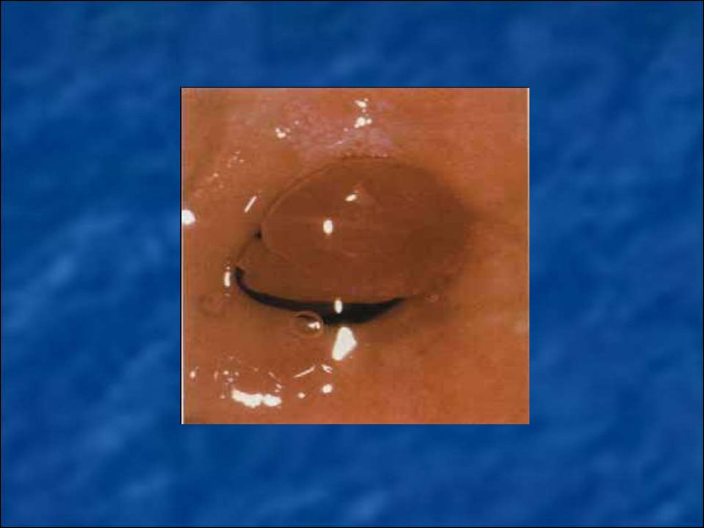
The polyps of mucous membrane of cervical canal are created
from the mucous of the external os, middle or upper third part of
endocervix. They can have a pedicle or wide base. Depending on the
dominance in their structure of grandular or connective tissue
grandular, grandular-fibrose and adenomatous polyps are distinguished.
23.
24.
25.
26.
27.
28.
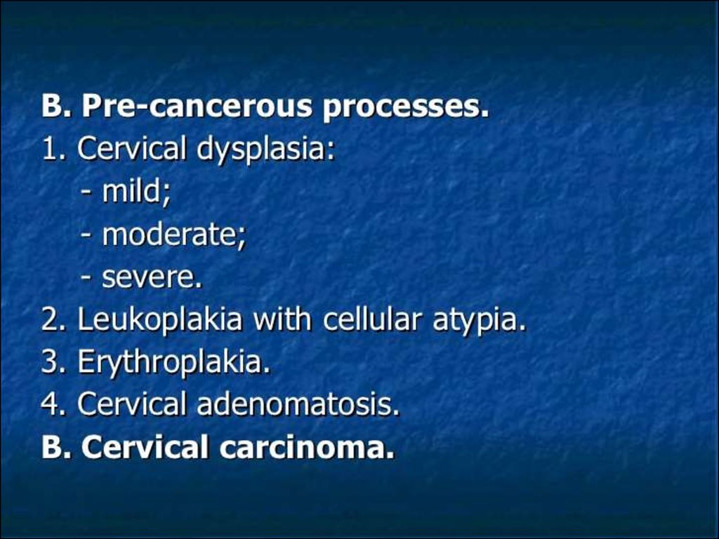
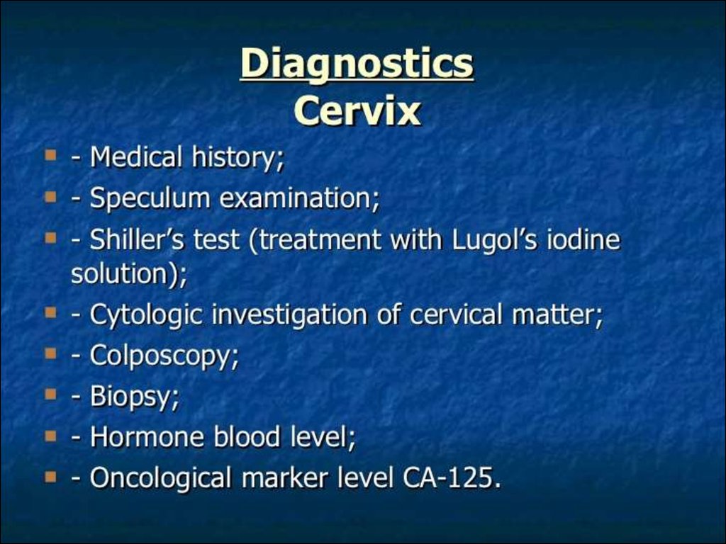
Precancerous cervical deseases(dysplasia, CIN, cervical
intraepithelial neoplasia) - is the
proliferation of cervical tissue with
the phenomena of cell atypia.
Thanks to colposcopic examination
dysplasia were distinguished on
simple leukoplakia, areas of
dysplasia, papillary zone of dysplasia,
precancer transformation zone,
condylomas and precancerous
polyps.
29.
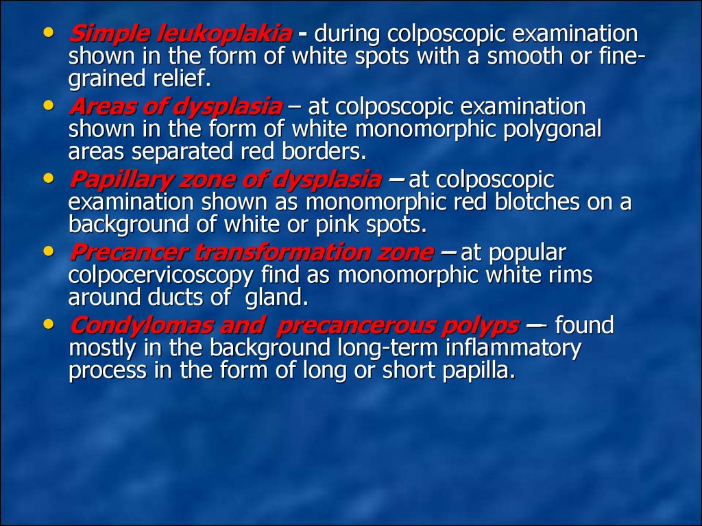
• Simple leukoplakia - during colposcopic examinationshown in the form of white spots with a smooth or finegrained relief.
Areas of dysplasia – at colposcopic examination
shown in the form of white monomorphic polygonal
areas separated red borders.
Papillary zone of dysplasia – at colposcopic
examination shown as monomorphic red blotches on a
background of white or pink spots.
Precancer transformation zone – at popular
colpocervicoscopy find as monomorphic white rims
around ducts of gland.
Condylomas and precancerous polyps –- found
mostly in the background long-term inflammatory
process in the form of long or short papilla.
30.
31.
32.
33.
34.
35.
36.
• Methods of treating precancerous cervical deseaseslare determined by the nature and degree of dysplasia
and divided into:
1. Conservative
- Anti-inflammatory therapy - purposeful antibacterial,
antimycotic, antiviral, antiseptic therapy intended to
normalize biocenosis vagina.
- When papilloma viral infection using interferon drugs.
- Hormone therapy
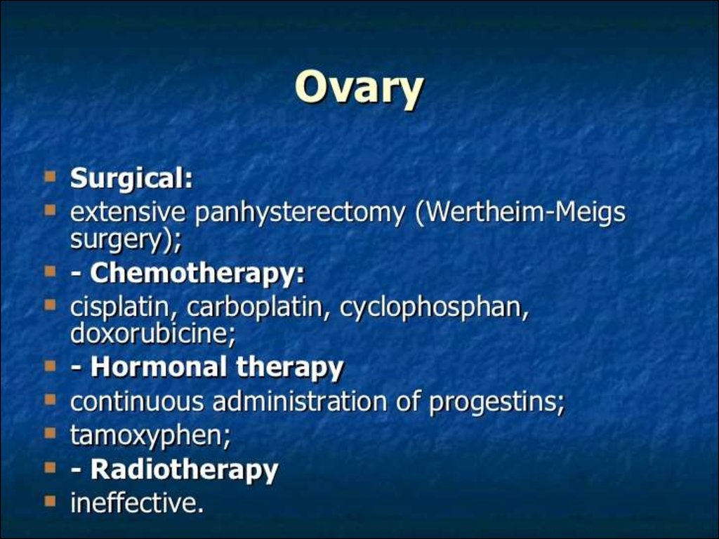
2. Surgical
- Local destruction (chemical coagulation, diathermocoagulation, cryodestruction, laser destruction, radiowave method);
- Radical operative intervention (excision of cervical,
cervical amputation, hysterectomy)
3. Combined
37.
38.
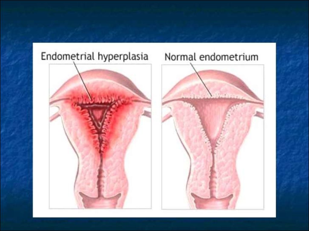
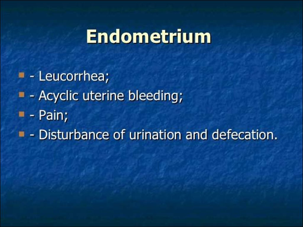
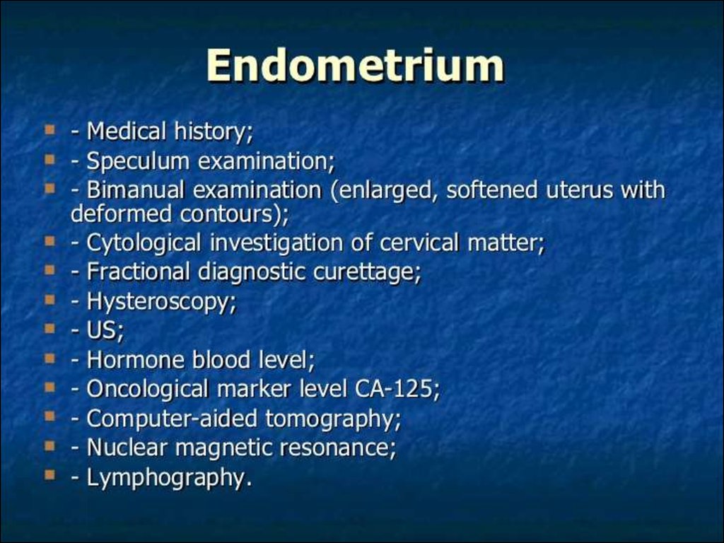
Endometrial hyperplasia - benign pathologyof mucous membrane of uterine and a
pathological proliferation of endometrium
glands, which characterized by progressing
clinical and morphological manifestations from
simple and complex hyperplasia to atypical
precancerous states of endometrium and
developing on a background of absolute or
relative hyperestrogenia.
39.
40.
41.
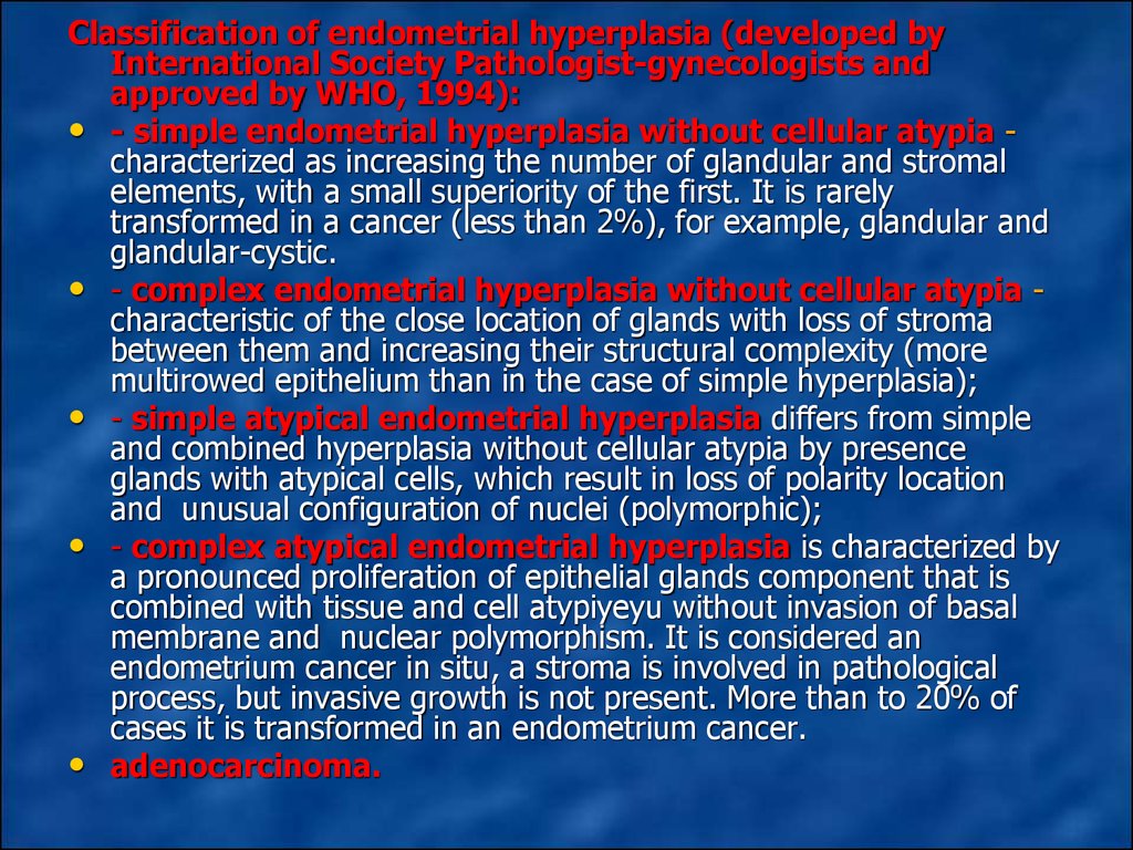
Classification of endometrial hyperplasia (developed byInternational Society Pathologist-gynecologists and
approved by WHO, 1994):
• - simple endometrial hyperplasia without cellular atypia characterized as increasing the number of glandular and stromal
elements, with a small superiority of the first. It is rarely
transformed in a cancer (less than 2%), for example, glandular and
glandular-cystic.
• - complex endometrial hyperplasia without cellular atypia characteristic of the close location of glands with loss of stroma
between them and increasing their structural complexity (more
multirowed epithelium than in the case of simple hyperplasia);
• - simple atypical endometrial hyperplasia differs from simple
and combined hyperplasia without cellular atypia by presence
glands with atypical cells, which result in loss of polarity location
and unusual configuration of nuclei (polymorphic);
• - complex atypical endometrial hyperplasia is characterized by
a pronounced proliferation of epithelial glands component that is
combined with tissue and cell atypiyeyu without invasion of basal
membrane and nuclear polymorphism. It is considered an
endometrium cancer in situ, a stroma is involved in pathological
process, but invasive growth is not present. More than to 20% of
cases it is transformed in an endometrium cancer.
• adenocarcinoma.
42.
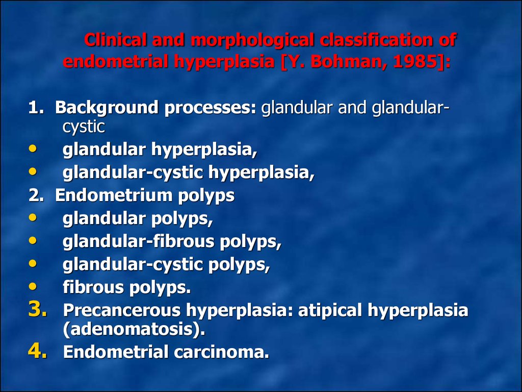
Clinical and morphological classification ofendometrial hyperplasia [Y. Bohman, 1985]:
1. Background processes: glandular and glandularcystic
• glandular hyperplasia,
• glandular-cystic hyperplasia,
2. Endometrium polyps
• glandular polyps,
• glandular-fibrous polyps,
• glandular-cystic polyps,
• fibrous polyps.
3. Precancerous hyperplasia: atipical hyperplasia
(adenomatosis).
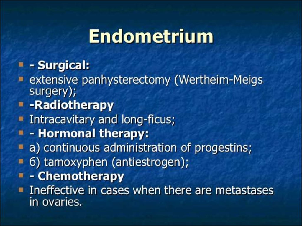
4. Endometrial carcinoma.
43.
44.
45.
46.
47.
48.
49.
50.
51.
52.
53.
54.
55.
56.
57.
58.
MALIGNANT NEOPLASMS OFFEMALE GENITAL ORGANS.