


 medicine
Similar presentations:


Oncology. Tumor
1.
LECTURE:ONCOLOGY
By A. Drevetnyak
2.
Tumor(s). /Lat., neoplasm, newgrowth/, pathological blastoma
formation, spontaneously originating in the
different organs, differing by polymorphism
of the structure, atipicity (that is difference of
tumor from initial tissues by the structure,
location and interrelation of the cells),
isolation (autonomy) and progressive
limitless growth.
Tumors may be:
a) benign and; b) malignant.
3.
Benign tumorspossess expensive growth, resulting at
that surrounding tissues move apart,
sometimes are compressed and
undergo atrophic changes. Clear
borders between the tumor and
surrounding it tissues in expansive growth
imitate the formation of capsule, though it
has no true capsules resembling inner
organs.
4.
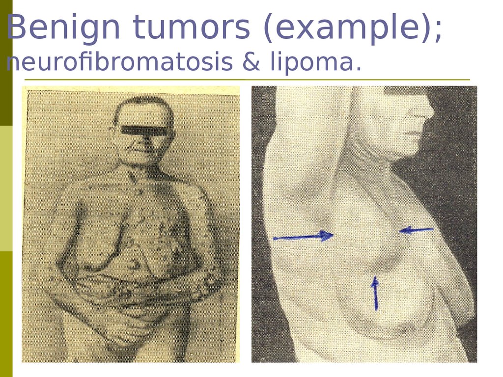
Benign tumors (example);neurofibromatosis & lipoma.
5.
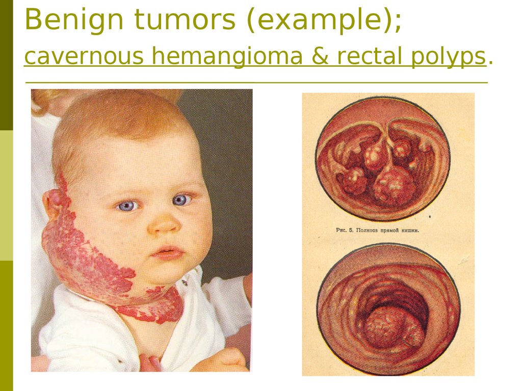
Benign tumors (example);cavernous hemangioma & rectal polyps.
6.
Malignant tumors• infiltrate and destroy
surrounding tissues.
Infiltrative (invasive) growth is
the main criterion discriminating
malignant tumors from benign
ones. Ability to metastatic
apeading is characteristic
feature of malignant tumors as
well.
7.
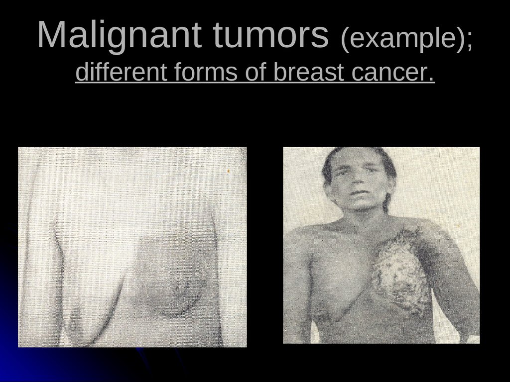
Malignant tumors (example);different forms of breast cancer.
8.
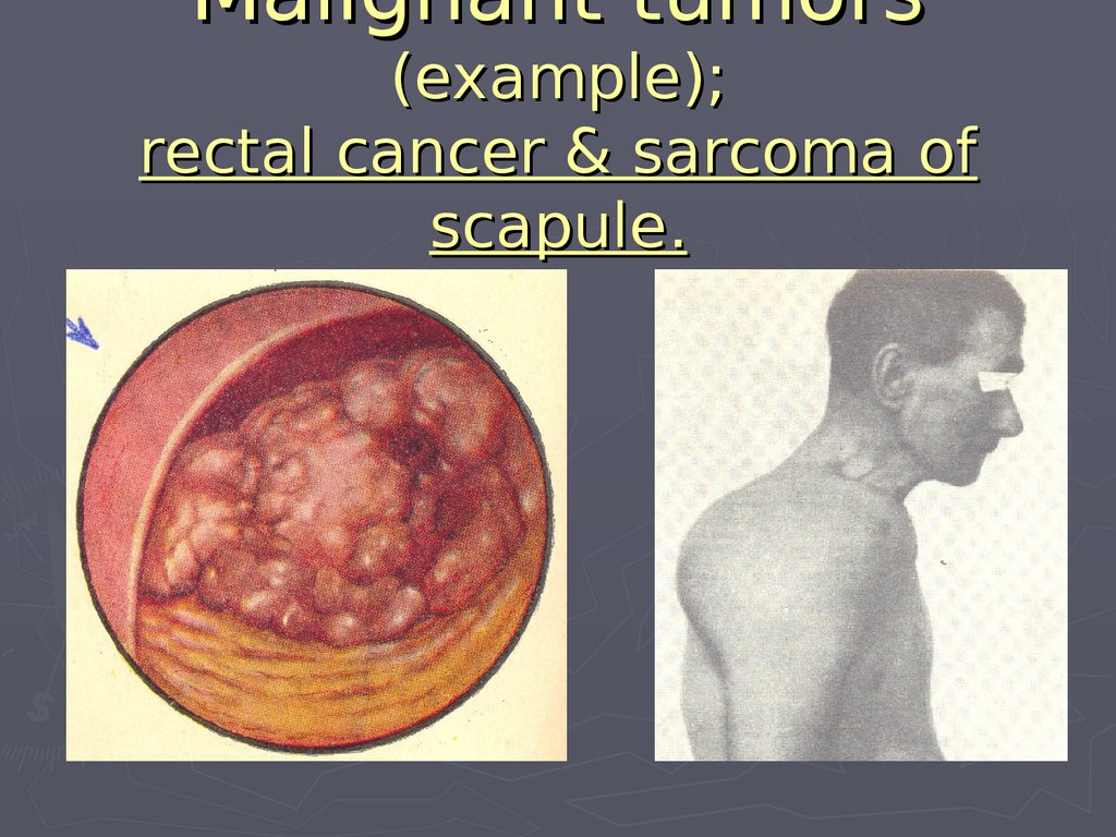
Malignant tumors(example);
rectal cancer & sarcoma of
scapule.
9.
Annually6 mln people fall ill with tumors
and 5 mln die from them. In the
developed countries among the
causes of death malignant
neoplasms take the 2nd place
after cardio-vascular pathology.
10.
Inmales the most part from all cases
of malignant tumors makes cancer of
the lung, stomach, prostate, colon and
rectum, skin.
In females the most part of all
malignant tumors takes cancer of
mammary gland, stomach, uterus,
large and small intestine. Males fall ill
with tumors 2 times as frequently, as
females.
11.
1)ETIOLOGY
Vihrov’s theory of
irritation.
2) Kangeim’s theory of
embryonic germs.
3) Fisher-Vazels’
regeneration-mutation
theory.
4) Zilber’s viral theory.
5) Immunological theory.
12.
At present tumors areconsidered to be polyetiologic
diseases.
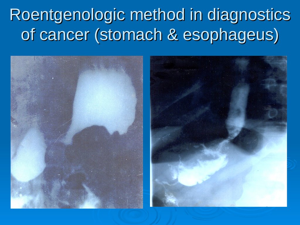
In the base of their development is:
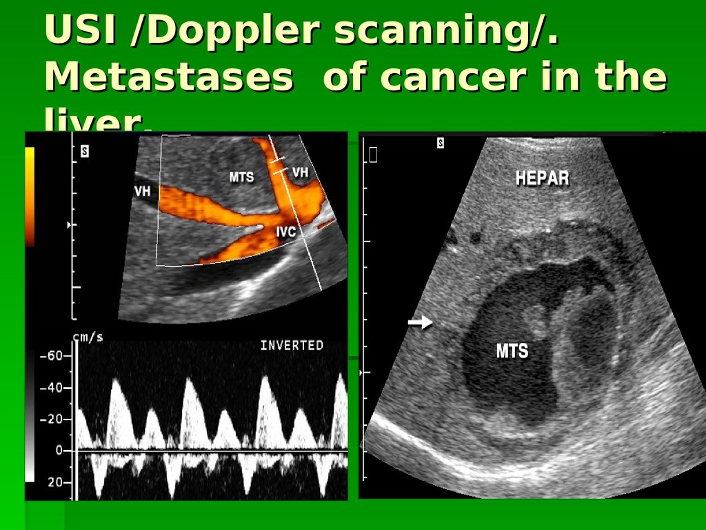
a) chemical carcinogen substances
(asbestos, polycyclic aromatic
carbohydrates – benzipiren, benzidin and
others);
b) physical (radiation: ionizing radiation,
UV-radiation);
c) biological (certain viruses);
c) frequent, repeated traumatism of tissues
with subsequent regeneration.
13.
For the origin of tumor it is ofnecessity the presence of internal
causes: genetic predisposition
and definite condition of immune
and neuro-humoral systems.
At that, oncogenic effect may be reinforced by
non- oncogenic agents and modified by
different factors.
Practically all chemical oncogenic
substances in the organism are undergo
different intricate transformations before
they gain the ability to excite oncogenic effect.
14.
Various etiological factors predominate intumours ethiology with different
localization-I.
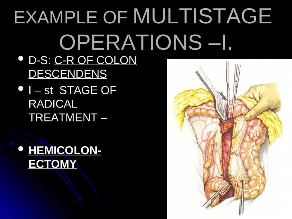
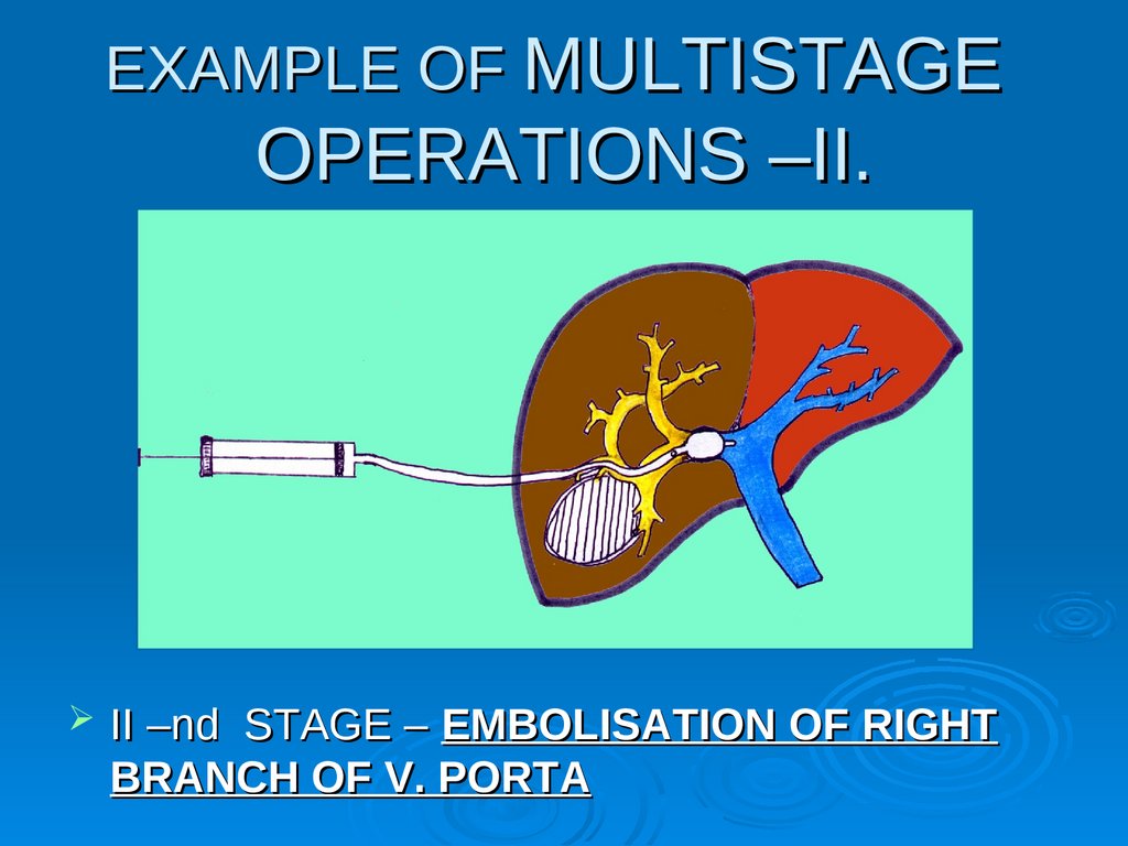
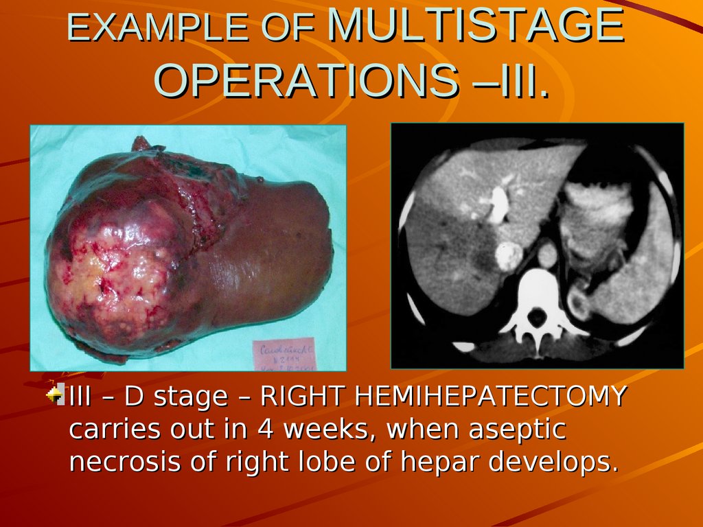
• One of the main causes of skin cancer is
UV radiation.
• Among the causes of cancer of the
larynx and cancer of the lung the most
importance is given to inhalation of
oncogenic substances /smoking/ &
chronic bronchitis play an important
role.
15.
Various etiological factorspredominate in tumours ethiology
with different localization-II.
• Tumors of the body of uterus, mammary,
prostate, thyroid glands, hypophysis,
adrenals - occur during dishormonal shifts
in the organism.
• Immunodepressive factors /in the usage of
powerful immunodepressants with the aim to
suppress the reaction of tearing away during
transplantation of organs and tissues/ the
frequency of tumor (e.g., skin cancer, cancer
of the cervix of the uterus) is 10-100 as
higher than in the same age group of
population.
16.
PATHOGENESIS-I.Arise of cancer is always separated
from the moment of etiological factor
action, so-called “latent period”, during
which clinical signs of tumor are entirely
absent.
Mechanisms
of reorganization of the
normal cell into the tumor one have
been not yet clear.
17.
PATHOGENESIS-II.At that, the cell as a result of the genome modification
gains new heritable features:
- steady reproduction;
- incomplete maturing;
- loss of the normal contact with surrounding cells
and issues;
- biological and biochemical cell organization is
changing;
- tumor cell excretes specific antigens and the
organism as a whole begins to take it in as a foreign
one, dangerous for the organism.
18.
PATHOGENESIS-III.The increase of immune exertion is a result of
the process mentioned above. In sufficient
mobilization of immune protection this
“dangerous” cell dies, tumors do not arise.
Such a process in the organism takes
place continuously!
Occasionally it happened, that cancer cell
occurs in the place, inaccessible for the action
of immunity mechanisms (in the focus of
chronic inflammation), where the lymphocytes,
antibodies can’t penetrate. In such a case
malignant cell becomes viable and begins
to reproduce.
19.
PATHOGENESIS-IV.The main components, predetermining the
origin of tumor are:
a) local preparedness of tissues;
b) general predisposition in the organism
to the occurrence of malignant tumorous
process;
c) “starting mechanism” /that is etiological
source, impulsing the development of the
process in the definite place in predisposed to
tumor individual/.
20.
PATHOGENESIS-V.Presence of precancerous condition of the
organ or tissue is the main of local factors,
predisposing to malignant tumor (“There is
no cancer without precancer, but not every
cancer turns into cancer”).
Therefore,
it is very important for
clinician /though not always it is
possible/ to fix and diagnose
precancerous condition.
21.
PATHOGENESIS-VI.Common factors predisposing to the
development of malignant process:
1) genetic predisposion;
2) changes of endocrine system function;
3) middle age and old age.
By the facts of American researchers: among individuals aged 40 years old 60 mlns
per 10 000 population die annually from cancer;
among those aged 60 years old – 400
individuals; among those aged 80 years old –
1 400 individuals.
22.
Influence of benign tumors onthe organism.
Despite of the fact that benign tumors grow
relatively slowly & don’t arise (after radical
excision), don’t metastise, they may result
in severe destructions in the organism due
to the compression of the vitally
impor-tant organs and structures, as:
1) compression of the brain by a benign
tumor of the meningeal tunic; 2) compression of large vessels; 3) compression of the
bronchus, and so on.
23.
Influence of malignant tumorson the organism (in common).
Two interrelated forms of systemic action
on the organism, common for all malignant
tumors are distinguished as:
1) competition with the organism tissues for
the vitally significant metabolites, &
2) the influence on the biological features of
different tissues resulting in disorder of
their differentiation and the weakening of
regulating influence from the side of the
organism.
24.
Influence of malignant tumors onthe organism-I.
In carbohydrate metabolism.
- In malignant tumors glucose isn’t produced.
On this account tumors can “pump” glucose
from the blood. Reserve recourses of
glycogen from the liver and mussels are
mobilized and spent. The main process, that
allows to exceed /compensate/ decrease of
glucose is glucogenesis.
-In the cases when glucogenesis has not
been stimulated or tumor has great sizes, a
pronounced hypoglycemia begins.
25.
Influence of malignanttumors on the organism-II.
In aminoacide metabolism.
Tumor tissue is a peculiar trap for nitrogen,
as entering by the alimentary path as
released in the decay of proteins and
nucleonic acids.
Introduction of glucose into the organism
saves nitrogen, prevents arise of negative
nitrogenous balance, weakens catabolism
of tissue, specifically muscular proteins.
26.
Influence of malignant tumorson the organism-III.
In fatty /lipids/ metabolism.
Growth of tumor leads to the intensive
mobilization of lipids /fatty depot and muscles/.
This process is accompanied by hyperlipidemia.
Some portion of lipids assimilates by tumor to
form membranes of the growing tumor cells.
Mobilization of lipids is considered as a
compensatory reaction of the organism on the
hypoglycemic influence of tumor, allowing the
tissues to use oxidation of fatty acids in the lack of
glucose as the additional source of energy.
27.
Nomenclature, structure& classification of tumorsI.
Histogenetic and histological principles
are in the base of the nomenclature and
classification of tumors. Their cellular and
tissue characteristic is reflected in the
names of tumors.
The names of the most tumors consist
of two parts; the 1st part includes
indication on the source of the tumor
development (cells, organ, tissue), the
second part is suffix “oma”, denoting
“tumor”. E.g., tumor developing from the
fatty tissue is called “lipoma”, from
cartilaginous tissue – “chondroma”, from
muscular tissue – “myoma”, and so on.
28.
structure &classification of
tumors-II.
In the name of tumor besides its histological
features, there is indication on its
connection with one or another organ (e.g.,
adenoma of thyroid gland), or anatomical
area (e.g., lipoma of the thigh).
In the building of some tumors’ names we
meat deviations from the indicated
principle: e.g., epithelial tumor of the liver is
called hepatoma, tumor of the brain
membranes – meningeoma, tumor of the
thymus gland – thymoma, etc.
29.
Nomenclature, structure& classification of tumorsIII.
Quite often in the name of tumor its cellular
content is stressed; e.g., tumor formed by
histiocytes - histiocytoma, -//- from Leidig’s
cells – leidihoma, from Sertoli’s cells –
sertolioma etc. Tumors arising from the
elements of hematogenic system –
lymphomae.
Tumor structure may resemble tissues or
organs; then they say about histioid or
organoid tumors. In the case of revealing in
tumor the elements of embryonal tissues,
such tumors are called “teratomae”.
Frequently the question about malignancy of
these tumors is difficult to solve. In these
30.
structure &classification of
tumors-IV.
Under the conception of possible
sources of development, tumors are
subdivided by the main kinds of
tissues:
1) epithelial;
2) connective (-tissue);
3) muscular;
4) vascular;
5) nervous;
31.
Nomenclature, structure& classification of
tumors-V.
Malignant connective (-tissue) tumors
have got the common name sarcomae. This
term is applied also for determining
malignant tumors of the muscular, vascular
and nerve tissues.
The term “sarcoma” is as a rule added by
the indication on its tissue source
(liposarcoma, chondrosarcoma). If a
tissue source of sarcoma is muscular tissue,
depending on its character (smooth, crossstriated (cross-striped), it is spoken about
32.
Nomenclature, structure &classification of tumors-VI.
Technologically various is indication of tumor
originating from epithelium. So, in the names of
benign epithelial tumors not only initial
epithelium, but especially peculiarities of tumor
tissue are taken into consideration. For
example, tumors originating from multi-layer
pavement or transitional epithelium are called
papillomae; tumors forming glandular-like
structures, originating from cylindrical
epithelium - adenomae (polyps). Malignant
tumors developing from epithelium, are called
cancer (carcinoma); for example,
“adenocarcinoma” (malignant tumor from
gland-like glandular-like structures).
33.
Nomenclature, structure &classification of tumors-VII.
CANCER /from Lat./ - malignant tumor,
developing from epithelial tissue.
Cancer –is possessing autonomous
progressive irreversible character
pathological overgrowth of atypical
epithelial cells, replacing and infiltrating
normal tissues.
34.
Nomenclature, structure &classification of tumors-VIII.
However not all mentioned features of cancer
may be considered as absolute ones. So, there
are forms of cancer, the growth of which is not
autonomous, but depends on the definite
hormones or the other factors of the organism.
The notion “progressive” also doesn’t spread on
all cases of cancer. Some kinds and stages of
cancer may exist for a long time at the level,
that has been registered in the primary
diagnostic examination without manifestating
any signs of local growth and without giving
metastases. Even “irreversibility” can’t be
considered as an absolute feature of cancer,
since occasionally in animals and human
beeings cancer may spontaneously regress.
35.
Nomenclature, structure &classification of tumors-IX.
Macroscopic view of tumor is various
one. Tumor may be have a shape of
a rounded or oval node, and may
resemble mushroom or cauliflower.
Tumor surface may be smooth, or
hilly, or rough.
36.
Nomenclature, structure &classification of tumors-X.
Relating
to the lumen of the organ, tumor
may be:
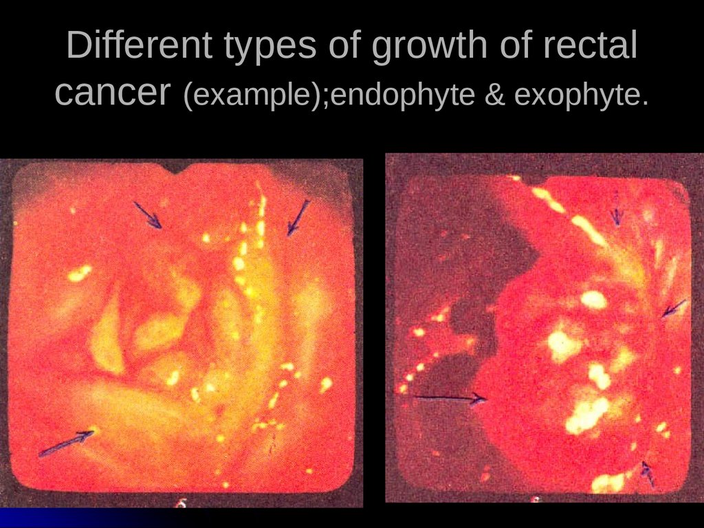
a) endophytic (tumor is growing through
the wall, has in great extent infiltrative,
“prostrate” character); or
b) exophytic (is growing through, the
lumen of the cavity, as of the stomach,
intestine, pharynx, urinary bladder and so
on).
37.
Different types of growth of rectalcancer (example);endophyte & exophyte.
38.
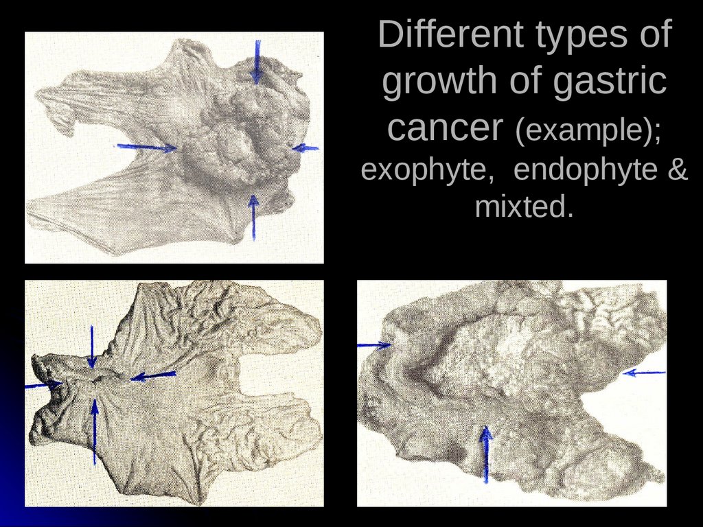
Different types ofgrowth of gastric
cancer (example);
exophyte, endophyte &
mixted.
39.
Nomenclature, structure &classification of tumors-XI.
Sometimes
the tumor as if infiltrates the
whole organ diffuse-like. Macroscopically
the border between the tumor and
normal tissue is not always
distinguishable.
In the cases, when the border is distinctly
distinguishable, they say about presence
of tumor “capsule» though tumors do not
possess true capsules.
40.
Nomenclature, structure &classification of tumors-XII.
The base for diagnosis of tumor in histological
investigation is the presence of structural
atypicity of forming it cells. Normal cells and
tissues serve as the standard for determining
the degree of atypicity for comparison.
One of the main signs of atypicity tumor tissue
is the absence of completeness of cycles of
cells and tissues development.
41.
Nomenclature, structure &classification of tumors-XIII.
Microscopic investigation of tumor displays the
degree of their differences from initial tissues,
gives the concept of the degree of their
differentiation.
The criterion of their malignance is also ability
to metastize. Usually metastases have the
structure of initial tumor, but may considerably
differ from it by the degree of differentiation
(may be less differentiated). Therefore,
recognition of the primary focus in
morphological investigation of metastases is
not always possible.
42.
Nomenclature, structure &classification of tumors-XIV.
Structural atipicity of tumor spreads over all its
components – and the main cellular one
(parenchyma of tumor), and stroma /base of
tumor/ (connective tissue, including interstitial
substance, vessels and even nerve elements).
On the assumption of relationships between
stroma and parenchyma conceptions of the
“encephaloid cancer” /soft cancer/ (poor with
stroma) and scirrhus (with sharp prevalence of
connective tissues over parenchyma).
43.
Nomenclature, structure &classification of tumors-XV.
Structure and classification of
cancer.
The variety of macroscopic kinds of
cancer is conditioned by the tumor
character, type of its growth, as well
as by the peculiarities of the structure
of tissues and organs in which this
growth takesplace.
44.
Nomenclature, structure &classification of tumors-XVI.
In benign tumors of epithelial nature
the character of tumor is usually as
follows:
1) expansive (that is with pressing
back and compression of surrounding
tissues) and
2) exophytic (that is with eminence
over epithelial covering or pavement
of the hollow organ).
45.
Nomenclature, structure &classification of tumors-XVII.
For the cancer in which tumor complexes or
individual tumor cells may infinitely
penetrate surrounding tissues, various
layers and zones (as of the initially affected
organ as of the adjusting organs and
tissues) invasive or infiltrative growth are
of character. Tumor roots itself into lymph
and blood vessels, its cells spread over the
whole organism, giving the origin of the
secondary tumor nodes /or metastases/.
46.
Nomenclature, structure &classification of tumors-XVIII.
Cancer, having endophytic growth
spreads mainly in the thickness of the
hollow organ wall, without projecting into
its lumen. Quite often these both types of
cancer growth (exo- and endophytic) are
combined; at that tumor has hemispherical
or mushroom shape.
47.
Nomenclature, structure &classification of tumors-XIX.
In
some kinds of cancer due to the lack of
correspondence between the amount of tumor
mass and the level of its blood supply
pronounced secondary changes as
inflammatory-necrotic processes develop. This
leads to ulceration of tumor, and consequently
it gets the shape of so-called “saucer-shaped
cancer”.
48.
Nomenclature, structure &classification of tumors-XX.
Quite often the base for indication the
tumor kind is the presence of the
substance or structure, producing by its
cells, e.g.: mucous, colloid, cricoidcellular, pseudomucinous cancer and
so on. All of them belong to the glandular
cancers, most of occur in the stomach or
large intestine, and differ in a high intraor extra-cellular production of mucus.
This kind of cancer may lose glandular
structure, and tumor cells are located in
mucous masses (colloid cancer).
49.
Nomenclature, structure &classification of tumors-XXI.
In
intracellular production of mucus cancer
cells, overfull with mucoid substance lie
separately in fibrous stroma (cricoidcellular cancer).
Mucous cancer is one of the most
malignant form and apt to the early
metastatic spread.
50.
classification of tumors-XXII.Some kinds of cancer have the ability to
produce specific structures characteristic
for tissues with which they are connected
histogenetically. So, in squamous cell
carcinoma corneous substance in the kind
of so-called “cancer pearls”, producing by
epidermis of normal skin frequently forms.
51.
Nomenclature, structure &classification of tumorsXXIII.
• At the same time epithelium of some
organs, in norm not forming corneous
masses, acquires similarity with skin
epithelium and begin to produce keratin
after the tumor transformation. In this
connection such forms of cancer, arising
in trachea, brochi, stomach, and ovaries
are called epidermoid cancer.
Epidermoid cancer of the stomach is
often indicated by the term “cancroid”.
52.
Nomenclature, structure &classification of tumors-XXIV.
Stromal component /besides epithelial
component/ is the important element of
tumor. It is represented by connective
tissue, vessels and nerves. The amount and
character of this component also finds its
reflection in the names of cancer forms. F.
e., some forms of adenocarcinomas are
characterized by a considerable prevalence
of tumor parenchyma over the stroma. The
latter gives them very soft consistence and
macroscopic similarity with the brain tissue
(medullary or encephaloid cancer).
53.
Nomenclature, structure &classification of tumors-XXV.
The other forms of cancer in the low level of the
stromal component development, microscopically
preserves similarity with its histogenetic source,
that has place in hepatocellular cancer.
Morphological variant of cancer with developed
stromal component in which epithelial tumor
elements are represented in the kind of separate
cells or small complexes, are called scirrhus
(Greek skirros – hard) or fibrous cancer.
54.
Nomenclature, structure &classification of tumors-XXVI.
On
the ground of histological structure, by the
extent of deviation from the normal tissue
structures there are distinguished: highly-,
moderately-, low-differentiated forms of
cancer.
There are also distinguished non-keratinizing
and more differentiated keratinizing cancer.
The latter represents histogenetically more
mature tumor and contains laminated
formations consisting of corneous scales.
55.
Nomenclature, structure &classification of tumors-XXVII.
It is International system of clinical
classification of cancer (TNM) that has got a
wide prevalence. This classification provides
determination of tumor process spread by
three criteria: the size of tumor itself, the
presence of metastases into the regional
lymph nodes, and distant metastases.
56.
Nomenclature, structure &classification of tumors-XXVIII.
• Spread of the primary tumor node is indicated
by symbol T (tumor): T1 – tumor of small sizes,
occupying a portion of the organ; T2 – tumor of
great sizes, but not exceeding the bounds of
the organ, T3 – tumor exceeding the bounds of
the organ and involving into the process
adjusting organs and tissues. Sometimes there
is distinguished the stage To (primary tumor is
not defines, but there are metastases) and
stage T is (to determine cancer in situ, that is
intraepithelial cancer). For some locations of
cancer stage T4 is provided (tumor exceeds the
bounds of the organ, causing destruction of
adjusting organs).
57.
Nomenclature, structure &classification of tumors-XXVIX.
For mammary gland tumor the gradation is
fulfilled by the sizes of tumor (in cm), for cancer
of the stomach – by the extent of growing
through the wall and spread on its portions
(cardia, body, output unit) and so on. Special
attention is paid to cancer in situ (cancer in
place). At this stage the tumor is located in
epithelium only, doesn’t grow through basal
membrane (so doesn’t grow through blood and
lymph vessels). At this stage malignant tumor is
not yet lacking in infiltrative growth and can’t
give hematogenic and lymphogenic metastatic
spreading.
58.
Nomenclature, structure &classification of tumors-XXX.
Symbol N (nodulus) is used to designate metastases
into the regional lymph nodes: Nx – there is no
information about the presence or absence of
metastases in the regional lymph nodes (the patient
has been observed incompletely, has not been
operated on); N0 – absence of metastases; N1 –
presence of metastases (in lymph nodes – collectors
of the first order). In some tumor locations depending
on the group of lymph nodes designations may vary
from N1 to N3 : N2 - there are metastases in lymph
nodes – collectors of the second order; N3 - there are
metastases in lymph nodes – collectors of the third
order).
59.
Nomenclature, structure &classification of tumors-XXXI.
Symbol M (metastases) indicates the
presence (M1 or M+) or absence (M0) of
metastases in the distant organs and
tissues.
60.
classification of tumors Index G (grade) XXXII.– defines the degree of
malignancy (degree of cells
differentiation) and is introduced into
diagnosis only after histological
estimation of tumor. Three groups of new
formations are distinguished: G1 – tumors
with low degree of malignancy (highdifferentiated tumors); G2 – tumor with
middle degree of malignancy (lowdifferentiated); G3 – tumors with high
61.
Nomenclature, structure &classification of tumorsXXXIII.
Index
P (penetration) is introduced
only for tumors of the hollow organs
and indicates te degree of growing
through their walls: P1 – tumors in
the limits of mucous membrane only;
P2 – tumors growths into submucous
layer; P3 – tumor growths through
the muscular layer; P4 – tumor
exceeds the bounds of the organ.
62.
Nomenclature, structure &classification of tumors-XXXIV.
Classification by TNM system is considered to
be convenient one, since it characterizes in
detail all sides of malignant process. It allows to
compare the results, obtained by specialists of
different countries. At the same time it doesn’t
give generalized information about the process
severity and opportunity to cure from tumor.
With this purpose clinical classification of
tumors is used.
63.
Nomenclature, structure &classification of tumors-XXXV.
According to clinical classification four stages
of tumors are distinguished:
stage I– the tumor is localized one, takes a
restricted portion, doesn’t grow through, the
organ wall, metastases (as into regional as into
distal lymph nodes) are absent;
stage II– tumor has moderate sizes, doesn’t
exceed the bounds of the organ, may be
presence of individual metastases into regional
lymph nodes;
64.
Nomenclature, structure &classification of tumors-XXXVI.
stage III– tumor has great sizes, with resolution,
grows through the whole wall of the organ / or
tumor of less sizes with multiply metastases
into regional lymph nodes/;
stage IV – growth of tumor through surrounding
organs, including those, unremovable (aorta,
vena cava, and soon), or any tumor with distant
metastases.
Stage 0 – cancer in situ also exists.
65.
Nomenclature, structure &classification of tumors-XXXVII.
For every location of malignant tumor
correspondence of clinical stages with the
stages by TNM system has been worked
out.
So, for cancer of the large intestine it looks as
follows:
stage 0 – Tis N0 M0; stage I – 1-2 N0 M0;
stage II – T 3-4 N0 M0; stage III – any T, N 1-2
M0; stage IV – any T, any N, M1.
66.
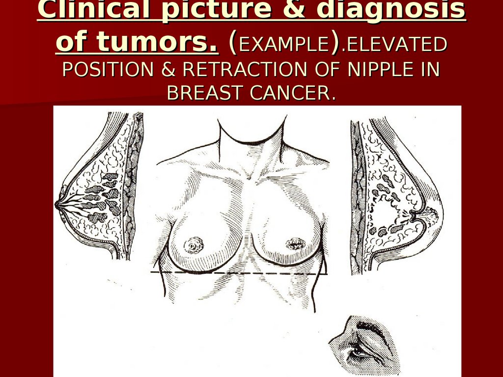
Clinical picture & diagnosis oftumors-I.
Diagnosis of benign tumors is based only on the
local symptoms, signs of the presence of the
tumor itself. At that tumor increase slowly in size
do not hurt, have a rounded shape, smooth /more
rarely – lobate/ surface, distinct border with
surrounding organs. Mainly there is prevalence of
cosmetic aspect of the disease (the presence of
the tumor itself).
Occasionally the signs of the organ’s functional
disorder appear (polyp of the intestine results in
obturative intestinal obstruction; benign tumor of
the brain, compressing surrounding portions leads
to the appearance of neurological
symptomatology and so on).
67.
Clinical picture & diagnosisof tumors. (EXAMPLE).ELEVATED
POSITION & RETRACTION OF NIPPLE IN
BREAST CANCER.
68.
Clinical picture & diagnosis oftumors-II.
In clinical pictures of malignant
tumors 4 principle syndromes are
distinguished:
- syndrome “plus-tissue”;
- syndrome of pathological
excretions;
- syndrome of the functional
disorder;
- syndrome of small signs.
69.
Сlinical picture ofmalignant tumors-I
Syndrome
“plus-tissue”. Tumor may be founded
directly in the area of its location as the additional
tissue (“plus-tissue”). This syndrome is revealed
during examination and palpation of superficial tumors
(in the skin, subcutaneous cellular tissue, muscles), as
well as on the extremities. Sometimes they succeed in
definition by palpation of the tumor, located in the
abdominal cavity. In addition to it syndrome “plustissue” is revealed by means of additional methods of
investigation: endoscopic, USI, rentgenography and
others.
70.
Сlinical picture ofmalignant tumors-II
Syndrome
of pathological discharge. In
the presence of malignant tumor due to its
growth through blood vessels a blood-stained
discharge and bleedings are often observed.
So, cancer of the stomach may cause stomach
bleeding; cancer of the lung - blood-spitting
(haemoptysis;) mammary gland cancer –
serous-blood-stained discharge from the
mammilla; cancer of the rectum – intestinal
bleedings; cancer of the kidney – hematuria;
cancer of uterus - blood-stained discharge from
genital ducts. If inflammation develops around
the tumor or mucous-making shape of cancer
is revealed, mucous (pyo-mucous) discharge
71.
Сlinical picture ofmalignant tumors-III
• Syndrome of the functional disorder.
Manifestations of this syndrome are various
ones and depend on the tumor location and
functions of the organ, in which the tumor is
located. So, in tumor of the intestine (especially
of the lest sections of the colon) the signs of
intestinal obstruction are of character.
Dyspeptic disorders (nausea, heartburn,
vomiting) are peculiar for cancer of the
stomach. In patients with cancer of esophagus
the leading symptom is disorder of the act of
food swallowing (dysphagia), and so on.
72.
Сlinical picture of malignant tumors-IVSyndrome of small signs. The patients with malignant new
formations frequently report not quite explainable /”groundless”/
complaints. There are mentioned weakness, fatigability,
increase of the body temperature, loss of weight and appetite
(aversion of meet food), anemia, increased ESR. All mentioned
above signs have been described by A.I.Savitsky and are
named “syndrome of small signs”. In some cases this syndrome
appears at the early stages of tumor and may be its only
manifestation. Sometimes it appears later on and is the
evidence of a manifest cancerous intoxication. At that the
patients have a specific “oncological” appearance they are
thinning, the turgor of tissues is reduced, the skin - pale with
grayish or icteric shade, the hair dull, the eyes – sunken. Such
an appearance of the patient says about presence in their
organism of neglected oncological process.
73.
Diagnostics of tumors-IThe
base of tumor diagnosis is their timely
identification at the early stages of disease
(1st principle: early diagnosis) when applying of
radical methods of treatment is the most effective.
Great importance in the early definition of tumor
belongs to oncological vigilance of the doctor
(2nd principle). It includes: 1) knowledge of tumor
symptoms /especially at the early stages); 2) careful
examination of patients /even in minimum complaints/;
3) collective decision of questions in difficult for
diagnostics cases. In threatening of malignant tumors
in all doubtful cases it is usual to make the most
threatening diagnosis and undertake more radical
methods of treatment
(3rd principle: hyperdiagnostics).
74.
Diagnostics of tumors-IIDiagnostics
of tumors are subdivided into:
1) primary diagnostics, carried out under conditions of
policlinic or during prophylactic examinations; &
2) clarifying diagnostics, usually carried out in the
hospital. During the primary diagnostics clinical
methods (anamnesis & objective examination) allow to
suspect tumor & to draw a rational plan of applica-tion
instrumental methods of investigation. To define early
forms of cancer of the lung fluorography of the organs
chest organs is used; cancer of the stomach & cancer
of the colon – fibrogastroxcopy & fibrocolono-scopy
with aiming biopsy; cancer of the uterus cervix –
cytological investigation; mammary gland cancer –
mammography & puncture biopsy & s.o.
75.
Diagnostics of tumors-IIIClarifying
diagnostics in patients with
already revealed malignant tumor or
suspicion on it is aimed to estimate
individual features of the disease and the
patient’s state in order to choose the most
rational method of treatment.
At that it is necessary to reveal:
a) local & b) common criteria of the
disease.
76.
Diagnostics of tumors-IVLocal criteria include: 1) adjusted location
of the primary tumor; 2) anatomical
peculiarities of tumor gowth; 3) histological
structure of tumor; 4) degree of differentiation
of tumor issue;5) stage of the disease.
Common criteria include: 1) genetic
predisposition of the patient to one or another
tumor; 2) immunological status of the patient;
3) state of metabolism; 4) hormonal profile.
77.
Diagnostics of tumors-VAlong with estimation of the local and
common criteria in the hospital individual
features of the disease are cleared up
and the degree of operation risk are
clarified. At that accompanying diseases,
functional indexes and the patient’s age
are taken into consideration. To reveal
individual features of the disease various
diagnostic methods are used.
78.
Diagnostics of tumors-VIX-ray
investigation includes: 1) uncontrast &
contrast methods. Uncontrast methods
/roentgenoscopy/ /graphy/, tomography) are used
to estimate the state of the organs of the thoracic
cavity, extremities, neck. Contrast methods (with
natural contrast – air, more often specific
contrasts) are used by the specific indications,
more often to diagnose GIT organs. Wide spread
have got methods, allowing to obtain detailed
highly-exact visualization of “slices” of the human
body organs & tissues at any depth & any level,computer X-ray tomography (CT) & nuclearmagnetic & nuclear-resonance investigation
(MRI), as well as ultrasound investigation (USI).
79.
Roentgenologic method in diagnosticsof cancer (stomach & esophageus)
80.
USI /Doppler scanning/.Metastases of cancer in the
liver.
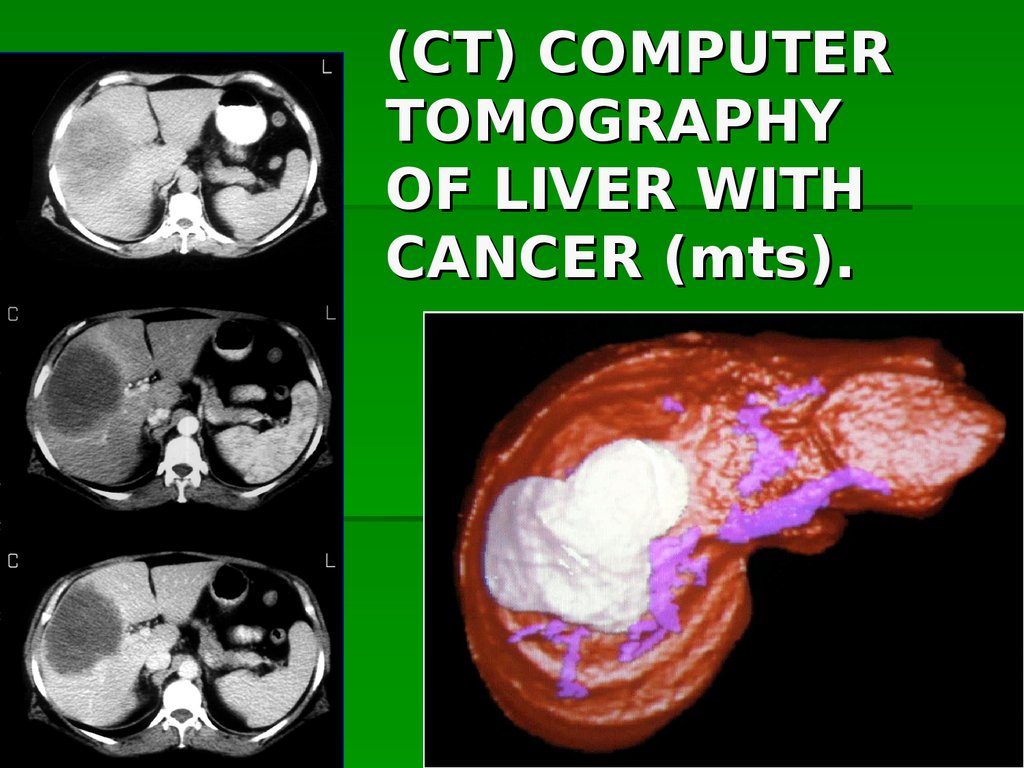
81.
(CT) COMPUTERTOMOGRAPHY
OF LIVER WITH
CANCER (mts).
82.
Diagnostics of tumors-VII• Endoscopic methods allow to carry out
investigation with the help of special
instruments, – endoscopes, - to
investigate hollow organs, abdominal and
thoracic cavities, intertissue space during
which to carry out
biopsy or take
material for histological investi-gation.
• Application of endoscopic methods allow
to diagnose early stage of tumor
(preinvasive cancer).
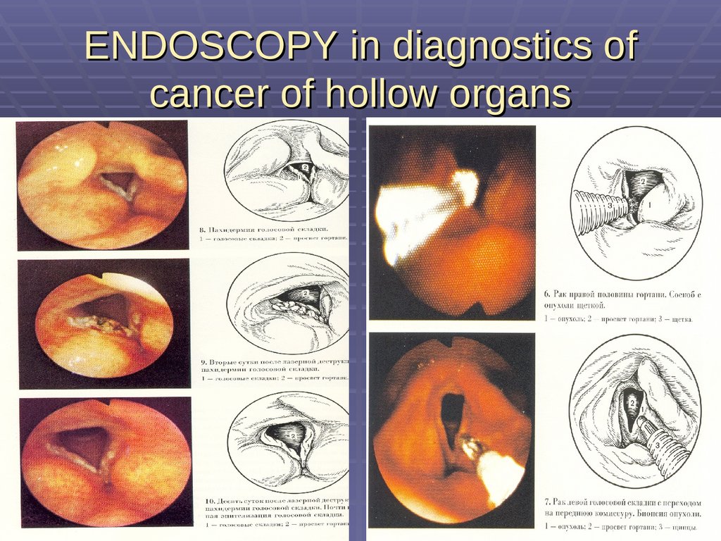
83.
ENDOSCOPY in diagnostics ofcancer of hollow organs
84.
Diagnostics of tumors-VIIIIn any case of tumor or in suspicion to
tumor morphological methods of diagnosis, in most cases it is biopsy, are used.
However, biopsy is not carried out in
suspicion to melanoma, since the
trauma may accelerate its growth.
Biopsy of osteogene sarcomas is carried
out only by the absolute indications.
85.
Diagnostics of tumors-IXRadioisotopic
methods of investigation are based
on the ability of many tumors & their metastases to
accumulate radioactive nuclides. These methods are
used to determine location of tumor, its borders,
presence of metastases, estimation of the results of
therapy, as well as revealing of functional shifts in
organs & systems, caused by the tumor & its metastases. For the diagnostic purpose radioactive
nuclide with a short period of half-disintegration is
inserted to the patient; distribution of nuclide is
caught in the patient’s organism by special counters.
86.
Diagnostics of tumors-XLaboratory
methods of diagnosis of malignant
tumors are less informative, since universal
laboratory tests for diagnosis of all kinds of
tumors are absent. Immunological tests are
used. With their help embryonic antigens,
typical for some tumors, as: alpha-fetoprotein
- in the primary cancer of the liver & teratoblastoma of the ovaries and testicle;
carcinoembryonal antigene – in the cancer of
intestine; alpha-2-H-fetoprotein - in
lymphoma, tumors of the brain and
neuroblastomas; prostate-specific antigene –
cancer of prostate gland and s.o.
87.
Diagnostics of tumors-XIIn the cases when the complex of
clarifying diagnostification methods is
found to be insufficient, the use of
diagnostic operation is indicated.
These operations are more often used
in suspicion for tumors of the
abdominal cavity organs.
88.
Treatment of tumors-ITreatment
of benign tumors - operative
excision of tumor /in individual cases it is
possible to use cryo- or laser- or
diathermo-destruction/.
Such an approach is grounded by the fact,
that benign tumors are precancerous
conditions along with ulcers, fistulas,
anacid gastritis, mastopathies, uterus
cervix erosions, pigmented birthmarks &
s.o.
89.
Treatment of tumors-IITreatment
of malignant tumors is
fulfilled by various means depending on:
a) character of tumor growth and its
histological shape; b) location of tumor;
c) clinical stage of tumor; d) the patient’s
age; presence of accompanying diseases.
Treatment of oncological patient may be:
a) radical (that cures the patient completely); 2) palliative (prolonging the
patient’s life); 3) symptomatic (alleviating only separate symptoms of the
disease).
90.
Treatment of tumors-IIIThe
main methods of treating of malignant
tumors are:
1) surgical treatment;
2) radioactive therapy;
3) medicinal treatment with the use of
antineoplastic remedies (chemotherapy).
* In the process of treating oncological patient it
is possible to combine the main methods of
treating malignant tumors. If two methods of
treating the patient are used, they say about
combined treatment, if all three methods are of
use – it is complex treatment.
91.
Treatment of tumors-IVSurgical
treatment is the main one in the most
tumors. It may be used either independently or
in combination with the other methods of
treatment. Surgical operation may be found as
a test one (in greatly developed tumor
process), radical or palliative. Palliative surgery
is for example applying of bypass anastomoses
in the tumors, that can’t be removed and cover
the lumen of GIT. Radical surgical operation
on account of malignant tumors is based on
the principle of complete removal of tumor
within the bounds of healthy tissues.
92.
SURGICAL TREATMENT OF LIPOMA93.
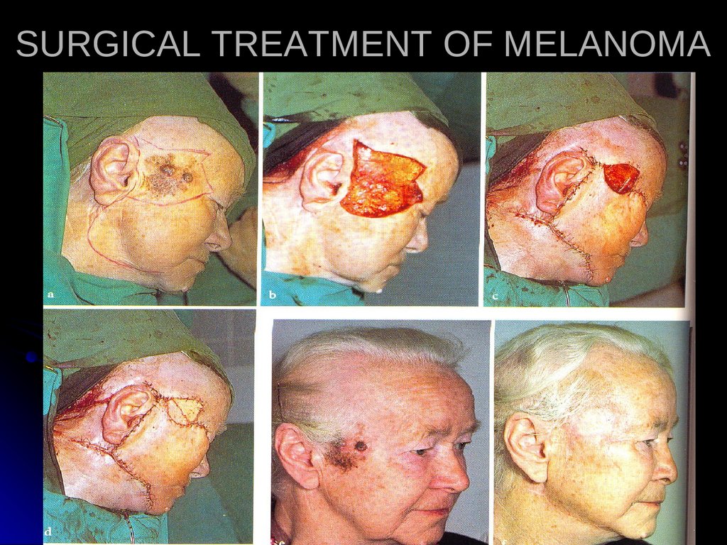
SURGICAL TREATMENT OF MELANOMA94.
Treatment of tumors-VDuring
the removal of malignant new
formation it is necessary to observe
so-called oncological principles:
1) ablastics;
2) antiblastics;
3) zoning;
4) saving «case» in process of
removing tumor.
95.
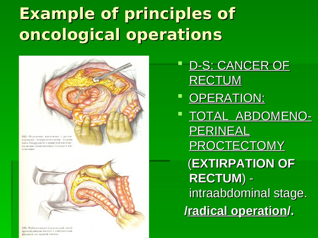
Example of principles ofoncological operations
D-S: CANCER OF
RECTUM
OPERATION:
TOTAL ABDOMENOPERINEAL
PROCTECTOMY
(EXTIRPATION OF
RECTUM) intraabdominal stage.
/radical operation/.
96.
Example of principles ofoncological operations
D-S:
CANCER OF
STOMACH (IV stage).
STENOSIS OF
ANTRAL /distal/ PART
OF STOMACH.
OPERATION:
applying of
GASTROENTEROANASTOMOSIS.
/palliative
operation/.
97.
-Treatment of tumors-VI
Ablastics – complex of measures directed at
prevention of tumor cells spread at the time of
operation.
For that: incision is performed only within the bounds of
certainly healthy tissues; to avoid mechanical injury of
the tumor tissue; aimed to ligate as quicker as possible
the vein vessels, branching from the formation; during
the operation on the hollow organ it is tying up with the
tape above & below the tumor (in order to avoid
migration of tumor cells along the lumen); the tumor is
removed as a single mass with cellular tissue and
regional lymph nodes; before manipulation with tumor
the wound is limited with a napkins; after the removal of
the tumor instruments & gloves are changed (or processed), limiting napkins are changed.
98.
Treatmentof
tumors-VII
- Antiblastics – complex of measures directed at
destruction at the time of operation of individual tumor
cells, detached from the basic tumor mass, which
may later on become the source of tumor or
metastases recurrence.
There are distinguished physical & chemical
antiblastics. The measures of physical antiblastics
include: using at the time of operation electric scalpel,
laser, cryodestruction, as well as irradiation of tumor
before operation & at the early postoperative period.
Chemical antiblastics includes: processing of the
wound after removing tumor with 70% spirit; i/v
injection of antitumoral chemical preparations on
operational table; regional perfusion of the area of
surgical operation with antitumoral chemical
preparations.
99.
Treatment of tumors-VIII-
Zoning. During the operation for
malignant tumor it is necessary to remove
not new formation only, but the whole of the
zone, where may be placed individual
cancer cells – this is the principle of zoning.
These cells may be situated in tissues
close to tumor, as well as in brunching from
it lymph vessels and regional lymph nodes.
100.
Example of principles ofoncological operations
D-S:
C-r of the right part of large bowel.
Volume of removing tissues.
101.
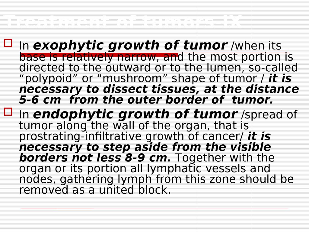
Treatment of tumors-IXIn exophytic growth of tumor /when its
base is relatively narrow, and the most portion is
directed to the outward or to the lumen, so-called
“polypoid” or “mushroom” shape of tumor / it is
necessary to dissect tissues, at the distance
5-6 cm from the outer border of tumor.
In endophytic growth of tumor /spread of
tumor along the wall of the organ, that is
prostrating-infiltrative growth of cancer/ it is
necessary to step aside from the visible
borders not less 8-9 cm. Together with the
organ or its portion all lymphatic vessels and
nodes, gathering lymph from this zone should be
removed as a united block.
102.
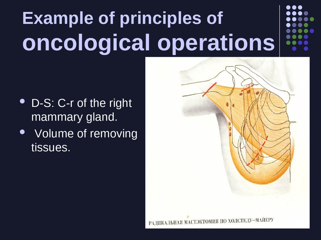
Example of principles ofoncological operations
D-S: С-r of the right
mammary gland.
Volume of removing
tissues.
103.
Treatment of tumors-X• (Saving/keeping/ «case»). Lymph vessels and
nodes, through wich is possible the spreading of
tumor cells, are located in the cellular space, divided
by fascial partitions. In this connection for the most
efficiency it is necessary to remove the cellular tissue
of the whole fascial sheath, advisable coupled with
fascia. As an example of observing the principle of
saving /keeping/ «case» is operation for cancer of
thyroid gland /at that extracapsular removal of thyroid
gland with visceral leaf of the IV fascia is used/In a
number of cases it is justification to perform combined
operations, when together with affected organ a partial
resection of the adjusting organs is done.
104.
Treatment of tumors-XI• Radical operations in cancer of any location
may be one- or multistage. For example,
cancer of the colon with great metastasis into
the liver, is not removable by means of
common resection of the liver. Hemicolectomy
will be the first stage. The second stage –
embolization of the branch of the portal veins
and hepatic arteries, which supply an affected
portion of the hepatic lobe. The third stage is
accomplished after 4 weeks, when aseptic
necrosis of the lobe begins; hemihepatectomy
105.
EXAMPLE OF MULTISTAGED-S:
OPERATIONS –I.
С-R OF COLON
DESCENDENS
I – st STAGE OF
RADICAL
TREATMENT –
HEMICOLON-
ECTOMY
106.
EXAMPLE OF MULTISTAGEOPERATIONS –II.
II –nd STAGE – EMBOLISATION OF RIGHT
BRANCH OF V. PORTA
107.
EXAMPLE OF MULTISTAGEOPERATIONS –III.
III – D stage – RIGHT HEMIHEPATECTOMY
carries out in 4 weeks, when aseptic
necrosis of right lobe of hepar develops.
108.
Treatment of tumors-XIIBesides classical surgical treatment laser- or
cryodestruction is performed. Cryodestruction
is used in cancer of the skin as well as in
neglected cases of cancer of the rectum,
esophagus with the aim to restore passability
and to control the pain, to stop bleedings from
tumor.
The results of surgical treatment depend
mainly on the kind and location of tumor and on
the stage of the disease. The best results have
been achieved in treatment of cancer of the
skin. In this location 5-years’ survival mounts to
90 %. At the same time in cancer of the
stomach even after radical operations 5 years’
and more live about 30 % patients.
109.
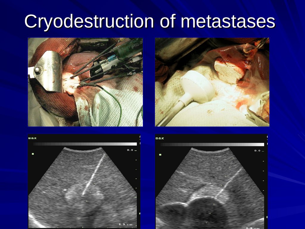
Cryodestruction of metastases110.
Treatment of tumors-XIIIRadiation therapy. Application of radiation
therapy for treatment of oncological patients is
based on the fact, that rapidly reproducing
tumor cells are more sensitive to the action of
ionizing radiation.
As the result of radiation damage of DNA &
RNA, inactivation of a number of ferments,
damage of cellular membranes - complicated
disorders of metabolic processes of tumor cells
arise. Consequently during of the mitosis
phases its (tumor’s) death begins.
Simultaneously with necrotic processes
regeneration of the connective tissue is
observed. Subjected to the necrosis tumor
tissue is replaced by cicatrical one.
111.
Treatment of tumors-XIVThe efficiency of the damage of tumor cells depend on the size of absorbed dose of radiation,
the factor of the time (in the cases of interrupted or continuous radiation), the state of oxygenation of tumor, its reproductive ability, the
degree of the cellular anaplasia, & the phase of
mitotic cycle. The most sensitive to radiation
are connective-tissue tumors with round-cellular structures (lymphosarcomas, myelomas),
some kinds of epithelial tumors (seminoma),
tumors with histological substrate of the integumentary epithelium (c-r of the skin, c-r of the
lip, c-r of the esophageus). Less sensitive are
glandular tumors (adenocarcinomas), highly
differentiated sarcomas (fibro-, osteosarcomas), as well as melanoblastomas.
112.
Treatment of tumors-XVExisting difference in radio-sensitivity of tumor
and normal tissue is causes the development
after radiation of the most expressive
destructive changes in tumor as compared with
normal tissues. This difference is called
radiotherapeutic interval. For the widening
this interval the methods possessing radiosensitizing action (radiation under conditions of
hyperbaric oxygenation, hyperthermia) are
used, preparations, oppressive restoration of
the damaged tumor cells are injected.
113.
Treatment of tumors-XVIDifferent kinds of electromagnetic or cor-
puscular irradiation are used for radiation
therapy, namely: X-ray, gamma-, bremsstrahlung (megavolt) radiations, streams
of neutrons, protons, electrons.
Depending on the place of being of the
radiation source three main kinds
(methods) of radiation therapy exist.
114.
Treatment of tumors-XVIIIn the external (distance) radiation
devises for X-ray therapy and
telegammatherapy (isotopes Co 60
and Cs 137) is used.
The method is the most effective in
superficially tumors. In deep-seated
tumors multifield (consisting of
several fields) cross impact or
rotatory radiation is applied.
115.
Treatment of tumors-XVIIIIntracaval radiation allows to move a
source of radiation near the place of tumor
location. The source of radiation through
the natural openings is inserted into the
urinary bladder, cavity of the uterus, oral
cavity – achieving the maximum dose of
radiation of the tissue tumor.
Intracaval radiation is often combined
with distance radiation therapy.
116.
Treatment of tumors-XIXApplique
’radiation is indicated
in the relatively superficial
tumors (of the skin, low lip, eye).
It is carried out with the help of
applicator, on the surface of
which radioactive Ra 224, Sr 90,
Co 60 and others are placed.
117.
Treatment of tumors-XXInterstitical radiation is carried out be
introducing into tumoral tissue radioac-tive
needles, hollow nylon tubes with isotopes or
granules Au 198. In some cases radiation of
tumor may be achieved by means of infiltration
of it with colloid solutions containing isotopes or
by means of their endolymphatic induce. In
some tumors, as in cancer of thyroid gland,
their features to accumulate selectively
radioactive iodine combinations are used.
118.
Treatment of tumors-XXIDepending on the purpose of radiation therapy /if it is
used independently/ is divided into: radical, palliative
and symptomatic.
Radical radiation therapy makes provision for radiation of zone
of the primary tumor and its regional metastases in medicinal
doses.
Palliative radiation therapy is aimed to the partial destruction
of tumor and stabilization of the process. At that, focal doses
are usually lower than in radical treatment.
Symptomatic radiation therapy is used at the later stages of
tumor process and is directed on the elimination of individual
symptoms, dominating in clinical picture of the disease (pain,
compression syndrome and so on). The effect achievable in the
result of symptomatic radiation therapy has a temporary
character.
119.
Treatment of tumorsXXIIRadiation
methods of treatment may be a
part of combined or complex treatment of
oncological patients. Preoperative radiation
therapy is indicated in the presence of tumors which
are more often recur and spread metastases, as in
melanoma, sarcoma of soft tissues, cancer of the
upper jaw, cancer of rectum, cancer of the mammary
gland, and others. At that, the principle task comes to
the lowering of tumor cells ability for implantation in
normal tissues and development of metastases or
local recurrence. Postoperative radiation therapy is
directed to the elimination of tumor cells in the
operative zone projection (in the case of insufficient
radical surgical operation).
120.
Treatment of tumors-XXIIIMedicinal treatment,or chemotherapy –
means the use of medicinal preparations,
having a damage effect on the tumor tissue.
In the most cases chemotherapy is one of the
components of complex treatment and is used
on a certain stage of the disease, supplying
abilities of surgical and radiation therapy.
However, in some oncological diseases
(hemoblastomas) chemotherapy is used as a
unique method of treatment.
121.
Treatment of tumors-XXIVAll
preparations used for medicinal treatment of
tumors are subdivided into two groups: hormonal
preparations and proper antitumoral preparations.
Hormones are applied for the treatment of tumors,
keeping the ability of the initial tissues to respond to
hormones, in norm regulating their growth (in so-called
hormone-depended tumors): cancer of prostate, cancer of
mammary gland, cancer of the uterus body, cancer of
thyroid gland). Antiestogenes are relative to hormones,
they block up the receptors of steroid hormones).
Corticosteroids are used in the schemes of hemoblastoses
treatment too.
122.
Treatment of tumors-XXVProper
chemotherapeutic preparations are
subdivided into:
1) cytostatics:
a) alkylating preparations; b) alkaloids;
2) antimetabolits;
3) antitumoral antibiotics;
4) platinum preparations;
5) immunomodulators.
123.
Treatment of tumors-XXVICytostatics
inhibit reproduction of tumor cells,
oppressing their mitotic activity. At that alkylating
preparations (cyclophosphan, thyo-taf, embyhin,
myelosan) attack nucleophilic group and form
covalent connections. Alkaloids (vincristin,
vindlastin), connecting with microtubes, blockade
metaphase of mitosis.
Antimetabolits (Metotrexat, 5-ftoruracil, mercaptopurin) influence on S-phase of mitosis by means
replacing normal metabolits and competition with
normal metabolits in the tying with certain (catalytic
and allosteric) centers of ferments and with cellular
ferments.
124.
Treatment of tumors-XXVIIAntitumoral
antibiotics (doxorubicin, bleomycin,
rubromycin, mitomycin) suppress DNA and RNA
synthesis, strengthen activity of cellular cycle
regulators.
Platinum compounds (cysplatin, oxaliplatin)
interact with DNA, forming interchain ties; are
connected with nuclear and cytoplasmic proteins.
Immunomodulators (interleukin-2, interferon) at
the expense of stimulation of immunity in some
tumor locations (cancer of the kidney) contributes
to the stabilization of oncological process even in
going too far stages of the disease.
125.
Treatment of tumors-XXVIIITaking into account up-to-date abilities of
chemotherapy all tumors may be conditionally
subdivided into 4 groups:
1. tumors, in which recovery with the help of
chemotherapy is principally possible
(chorionepithelioma of uterus, acute leucosis in
children, lymphogranulomatosis, seminoma of
testicle, Berkitt’s lymphoma);
2. tumors, in which chemotherapy gives clinical
efficiency (acute leucosis in adults, chronic
leocoses, lymphosarcoma, myeloma disease,
Yung’s tumor, cancer of mammary gland, cancer of
ovaries, Vilms’ tumor, angiogenic sarcomas);
126.
Treatment of tumors-XXIX3. tumors with low sensitivity to the action of
chemotherapy (cancers of GIT, melanoma, squamous
cell cancer (carcinoma) of the head and neck,
retinoblastoma, cancer of the urinary bladder,
insuloma, leuomyosarcoma, sinovial sarcoma,
osteogenic sarcoma);
4. tumors which are practically not sensitive to the up-
to-date chemotherapeutic preparations (cancer of the
cervix of uterus, fibrosarcoma).
127.
PROGNOSISPrognosis is determined first of all
by the location, stage & degree of
tumor differenti-ation, as well as by the
state of the initial immune patient’s
status. In the external tumor location &
timely treatment the prognosis is quite
favorable. Location of tumor in internal
organs, as well as presence of metastases in
regional lymph nodes (especially – of distant
metastases in the internal organs) greatly
worsen prognosis. In sharp suppression of
initial immune status (anti-tumoral
immunity) prognosis greatly worsens. In
low-differentiated cancer prognosis of