


 medicine
Similar presentations:


Lung cancer
1. LUNG CANCER
Department of oncologyCrimean State Medical University
Aliev K.A.
2. Lung cancer is the second most common malignancy
• Most lung carcinomas are diagnosed at anadvanced stage.
• The need to diagnose lung cancer at an early and
potentially curable stage is obvious.
3. EPIDEMIOLOGY Incidence
• Lung cancer incidence is increased in urban areas• The highest incidence in male is in Scotland, USA, Poland
• The lowest incidence in male is in Syria, Salvador,
Thailand
4. Now lung cancer is the first most common cause of death in men
5.
EPIDEMIOLOGYDeath rate
• The highest death rate in male is in Scotland (105,2),
Belgium (104,1), and in Netherlands (103,8).
• The lowest death rate is in Peru (7,3), Martinique
(12,2), and in Surinam (15,9).
6.
EPIDEMIOLOGYSex
• Lung cancer is more common in men than in
women.
• The incidence of lung cancer started to decline
among males in the early 1980s and has
continued to do so over past 20 years.
• By contrast, the incidence in women started to
increase in the late 1970s and only recently
reached a plateau.
7.
EPIDEMIOLOGYAge
• The probabilities of developing lung cancer
• among males: from birth to 39 years, 0.04%;
40-59 years, 1.24%; 60-79 years, 6.29%.
• among females: from birth to 39 years, 0.03%;
40-59 years, 0.92%; 60-79 years, 4.04%.
8.
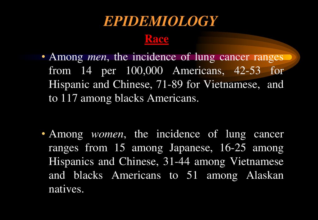
EPIDEMIOLOGYRace
• Among men, the incidence of lung cancer ranges
from 14 per 100,000 Americans, 42-53 for
Hispanic and Chinese, 71-89 for Vietnamese, and
to 117 among blacks Americans.
• Among women, the incidence of lung cancer
ranges from 15 among Japanese, 16-25 among
Hispanics and Chinese, 31-44 among Vietnamese
and blacks Americans to 51 among Alaskan
natives.
9.
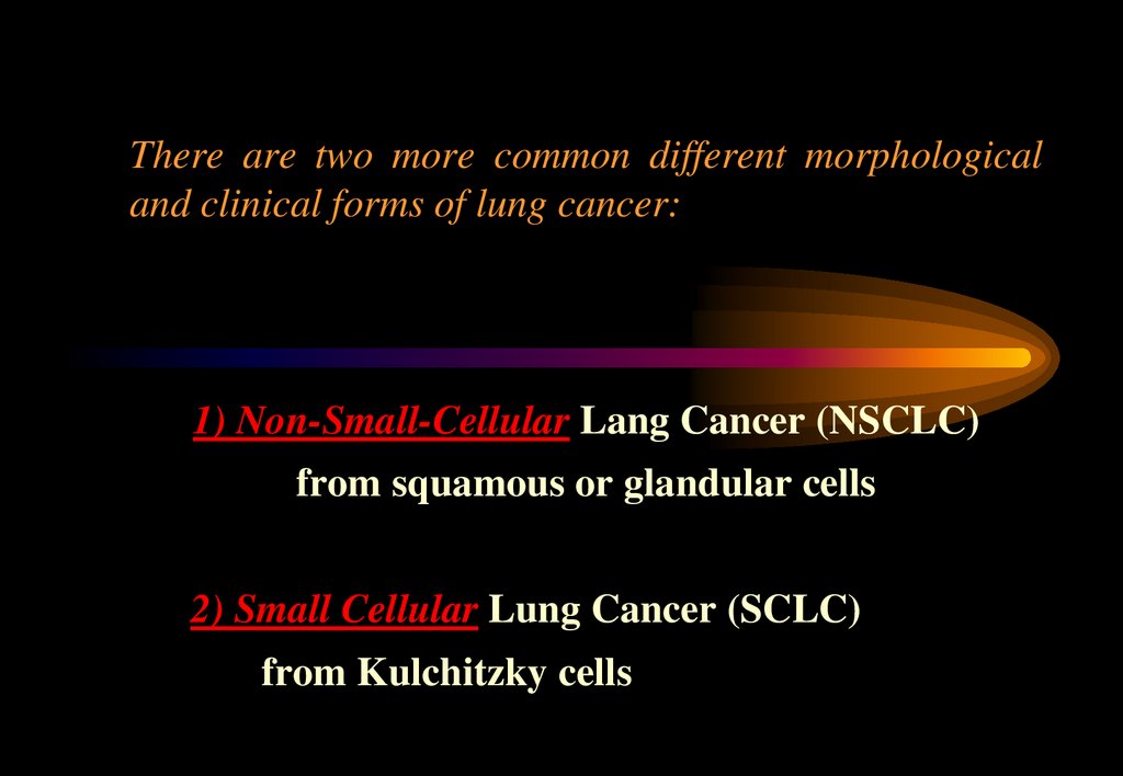
There are two more common different morphologicaland clinical forms of lung cancer:
1) Non-Small-Cellular Lang Cancer (NSCLC)
from squamous or glandular cells
2) Small Cellular Lung Cancer (SCLC)
from Kulchitzky cells
10.
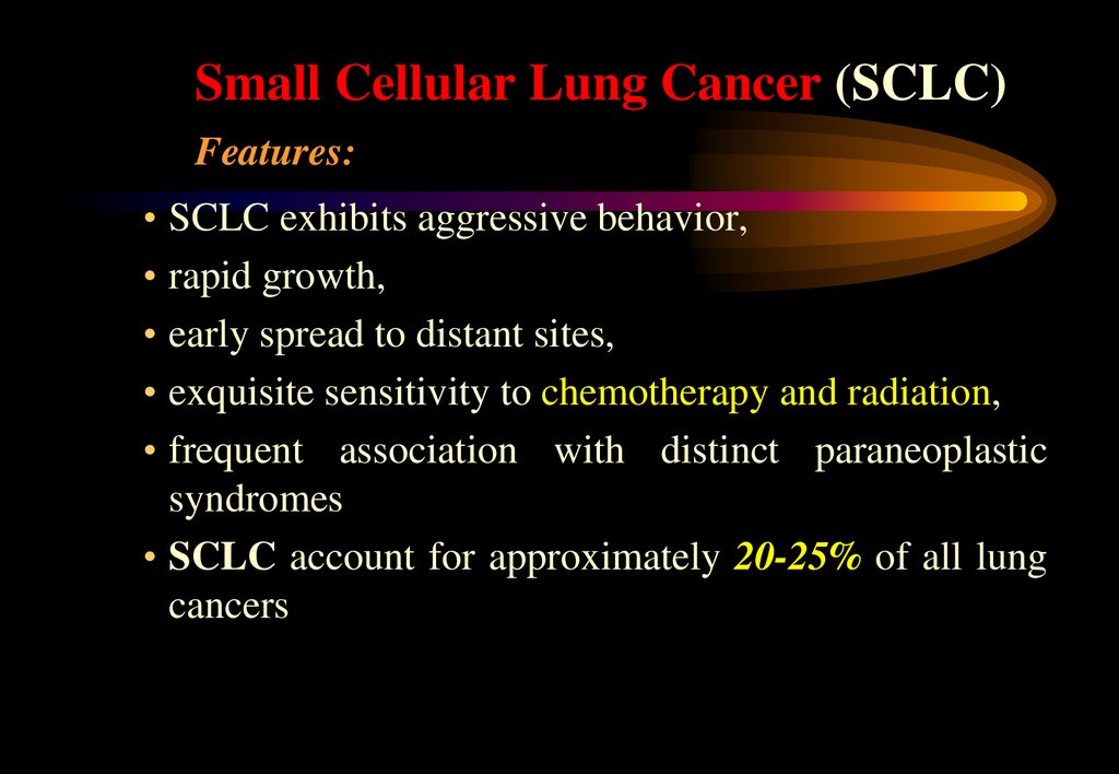
Small Cellular Lung Cancer (SCLC)Features:
• SCLC exhibits aggressive behavior,
• rapid growth,
• early spread to distant sites,
• exquisite sensitivity to chemotherapy and radiation,
• frequent association with distinct paraneoplastic
syndromes
• SCLC account for approximately 20-25% of all lung
cancers
11.
Non–small cell lung cancer (NSCLC)Features:
• accounts for approximately 75-80% of all lung cancers.
• non–small cell lung cancer requires meticulous staging,
because the treatment and prognosis vary widely
depending on the stage.
• in non–small cell lung cancer, surgical resection offers
patients the best chance for survival.
12.
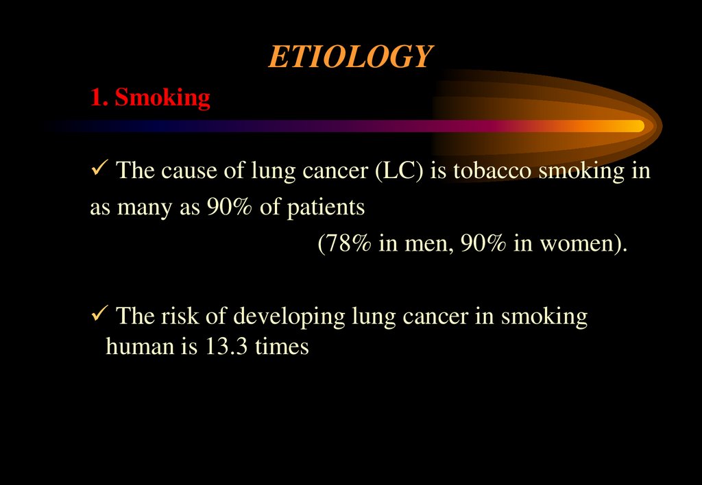
ETIOLOGY1. Smoking
The cause of lung cancer (LC) is tobacco smoking in
as many as 90% of patients
(78% in men, 90% in women).
The risk of developing lung cancer in smoking
human is 13.3 times
13.
ETIOLOGY2. History of interstitial lung disease
Concomitant chronic obstructive bronchitis,
tuberculosis, pneumosclerosis and pneumoconiosis are
a risk factor for LC.
3. Asbestos
Asbestos exposure increases the risk of developing LC
by as much as 5 times. The silicate type of asbestos
fiber is an important carcinogen.
Tobacco smoke and asbestos exposure act
synergistically.
14.
ETIOLOGY4. Radon
Approximately 2-3% of lung cancers annually are
estimated to be caused by radon exposure.
5. HIV (human immunodeficciency virus) infection
It is a 6.5-fold increase in lung cancer in patients
infected with HIV.
15.
ETIOLOGY6. Other environmental agents
Aromatic polycyclic hydrocarbons, chromium, and
diesel exhaust all have been implicated in causing LC.
16.
PATHOPHYSIOLOGY• The base of pathogenesis of central lung cancer is
the metaplasia of bronchial epithelium due to
irritant actions by smoke, and chronic
inflammatory processes.
• The base of peripheral lung cancer is the scars on
the lung parenchyma by tuberculosis, and
fibrosis.
17.
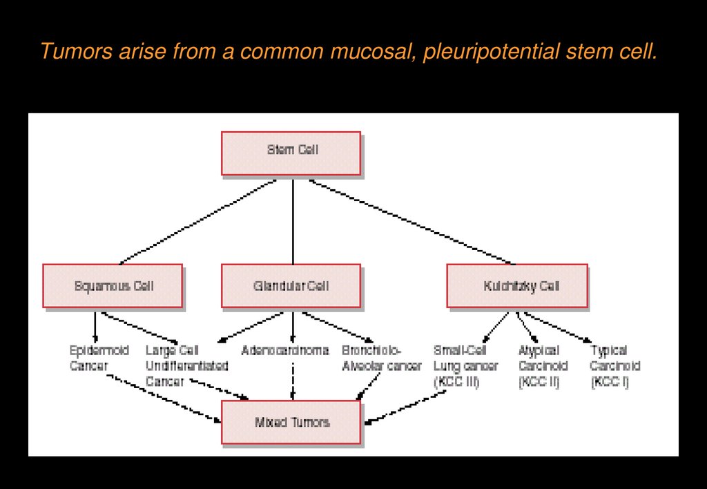
Tumors arise from a common mucosal, pleuripotential stem cell.18. HISTOPATHOLOGY
19.
HISTOPATHOLOGY• SCLC classified into 3 subcategories: 1) oat cell carcinoma,
2) intermediate cell type, and 3) combined cell carcinoma.
• NSCLC includes: 1) squamous cell carcinoma, 2) adenocarcinoma, and 3) large cell carcinoma. Sometimes, lung cancers can
exhibit 2 or more histologic patterns.
Site
• SCLC typically are centrally located, arising in peribronchial
locations. In other words, SCLC is most frequently considered to
be central lung cancer.
• NSCLC: Squamous cell carcinomas occur predominantly in a
central location, whereas adenocarcinoma presents as a peripheral
lesion.
20.
Stage grouping for lung cancer• Stage
TNM
————————————————
• IA
T1N0M0
• IB
T2N0M0
• IIA
T1N1M0
• IIB
T2N1M0 or T3N0M0
• IIIA
T1-3N2M0 or T3N1M0
• IIIB
T4 or T any N3M0
• IV
Any T any N M1
21.
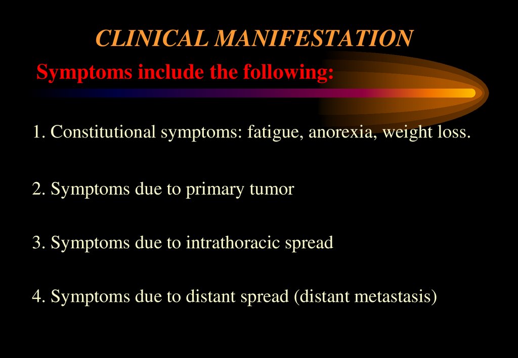
CLINICAL MANIFESTATIONSymptoms include the following:
1. Constitutional symptoms: fatigue, anorexia, weight loss.
2. Symptoms due to primary tumor
3. Symptoms due to intrathoracic spread
4. Symptoms due to distant spread (distant metastasis)
22.
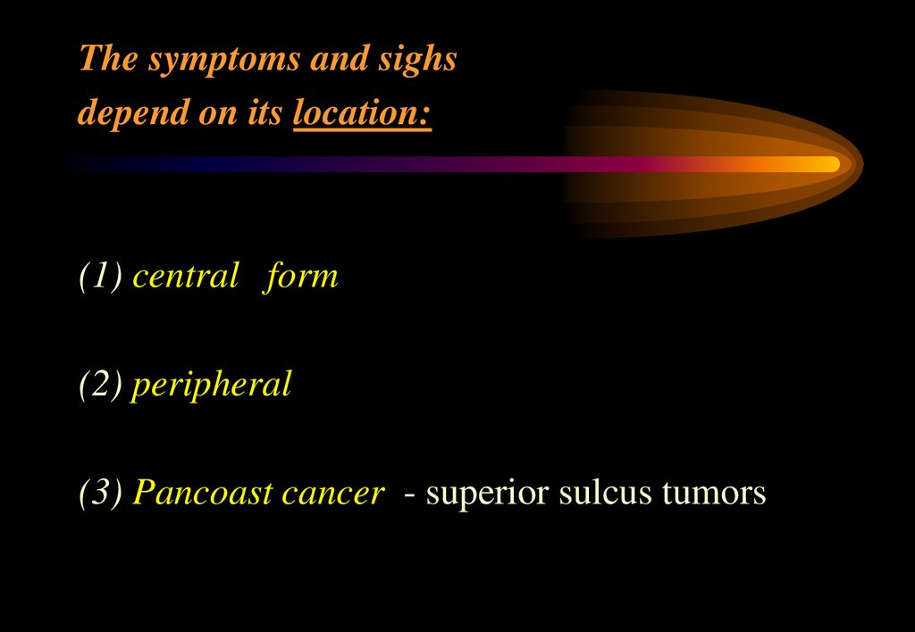
The symptoms and sighsdepend on its location:
(1) central form
(2) peripheral
(3) Pancoast cancer - superior sulcus tumors
23.
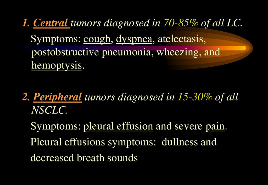
1. Central tumors diagnosed in 70-85% of all LC.Symptoms: cough, dyspnea, atelectasis,
postobstructive pneumonia, wheezing, and
hemoptysis.
2. Peripheral tumors diagnosed in 15-30% of all
NSCLC.
Symptoms: pleural effusion and severe pain.
Pleural effusions symptoms: dullness and
decreased breath sounds
24.
3. A Pancoast tumor is a rare form 1% that arises inthe superior sulcus of the lung apex and producing
shoulder pain.
Involvement of the lower roots of brachial plexus
cause arm pain and paresthesias in ulnar nerve
distribution.
The tumor may spread to the symptomatic ganglion,
leading to Horner syndrome: ipsilateral
enophthalmos, miosis, partial ptosis, and anhidrosis.
25.
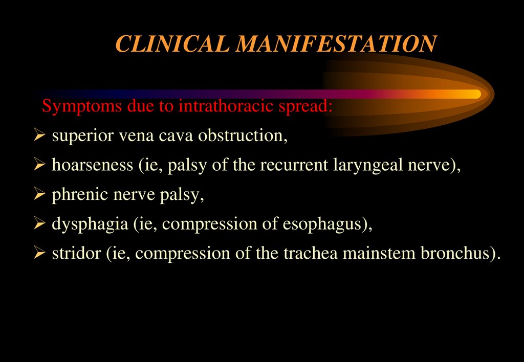
CLINICAL MANIFESTATIONSymptoms due to intrathoracic spread:
superior vena cava obstruction,
hoarseness (ie, palsy of the recurrent laryngeal nerve),
phrenic nerve palsy,
dysphagia (ie, compression of esophagus),
stridor (ie, compression of the trachea mainstem bronchus).
26.
CLINICAL MANIFESTATIONSymptoms due to distant spread:
neurological dysfunction (ie, brain metastasis, spinal cord
compression),
bone pain (bone metastasis),
abdominal/right upper quadrant pain (ie, liver metastasis).
27.
4) Paraneoplastic syndromes by ectopic hormoneproduction
Squamous cell carcinomas are more likely to be
associated with hypercalcemia due to
parathyroidlike hormone production.
Clubbing and hypertrophic pulmonary
osteoarthropathy and the Trousseau syndrome of
hypercoagulability are caused more frequently by
adenocarcinomas.
28.
DIAGNOSTICSDiagnostic strategy:
In the presence of a long history of smoking or other risk
factors for lung cancer, the presence of persistent
respiratory symptoms should prompt:
(1) chest X-Ray.
(2) histologic confirmation is necessary:
by sputum cytologic studies
by bronchoscopy
by CT-guided transthoracic needle biopsy
29.
DIAGNOSTICS1. Chest X-Ray (CXR)
A chest radiograph is usually the first test ordered
in patients.
On chest radiography, the findings of lung
carcinomas are varied and considered in the
differential diagnosis of many disorders.
30.
DIAGNOSTICS1. CXR findings in central form of lung cancer.
1). Bronchial stenosis of lung
Complete left lung collapse
secondary to bronchogenic
carcinoma of left mainstem
bronchus.
31.
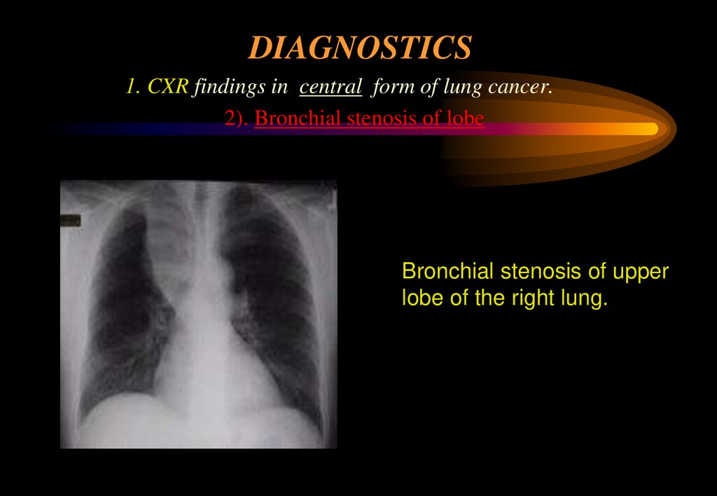
DIAGNOSTICS1. CXR findings in central form of lung cancer.
2). Bronchial stenosis of lobe
Bronchial stenosis of upper
lobe of the right lung.
32.
DIAGNOSTICS1. CXR findings in central form of lung cancer.
3). Patchy irregular or
homogeneous opacities in
a lobar or segmental
distribution.
4). Postobstructive
pneumonia in a segmental
or lobar distribution.
33.
DIAGNOSTICS1. CXR findings in central form of lung cancer.
5). Regional hyperlucency.
Partial stenosis of segmental bronchus leads to
hypoventilation of corresponding lung segment.
In partially atelectatic areas of the lung, hyperlucency
rather than opacity may be evident.
34.
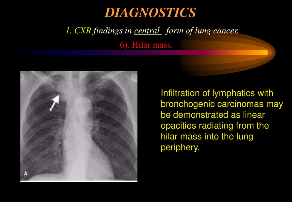
DIAGNOSTICS1. CXR findings in central form of lung cancer.
6). Hilar mass.
Infiltration of lymphatics with
bronchogenic carcinomas may
be demonstrated as linear
opacities radiating from the
hilar mass into the lung
periphery.
35.
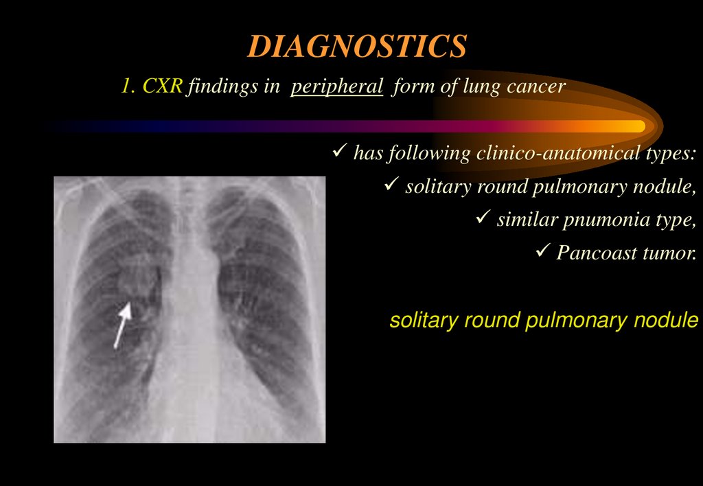
DIAGNOSTICS1. CXR findings in peripheral form of lung cancer
has following clinico-anatomical types:
solitary round pulmonary nodule,
similar pnumonia type,
Pancoast tumor.
solitary round pulmonary nodule
36.
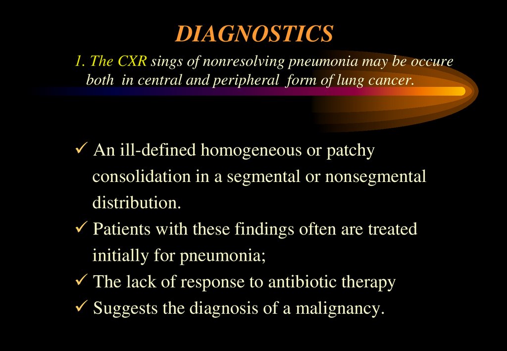
DIAGNOSTICS1. The CXR sings of nonresolving pneumonia may be occure
both in central and peripheral form of lung cancer.
An ill-defined homogeneous or patchy
consolidation in a segmental or nonsegmental
distribution.
Patients with these findings often are treated
initially for pneumonia;
The lack of response to antibiotic therapy
Suggests the diagnosis of a malignancy.
37.
DIAGNOSTICS1. Chest X-Ray (CXR).
Mediastinal lymph node enlargement:
Metastases to paratracheal,
tracheobronchial, peribronchial,
aortopulmonary, and subcarinal
lymph nodes.
38.
DIAGNOSTICS2. Sputum cytologic studies.
Sputum cytology can be a quick and inexpensive diagnostic test.
Sputum cytologic studies in the suspection of lung cancer can be
performed as obligatory method.
39.
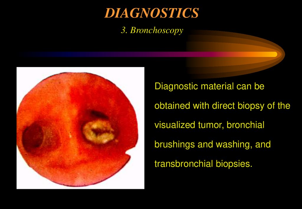
DIAGNOSTICS3. Bronchoscopy
Diagnostic material can be
obtained with direct biopsy of the
visualized tumor, bronchial
brushings and washing, and
transbronchial biopsies.
40.
DIAGNOSTICS4. Biopsy.
is preferred for tumors located in the periphery of the lungs.
41.
DIAGNOSTICSStaging workup
1. Ultrasaund or CT scan of the upper abdomen, including liver
and adrenals
2. Liver and kidney functions tests by electrolytes and renal
function studies.
3. A CT scan of the brain.
4. Bone scintigraphy.
5. Positron emission tomography.
6. Magnetic resonance imaging (MRI).
7. Mediastinoscopy and Thoracoscopy
42.
DIAGNOSTICSStaging workup
CT scans of chest (left) and abdomen (right)
Solitary pulmonary nodule in the
peripheral part of the right lung.
The adrenal glands are a common site for
metastatic small-cell lung cancer.
43.
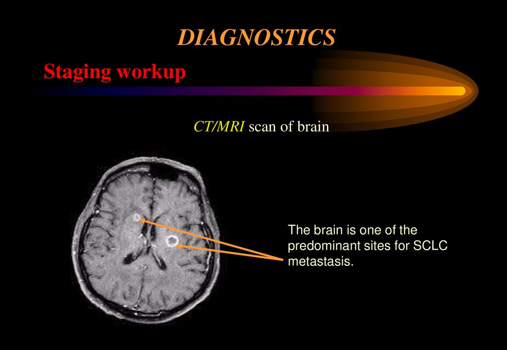
DIAGNOSTICSStaging workup
CT/MRI scan of brain
The brain is one of the
predominant sites for SCLC
metastasis.
44.
DIAGNOSTICSStaging workup
Positron emission tomography (PET)
Multiple hypermetabolic areas suggest
lymph-node metastatic disease in the
chest, abdomen, and right
supraclavicular region.
45.
DIAGNOSTICSStaging workup
Bone scan
Multiple abnormal areas of increased
radiotracer activity in the pelvis, spine,
ribs, and left scapula.
46.
DIAGNOSTICSStaging workup
Thoracentesis. Pleural effusions should be aspirated and examined
for malignant cells
Bone marrow aspiration is necessary in patients with myelophthisic
anemia by metastases.
47.
DIFFERENTIAL DIAGNOSESBronchogenic cyst
Neurogenic tumors
Teratodermoid tumor
Thymoma
Vascular aneurysm
Esophageal lesions
Lymphadenopathy from other malignant or benign lesions
48.
SURGICAL CARESurgical resection provides the best chance of long-term
disease-free survival and possibility of a cure.
The standard surgical procedures :
1. lobectomy (in peripheral tumors
without lymph node metastases)
2. lobectomy with mediastinal
lymph nodes dissection
(in peripheral tumors with lymph
node metastases)
metastases)
49.
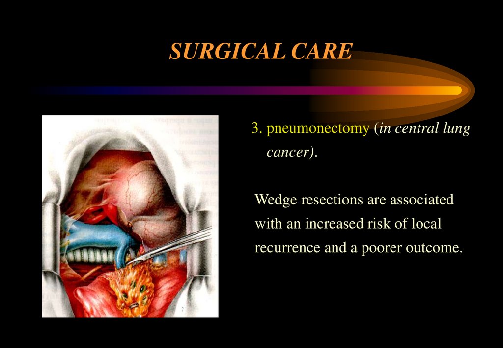
PRINCIPLES OF TREATMENTSURGICAL CARE
3. pneumonectomy (in central lung
cancer).
Wedge resections are associated
with an increased risk of local
recurrence and a poorer outcome.
50.
CHEMOTHERAPYAlone has no role in potentially curative therapy.
Is used alone in the palliative treatment of stage IIIB NSCLC
and stage IV.
- carboplatin-paclitaxel
- cisplatin-gemcitabine
- cisplatin-vinorelbine.
51.
RADIATION THERAPYReduces local failures in completely
resected stages (II and IIIA) NSCLC
But has not been shown to improve overall survival rates.
52.
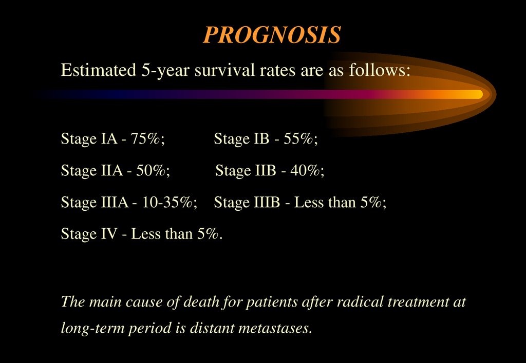
PROGNOSISEstimated 5-year survival rates are as follows:
Stage IA - 75%;
Stage IB - 55%;
Stage IIA - 50%;
Stage IIB - 40%;
Stage IIIA - 10-35%; Stage IIIB - Less than 5%;
Stage IV - Less than 5%.
The main cause of death for patients after radical treatment at
long-term period is distant metastases.