


 medicine
Similar presentations:


Gastric cancer
1. Gastric cancer
Valeriya Semenisty, MD2.
Gastric cancer encompasses a heterogeneous collectionof etiologic and histologic subtypes associated with a
variety of known and unknown environmental and
genetic factors.
It is a global public health concern, accounting for
700,000 annual deaths worldwide, and currently ranks
as the fourth leading cause of cancer mortality, with a
5-year survival of only 20%.
The incidence and prevalence of gastric cancer vary
widely, with Asian/Pacific regions bearing the highest
rates of disease.
3.
Approximately 3% to 5% of gastric cancers areassociated with a hereditary predisposition, including a
variety of Mendelian genetic conditions and complex
genetic traits.
4.
Gastric cancer has traditionally been subtypedpathologically according to Lauren’s1 classification
published in 1965 and revised by Carneiro et al.2 in
1995.
The four histologic categories include:
(1) glandular/intestinal,
(2) border foveal hyperplasia,
(3) mixed intestinal/diffuse, and
(4) solid/undifferentiated.
5.
More clinically relevant, the majority of gastric cancerscan be subdivided into intestinal type or diffuse type.
Diffuse gastric tumors frequently feature signet ring
cells
The intestinal subtype is seen more commonly in older
patients, whereas the diffuse type affects younger
patients and has a more aggressive clinical course.
6. ETIOLOGY
Environmental Risk Factorsdiet and lifestyle variables.
Infectious Risk Factors
H. pylori infection
Epstein-Barr virus
Genetics
7.
More than 70% of cases occur in developing countries, andmen have roughly twice the risk of women.
In 2008, estimates of gastric cancer burden in the United
States were 21,500 cases (13,190 men and 8,310 women) and
10,880 deaths. The median age at diagnosis for gastric cancer
is 71 years, and 5-year survival is approximately 25%.
Only 24% of stomach cancers are localized at the time of
diagnosis, 30% have lymph node involvement, and another
30% have metastatic disease. Survival rates are predictably
higher for those with localized disease, with corresponding 5year survival rates of 60%.
8. PATHOLOGY AND TUMOR BIOLOGY
Approximately 95% of all gastric cancers areadenocarcinomas.
9. PATTERNS OF SPREAD
Carcinomas of the stomach can spread by localextension to involve adjacent structures and can
develop lymphatic metastases, peritoneal metastases,
and distant metastases.
These extensions can occur by the local invasive
properties of the tumor, lymphatic spread, or
hematogenous dissemination.
10.
CLINICAL PRESENTATION ANDPRETREATMENT EVALUATION
Because of the vague, nonspecific symptoms that characterize gastric
cancer, many patients are diagnosed with advanced-stage disease.
Patients may have a combination of signs and symptoms such as weight
loss (22% to 61%)37; anorexia (5% to 40%); fatigue, epigastric discomfort,
or pain (62% to 91%); and postprandial fullness, heart burn, indigestion,
nausea, and vomiting (6% to 40%). None of these unequivocally indicates
gastric cancer. In addition, patients may be asymptomatic (4% to 17%).
Weight loss and abdominal pain are the most common presenting
symptoms at initial encounter. Weight loss is a common symptom, and its
clinical significance should not be underestimated.
Dewys et al. found that in 179 patients with advanced gastric cancer,
>80% of patients had a >10% decrease in body weight before diagnosis.
Furthermore, patients with weight loss had a significantly shorter
survival than did those without weight loss
11.
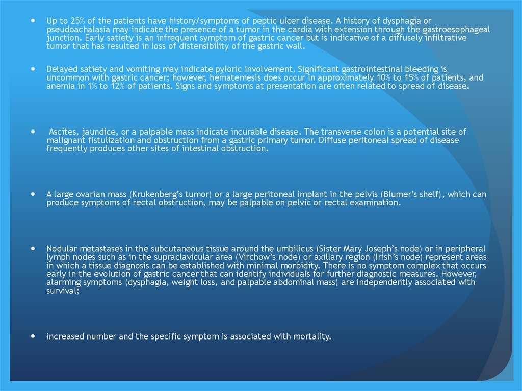
Up to 25% of the patients have history/symptoms of peptic ulcer disease. A history of dysphagia orpseudoachalasia may indicate the presence of a tumor in the cardia with extension through the gastroesophageal
junction. Early satiety is an infrequent symptom of gastric cancer but is indicative of a diffusely infiltrative
tumor that has resulted in loss of distensibility of the gastric wall.
Delayed satiety and vomiting may indicate pyloric involvement. Significant gastrointestinal bleeding is
uncommon with gastric cancer; however, hematemesis does occur in approximately 10% to 15% of patients, and
anemia in 1% to 12% of patients. Signs and symptoms at presentation are often related to spread of disease.
Ascites, jaundice, or a palpable mass indicate incurable disease. The transverse colon is a potential site of
malignant fistulization and obstruction from a gastric primary tumor. Diffuse peritoneal spread of disease
frequently produces other sites of intestinal obstruction.
A large ovarian mass (Krukenberg’s tumor) or a large peritoneal implant in the pelvis (Blumer’s shelf), which can
produce symptoms of rectal obstruction, may be palpable on pelvic or rectal examination.
Nodular metastases in the subcutaneous tissue around the umbilicus (Sister Mary Joseph’s node) or in peripheral
lymph nodes such as in the supraclavicular area (Virchow’s node) or axillary region (Irish’s node) represent areas
in which a tissue diagnosis can be established with minimal morbidity. There is no symptom complex that occurs
early in the evolution of gastric cancer that can identify individuals for further diagnostic measures. However,
alarming symptoms (dysphagia, weight loss, and palpable abdominal mass) are independently associated with
survival;
increased number and the specific symptom is associated with mortality.
12. PRETREATMENT STAGING
Tumor markers – CEA, CA19-9,CA125EUS
CT
MRI
PET-CT
Staging Laparoscopy and Peritoneal Cytology
13. STAGING, CLASSIFICATION, AND PROGNOSIS
14. TREATMENT OF LOCALIZED DISEASE
Stage I Disease (Early Gastric Cancer)Endoscopic Mucosal Resection
Limited Surgical Resection
Gastrectomy
15. Stage II and Stage III Disease
GASTRECTOMY16. Adjuvant Therapy
Adjuvant therapy indicates administration of atreatment following a potential curative resection of
the primary tumor and regional lymph nodes.
Therapy after resections that leave microscopic or gross
disease are not adjuvant treatment, but rather therapy
for known disease, which is palliative in nature.
Neoadjuvant chemotherapy involves the use of
systemic treatment before potentially curative surgery.
17.
There are several theoretical reasons for beginning adjuvanttherapy soon after operation (perioperative chemotherapy).
Studies have shown a rapid increase in cell growth of
metastases after a primary tumor has been removed related
to a decline in certain circulating factors, which serve to
inhibit angiogenesis or other cell-cycle promotors, once the
primary tumor is removed.
Perioperative or neoadjuvant chemotherapy has been studied
because the ability to perform a R0 resection in gastric
cancer is difficult. In addition, a substantial number of
patients undergoing gastrectomy have prolonged recovery.
18.
Neoadjuvant chemotherapy has a dual goal: allowing ahigher rate of R0 resections and treatment of
micrometastatic disease early in the course of
treatment.
19.
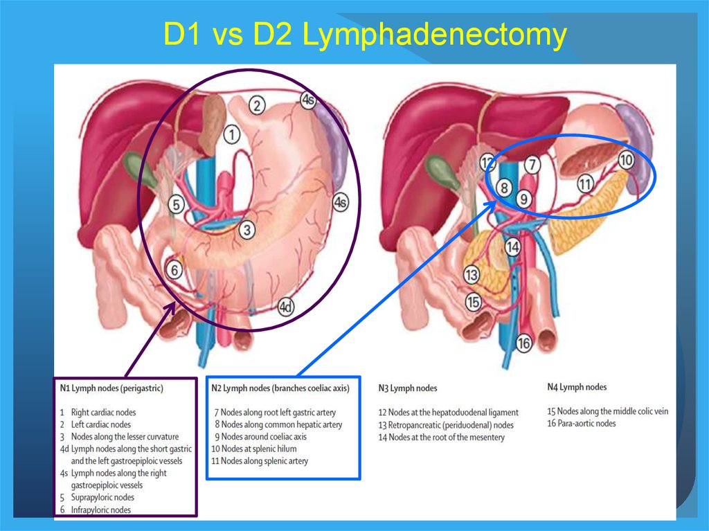
D1 vs D2 Lymphadenectomy20.
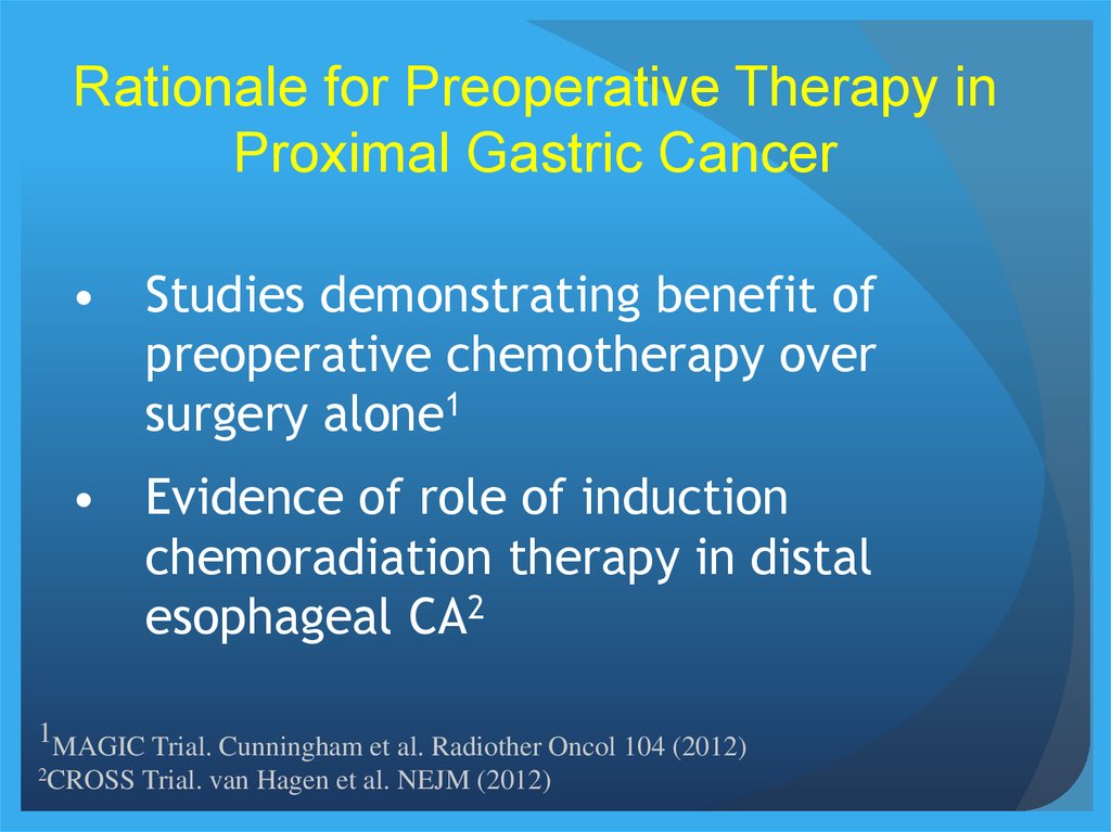
Rationale for Preoperative Therapy inProximal Gastric Cancer
• Studies demonstrating benefit of
preoperative chemotherapy over
surgery alone1
• Evidence of role of induction
chemoradiation therapy in distal
esophageal CA2
1MAGIC Trial. Cunningham et al. Radiother Oncol 104 (2012)
2CROSS
Trial. van Hagen et al. NEJM (2012)
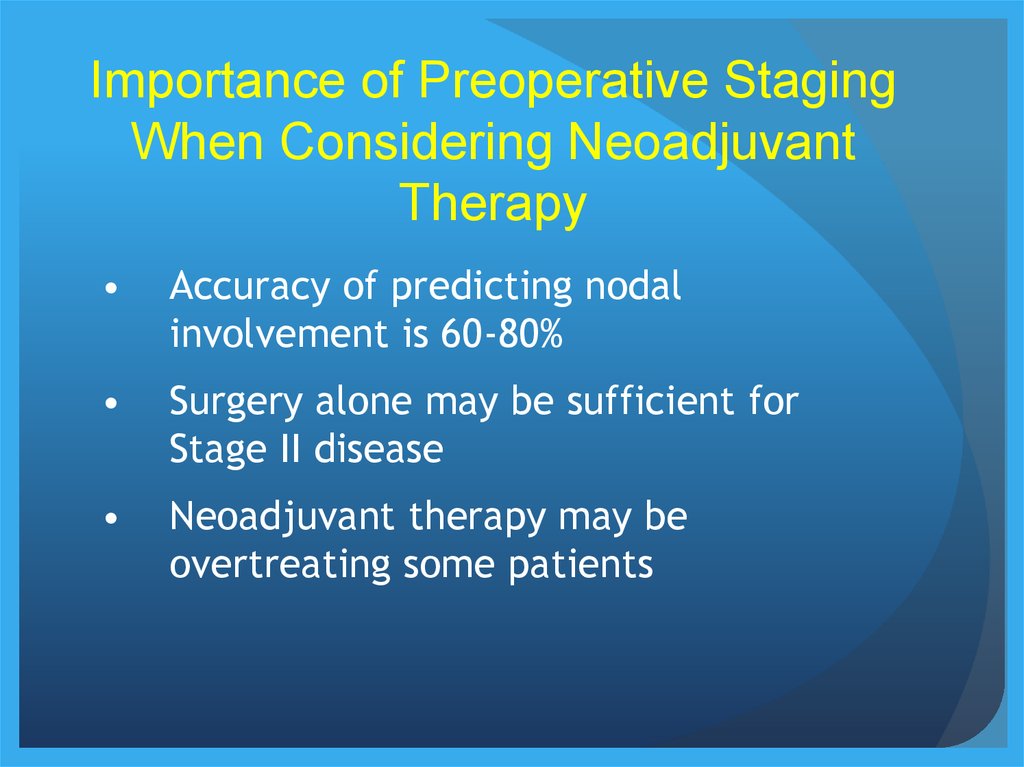
21.
Importance of Preoperative StagingWhen Considering Neoadjuvant
Therapy
Accuracy of predicting nodal
involvement is 60-80%
Surgery alone may be sufficient for
Stage II disease
Neoadjuvant therapy may be
overtreating some patients
22.
Rationale for Up Front Surgery inPatients With Gastric Cancer
Pathologic staging may result in more
appropriate choice of adjuvant therapy
(accurate stage II vs III, D1 vs D2,
margins).
Symptomatic patients may require initial
surgery.
In reality, gastrectomy is often performed
before MDT consultation.
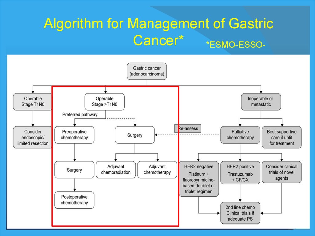
23.
Algorithm for Management of GastricCancer*
*ESMO-ESSO-
24.
Post-Operative Chemo vs Chemoradiation:ARTIST Trial
Samsung
University
458 patient RCT
D2 gastrectomy
~5% proximal CA
Postoperative
adjuvant Cap-Cis
± RT
Lee et al. JCO Jan 2012
No difference in
DFS
No difference in
locoregional rec
25.
Post-Operative Chemo vs Chemoradiation:Nanjing University
Recurrence-Free Survival
P=0.029
380 patients
Randomized trial
All D2
gastrectomy
~10% GE junction
Postoperative
adjuvant 5FU-LV ±
IMRT
Zhu et al. Radiother Oncol 104 (2012)
Improved RFS
with IMRT (50 vs
32 mo)
No difference in
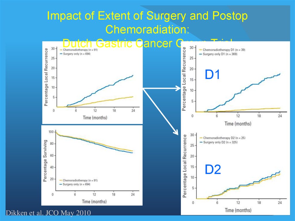
26.
Impact of Extent of Surgery and PostopChemoradiation:
Dutch Gastric Cancer Group Trial
D1
D2
Dikken et al. JCO May 2010
27.
Chemoradiation After Surgery Versus Surgery Alonefor Gastric and GEJ Adenocarcinoma
MacDonald et al. NEJM 2001
20% GE Junction
Criticized for inadequate
surgical radicality
28.
CRITICS StudyPreoperative
Chemotherapy
D1+ Surgery
3x ECC q 3 wks
3x ECC q 3 wks
R
Preoperative
Chemotherapy
Chemoradiotherapy
D1+ Surgery
3x ECC q 3 wks
2 weeks
3-6 weeks
45 Gy/25 fx
+ capecitabine
+ cisplatin
Within 4-12 weeks
29. Summary
Adjuvant Therapy for Proximal Gastric CancerSummary
1. While preoperative therapy may be preferred in
most cases, initial gastrectomy is being commonly
performed.
2. While R0 gastrectomy with D2 lymphadenectomy is
recommended, less radical surgery is common.
3. Chemoradiation appears to have a role in reducing
local recurrence.
4. Postoperative chemoradiation should be considered
when managing a post-op patient, particularly when
<D2 gastrectomy was performed.