


 x 200")


 medicine
Similar presentations:
")


")
Diseases of endocrine system
1. LECTURE. Diseases of endocrine system.
Волгоградскийгосударственный
медицинский
университет
Кафедра
патологической
анатомии
LECTURE. Diseases of
endocrine system.
2. Endocrine Pathology
Cell signaling systemSurface receptors
• cAMP and tyrosine kinase system
Cytoplasmic receptors
• Penetrate cell membrane
• Gene activation -> transcription ->
translation
Intranuclear receptors
• Gene activation -> transcription ->
translation
3. Endocrine Pathology
Too much hormone activityToo little hormone activity
• Autoimmune destruction
• Inflammatory destruction
• Tumor or vascular destruction
Space occupying lesions (tumors)
• Malignant
• Benign
4. The Basics
Anterior• Comes from GI
• Controlled by
hypothalmus
Posterior
• Hormones
orginate further
up.
5. Pituitary Vascular
Signaling proteinsare release in
hypothalmus.
Travel by blood to
anterior pituitary
Cause release of
many activating
hormones
System of
amplification
6. Pituitary Control
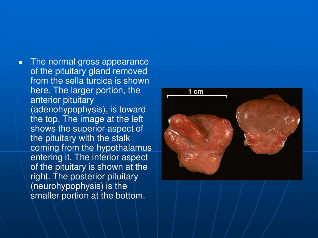
7.
The normal gross appearanceof the pituitary gland removed
from the sella turcica is shown
here. The larger portion, the
anterior pituitary
(adenohypophysis), is toward
the top. The image at the left
shows the superior aspect of
the pituitary with the stalk
coming from the hypothalamus
entering it. The inferior aspect
of the pituitary is shown at the
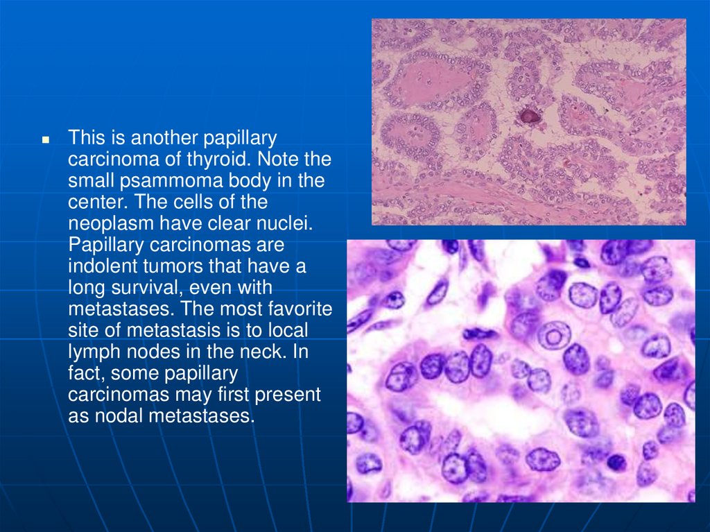
right. The posterior pituitary
(neurohypophysis) is the
smaller portion at the bottom.
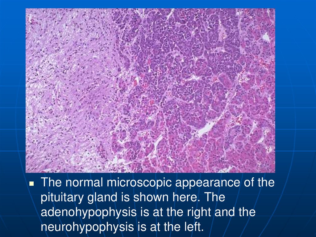
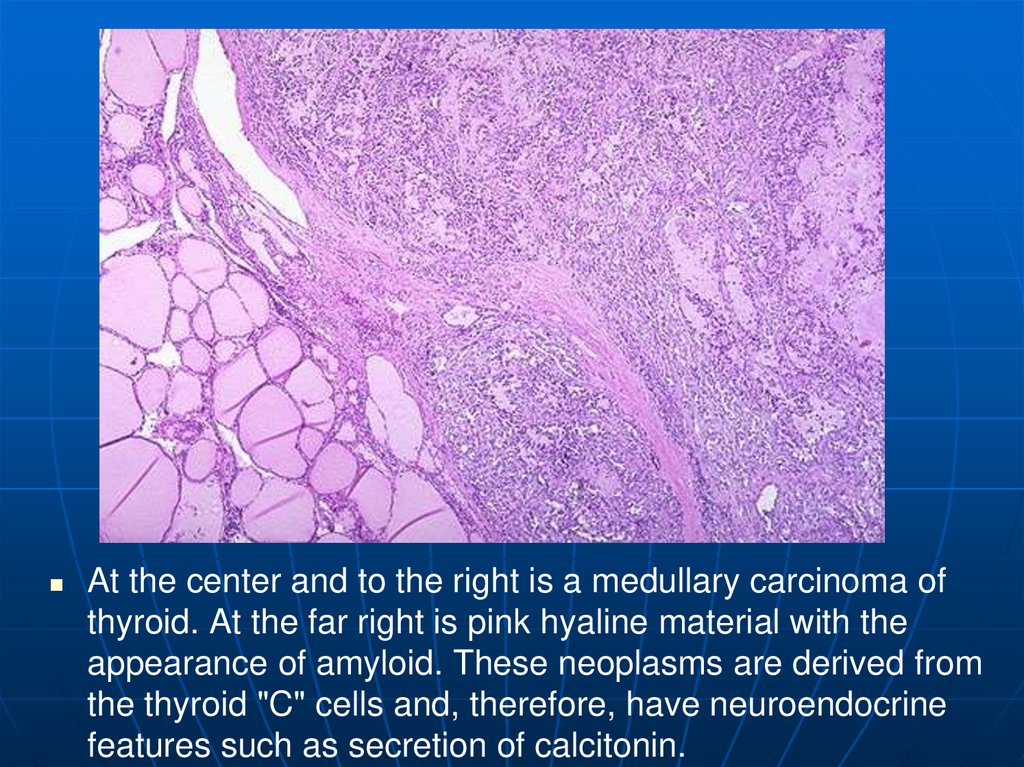
8.
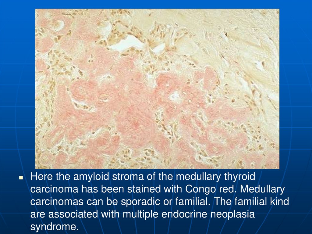
The normal microscopic appearance of thepituitary gland is shown here. The
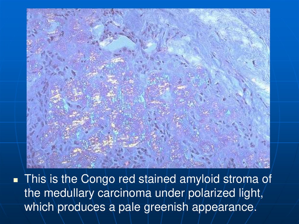
adenohypophysis is at the right and the
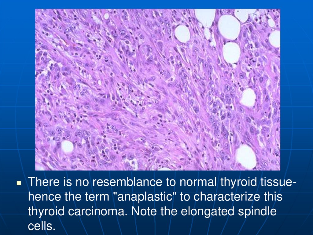
neurohypophysis is at the left.
9.
The normal microscopic appearance of the adenohypophysis is shown here. Theadenohypophysis contains three major cell types: acidophils, basophils, and
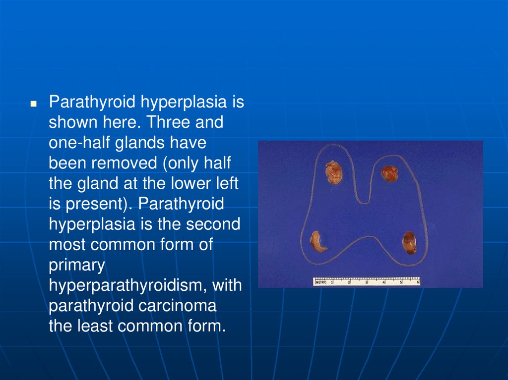
chromophobes. The staining is variable, and to properly identify specific hormone
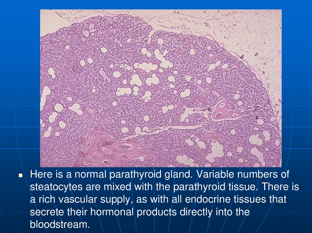
secretion, immunohistochemical staining is necessary. A simplistic classification is as
follows:
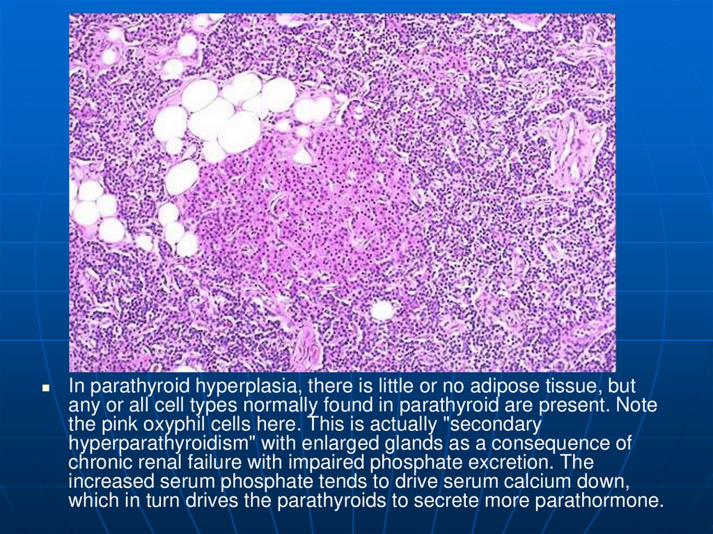
The pink acidophils secrete growth hormone (GH) and prolactin (PRL)
The dark purple basophils secrete corticotrophin (ACTH), thyroid stimulating hormone (TSH), and
gonadotrophins follicle stimulating hormone-luteinizing hormone (FSH and LH)
The pale staining chromophobes have few cytoplasmic granules, but may have secretory activity.
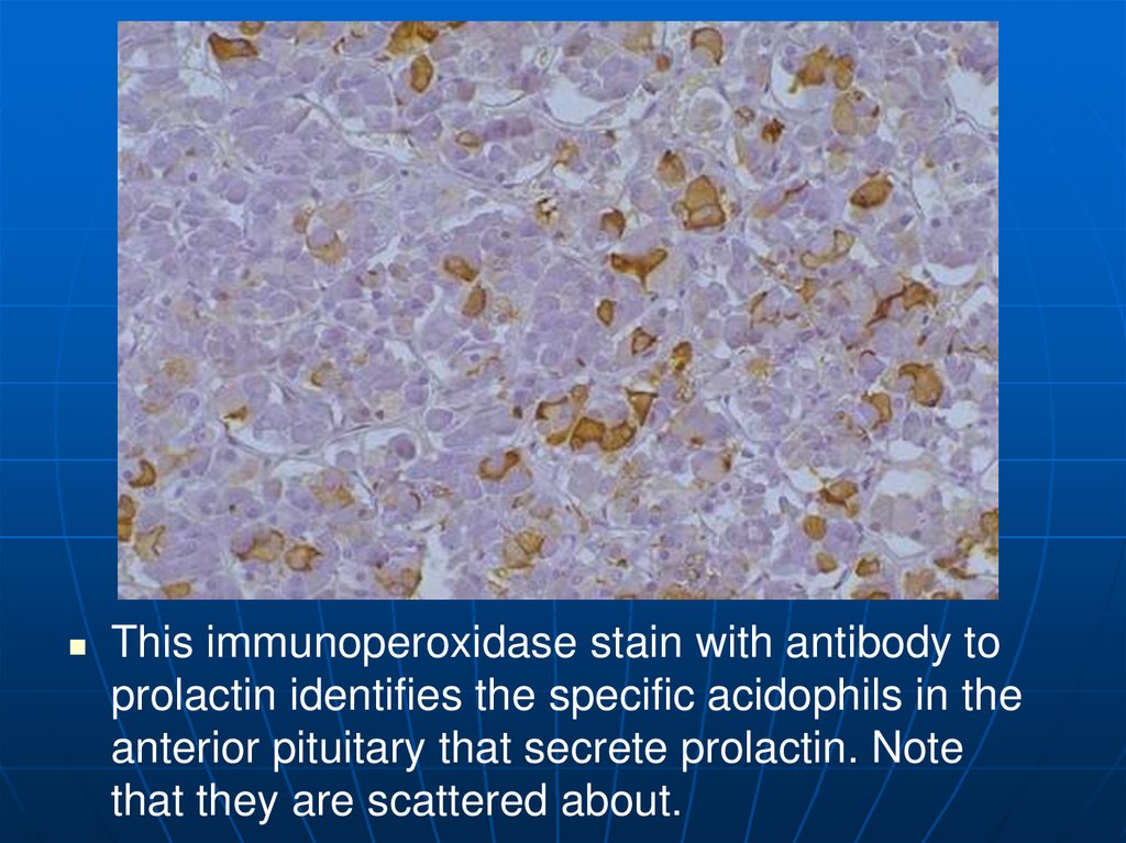
10.
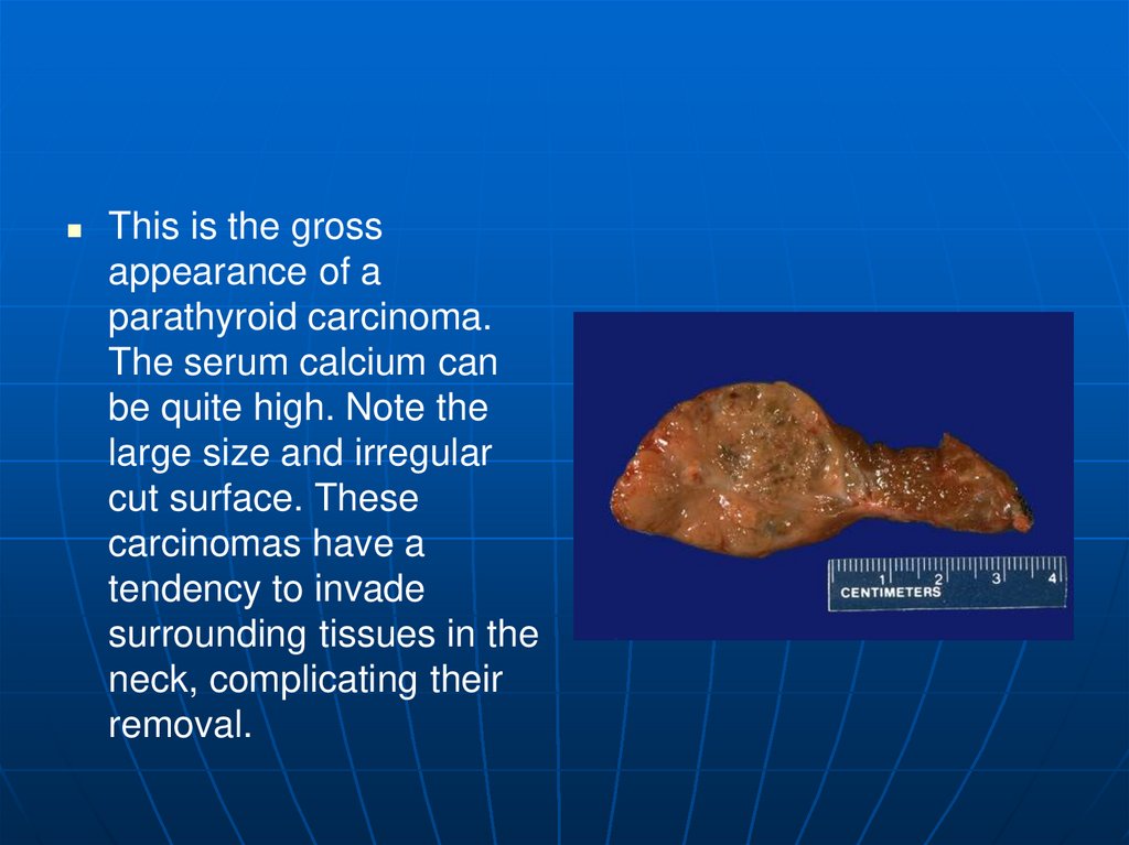
This immunoperoxidase stain with antibody toprolactin identifies the specific acidophils in the
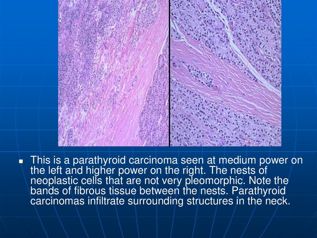
anterior pituitary that secrete prolactin. Note
that they are scattered about.
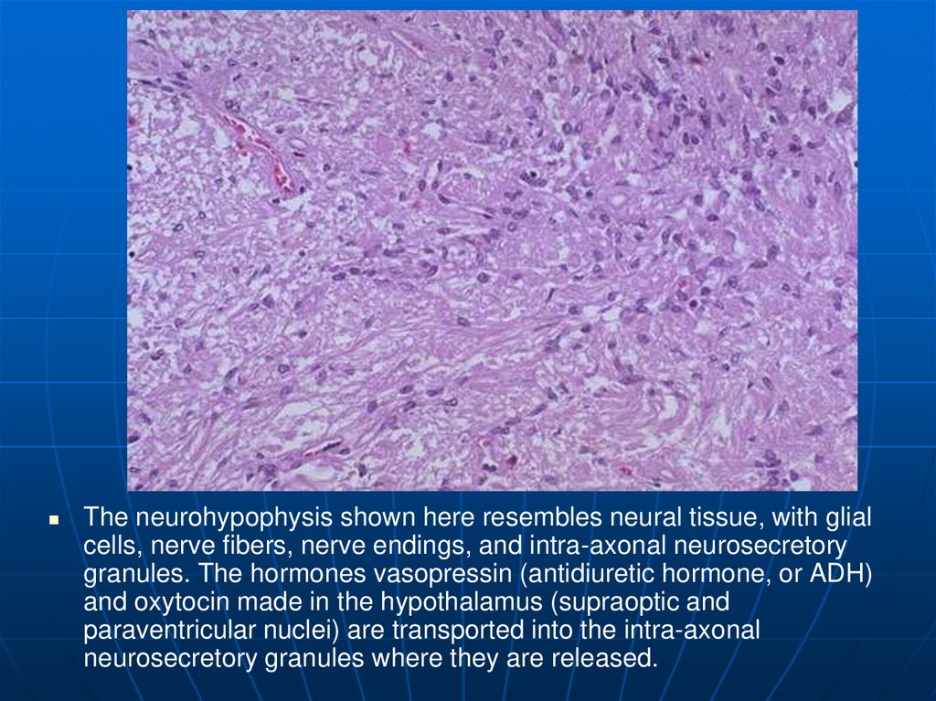
11.
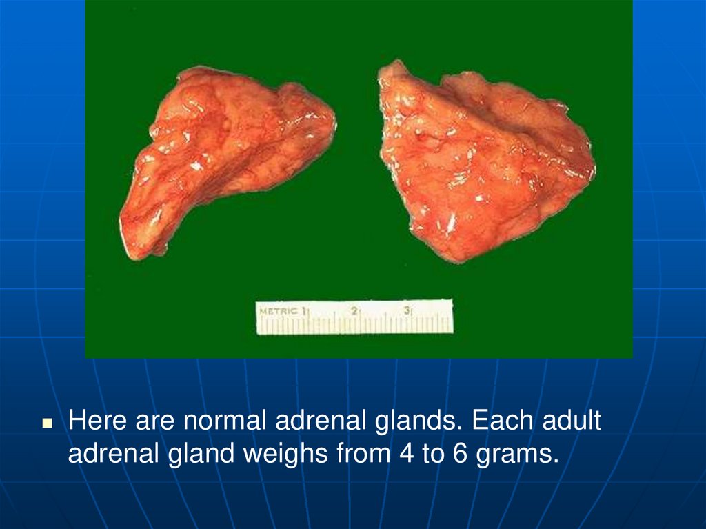
The neurohypophysis shown here resembles neural tissue, with glialcells, nerve fibers, nerve endings, and intra-axonal neurosecretory
granules. The hormones vasopressin (antidiuretic hormone, or ADH)
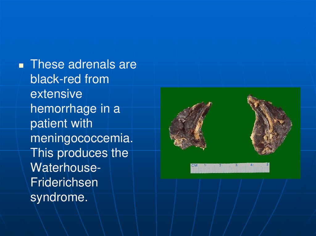
and oxytocin made in the hypothalamus (supraoptic and
paraventricular nuclei) are transported into the intra-axonal
neurosecretory granules where they are released.
12. Space Occupying Lesions
TumorsEmbryonic rests
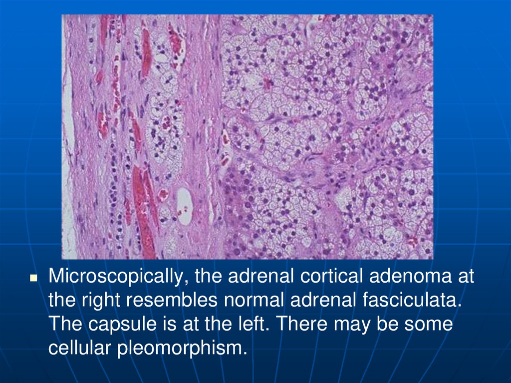
Squeeze gland
out of existence.
• Generalized failure
Visual field
changes
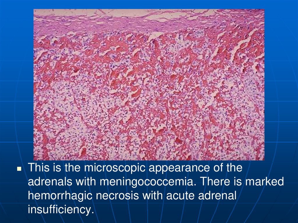
13. Visual Fields
Loss of temporalfields.
• Nasal retina
Damage to
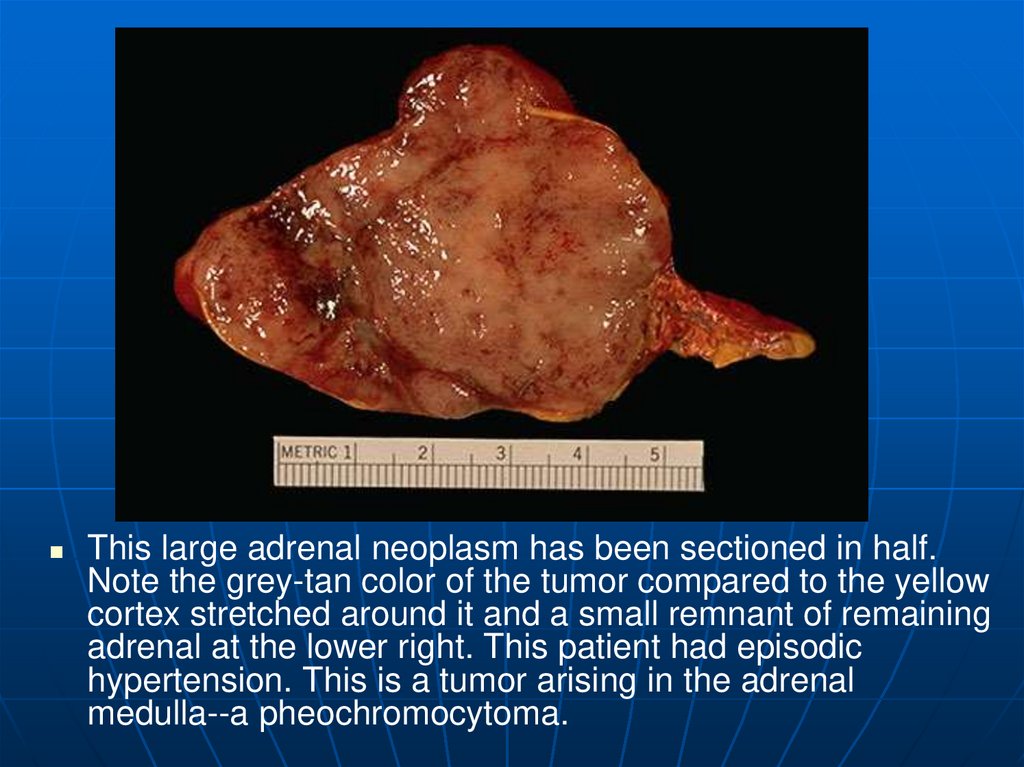
decusating optic
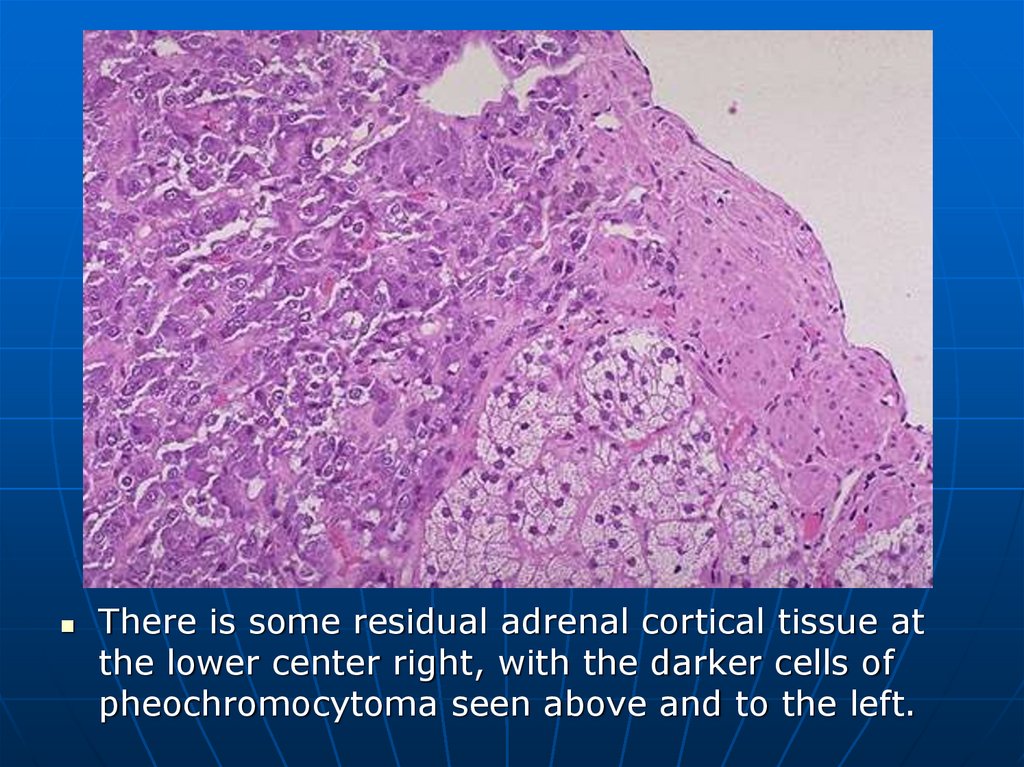
nerve fibers
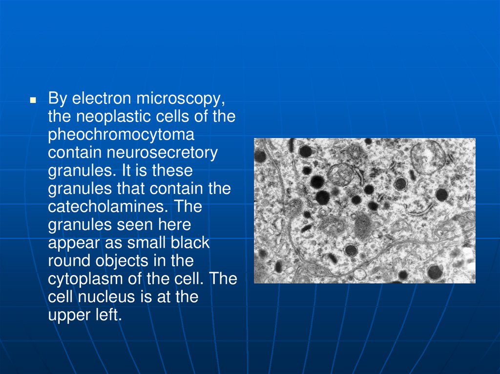
14. Pituitary Adenomas
RareMake nothing or
Prolactin
ACTH, GH,TSH are very rare
More often end up with pituitary
failure.
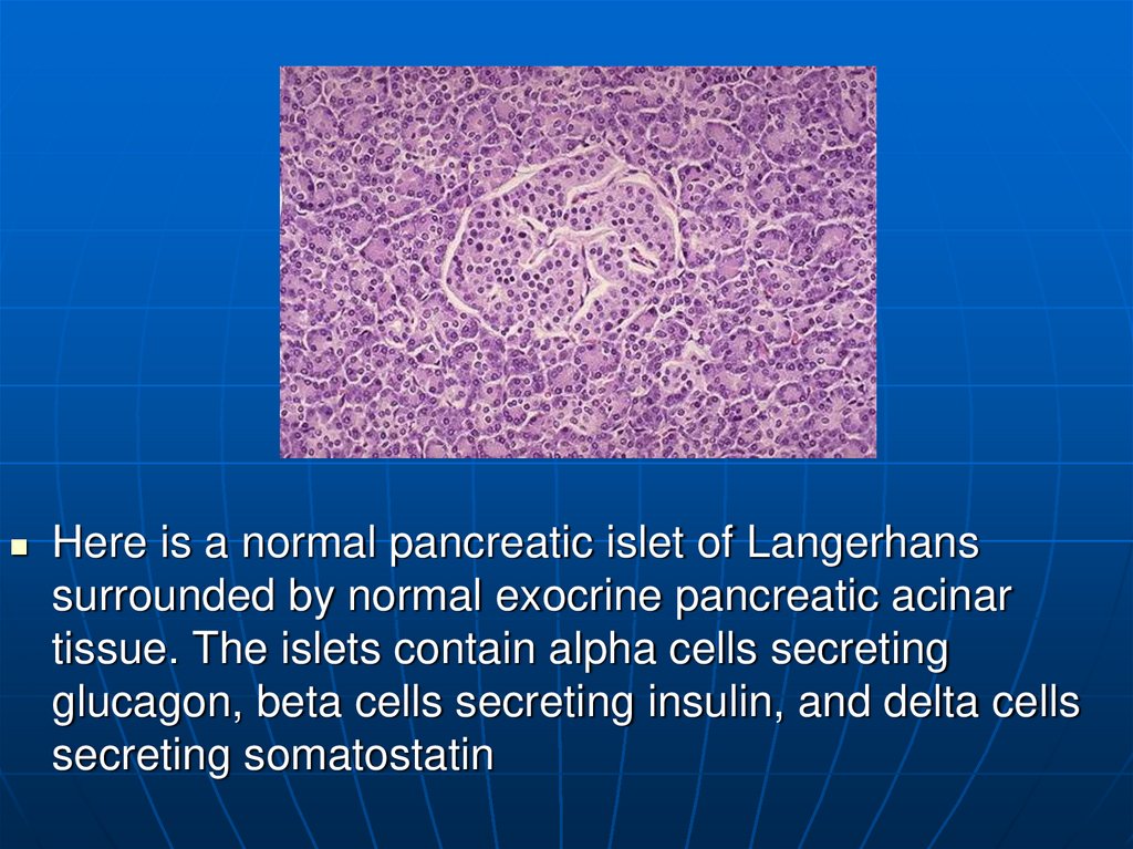
• Squeeze the daylights out of the
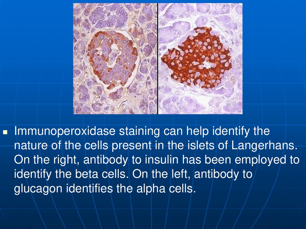
gland.
15.
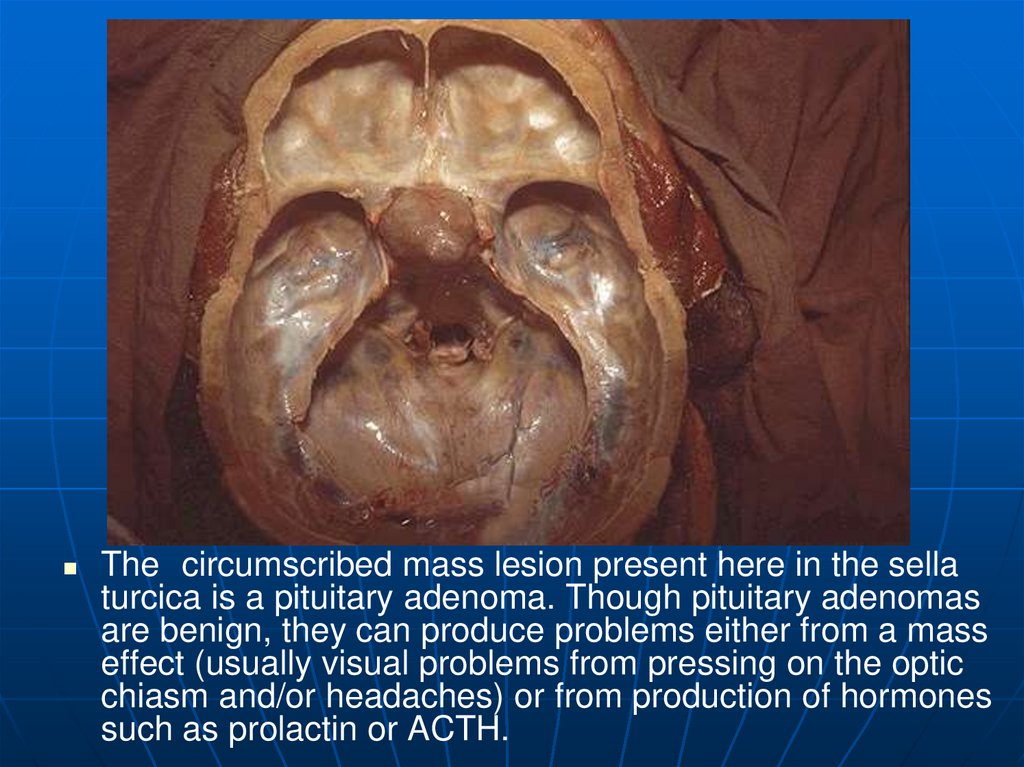
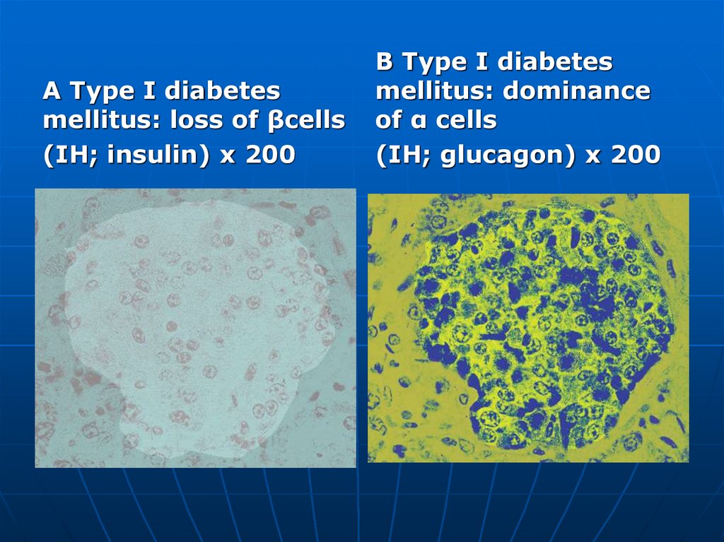
The circumscribed mass lesion present here in the sellaturcica is a pituitary adenoma. Though pituitary adenomas
are benign, they can produce problems either from a mass
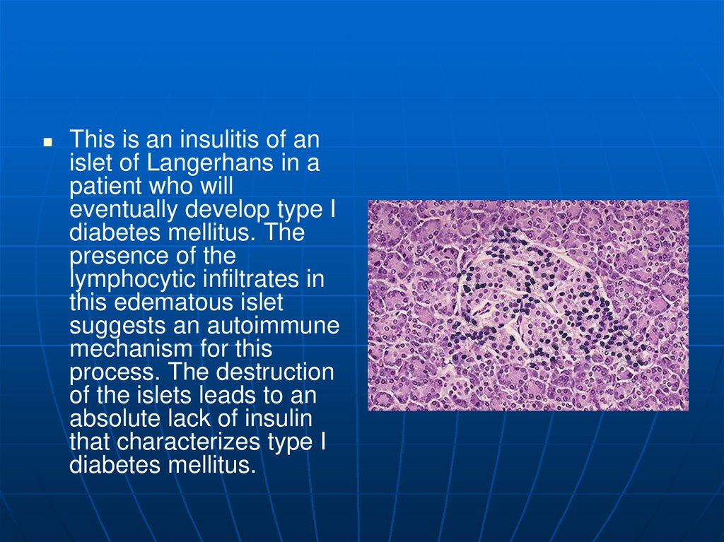
effect (usually visual problems from pressing on the optic
chiasm and/or headaches) or from production of hormones
such as prolactin or ACTH.
16.
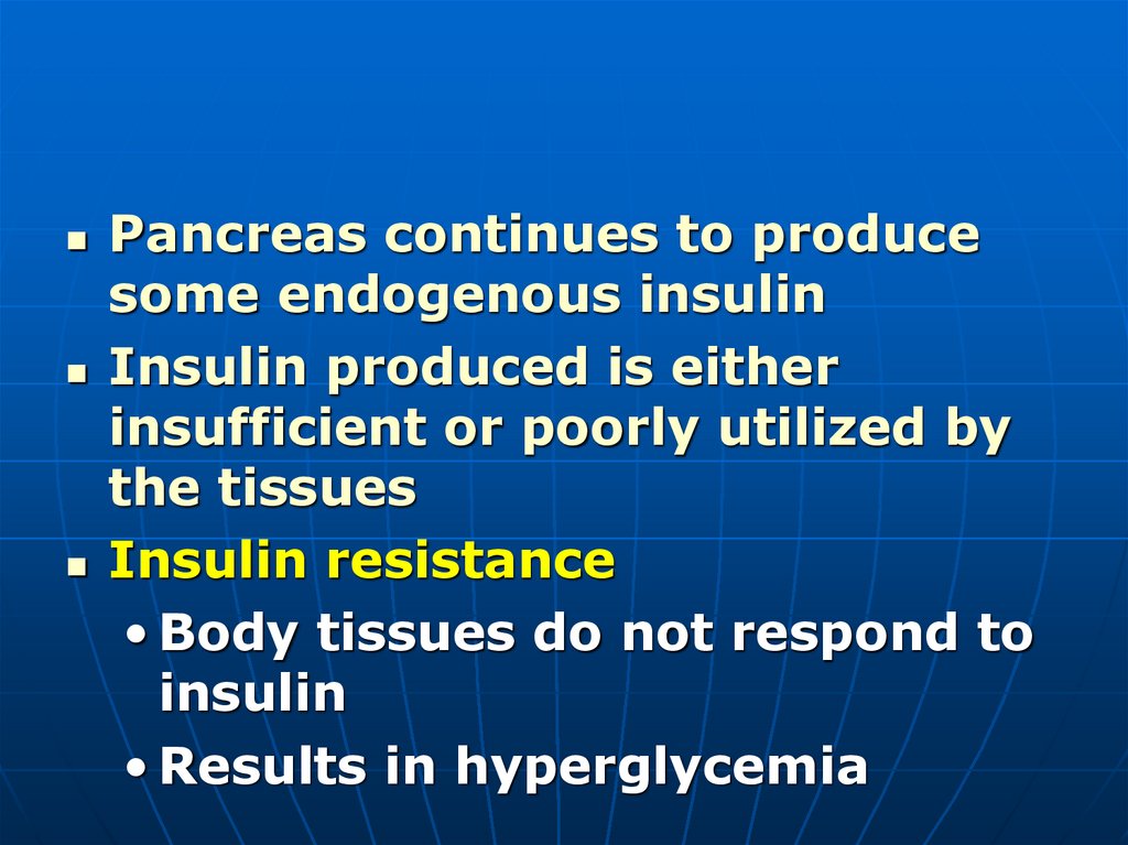
This is a microadenoma of the anterior pituitary.Such microadenomas may appear in 1 to 5% of
adults. These microadenomas rarely have a
significant hormonal output that leads to clinical
disease.
17.
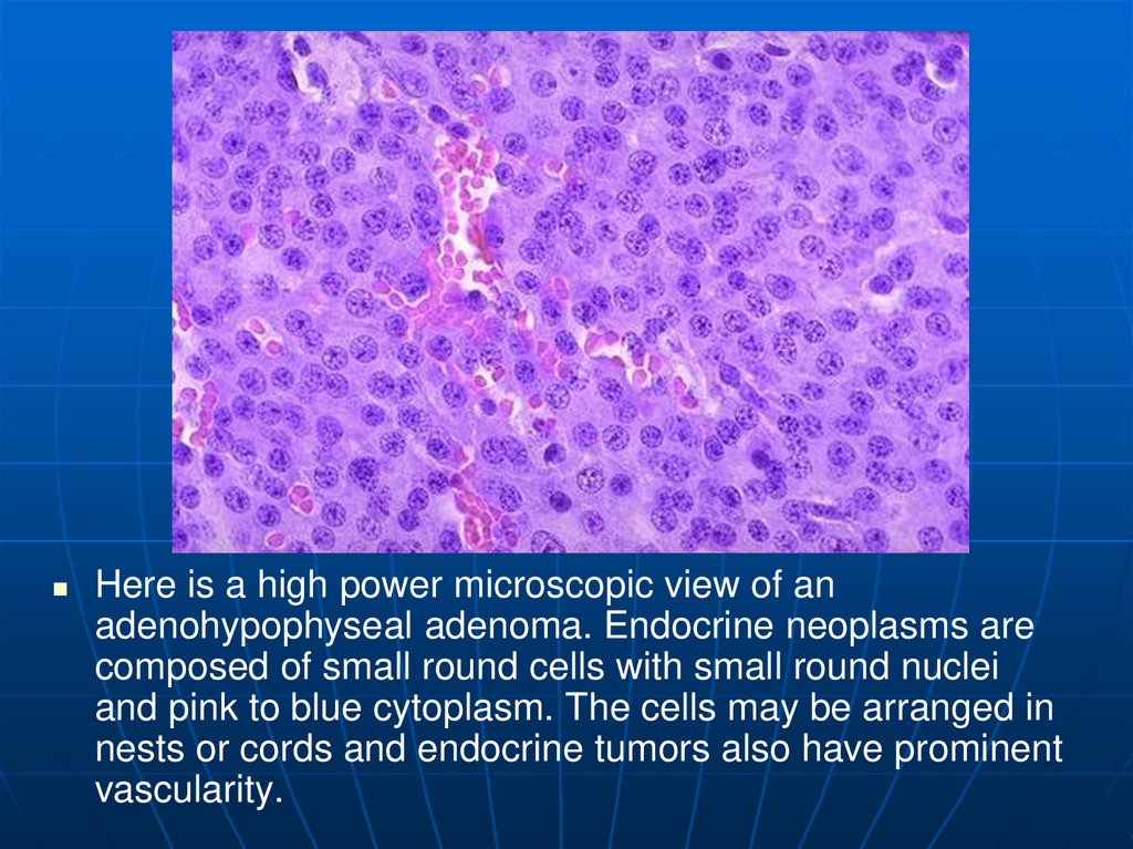
Here is a high power microscopic view of anadenohypophyseal adenoma. Endocrine neoplasms are
composed of small round cells with small round nuclei
and pink to blue cytoplasm. The cells may be arranged in
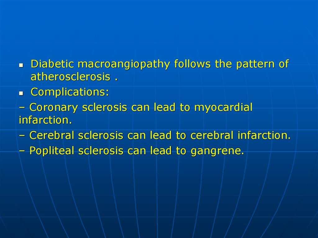
nests or cords and endocrine tumors also have prominent
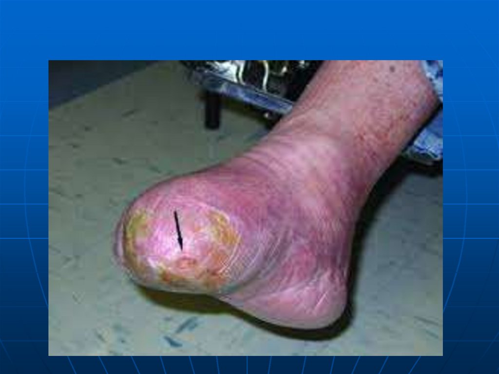
vascularity.
18.
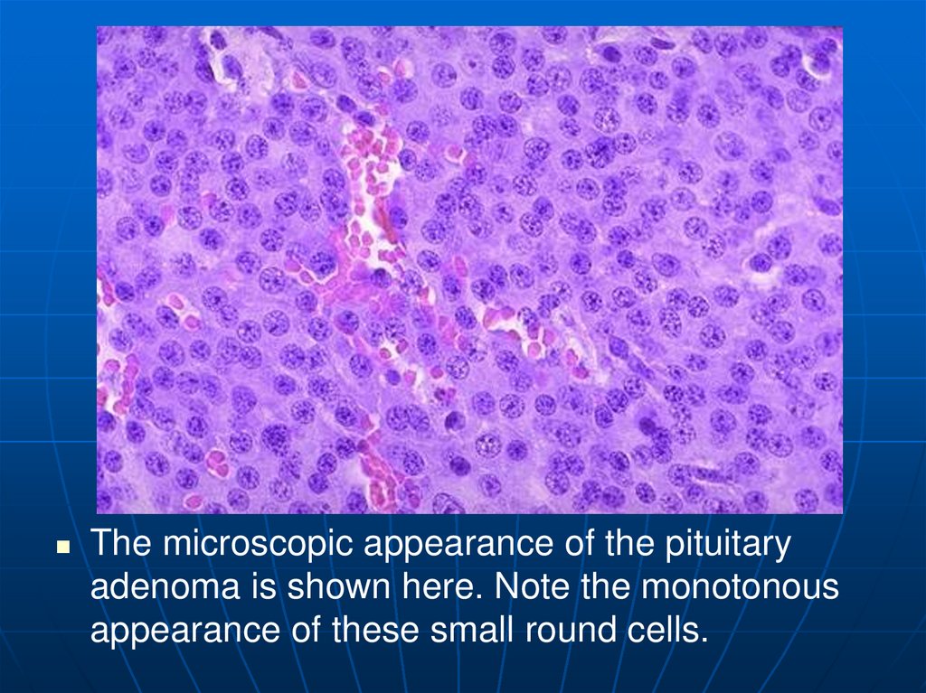
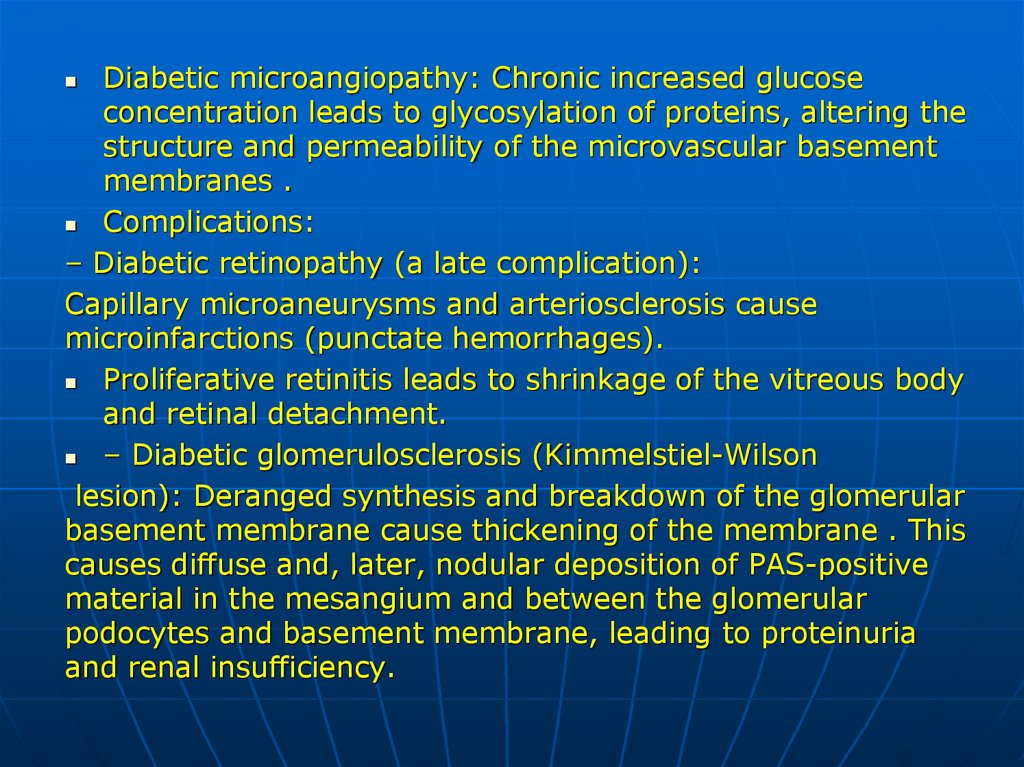
The microscopic appearance of the pituitaryadenoma is shown here. Note the monotonous
appearance of these small round cells.
19. Acromegaly
Growth hormoneexcess after
closing of
epiphyses.
Periosteal bone
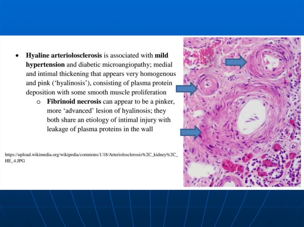
growth.
Diabetes
Prognathism
20. Hypopituitarism
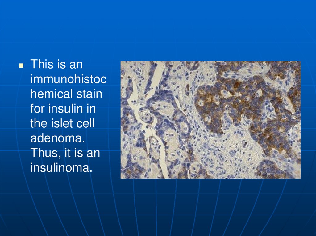
Destruction ofgland.
Ischemia
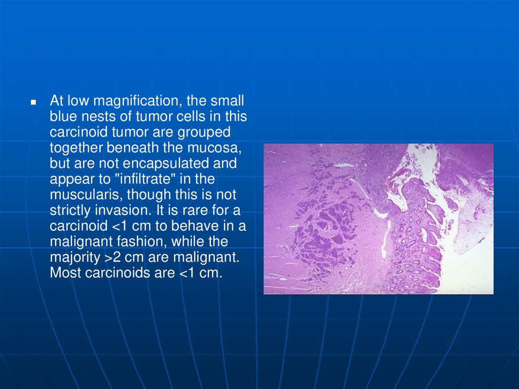
‘Benign’ adenoma
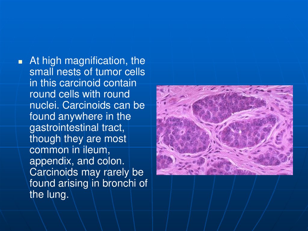
destroying gland
Craniopharyngioma
• Rathke’s pouch
remenant
• Benign cyst, but
really in the wrong
place.
21. Ischemic Destruction
Shehan’s syndromePost delivery
problem
No lactation
In time general
failure of
‘downstream’
systems
• Thyroid
• Adrenal cortex
• Ovulation
22.
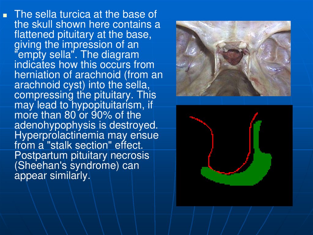
The sella turcica at the base ofthe skull shown here contains a
flattened pituitary at the base,
giving the impression of an
"empty sella". The diagram
indicates how this occurs from
herniation of arachnoid (from an
arachnoid cyst) into the sella,
compressing the pituitary. This
may lead to hypopituitarism, if
more than 80 or 90% of the
adenohypophysis is destroyed.
Hyperprolactinemia may ensue
from a "stalk section" effect.
Postpartum pituitary necrosis
(Sheehan's syndrome) can
appear similarly.
23.
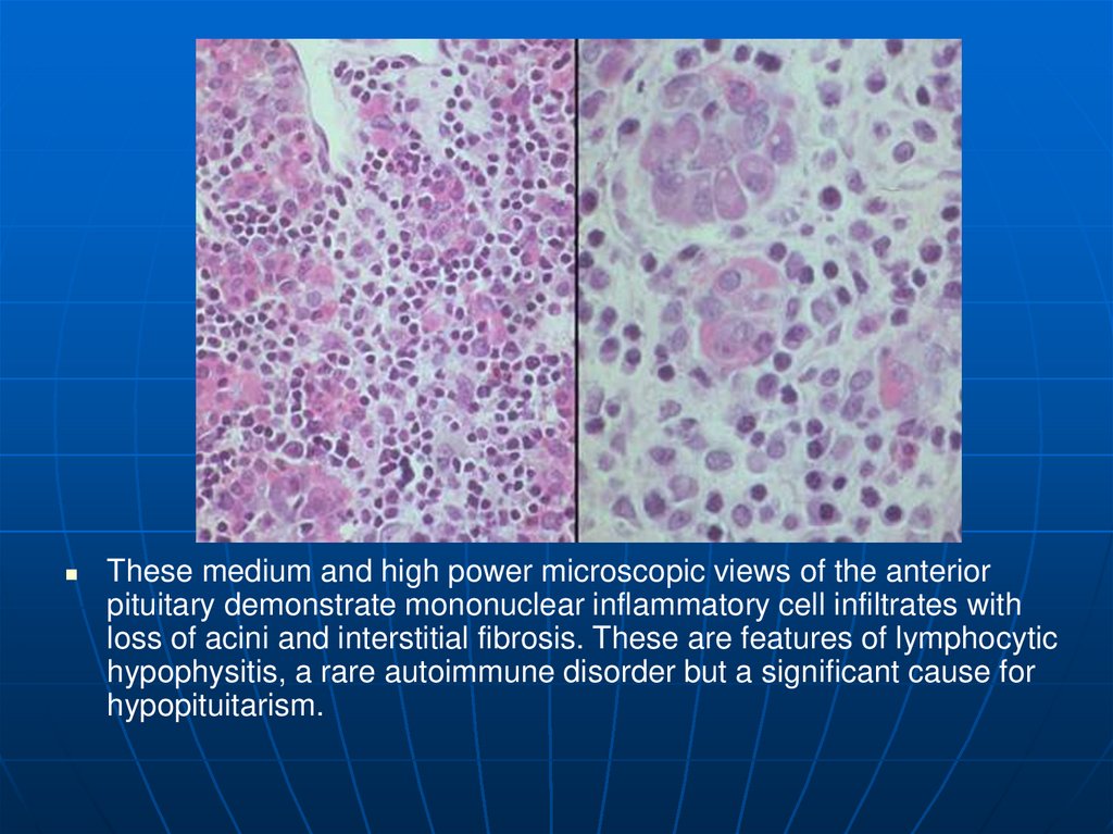
These medium and high power microscopic views of the anteriorpituitary demonstrate mononuclear inflammatory cell infiltrates with
loss of acini and interstitial fibrosis. These are features of lymphocytic
hypophysitis, a rare autoimmune disorder but a significant cause for
hypopituitarism.
24.
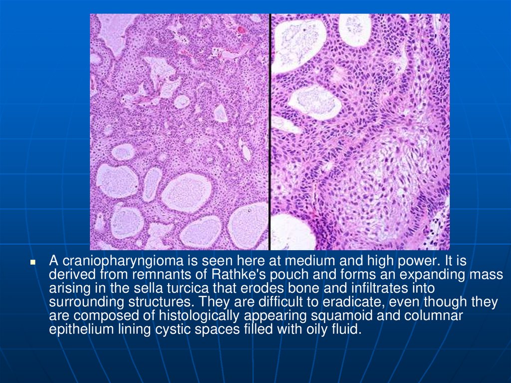
A craniopharyngioma is seen here at medium and high power. It isderived from remnants of Rathke's pouch and forms an expanding mass
arising in the sella turcica that erodes bone and infiltrates into
surrounding structures. They are difficult to eradicate, even though they
are composed of histologically appearing squamoid and columnar
epithelium lining cystic spaces filled with oily fluid.
25. Posterior Pituitary
Loss of ADH• Diabetes insipidis
• Dose not make concentrated urine
• Large volumes of dilute urine
Head injuries
Tumors of periventricular area
26. Control of Thyroid Hormone
HypothalmusPituitary
Thyroid
Tissue level
• Establishes
metabolic rate
for the whole
organism
27.
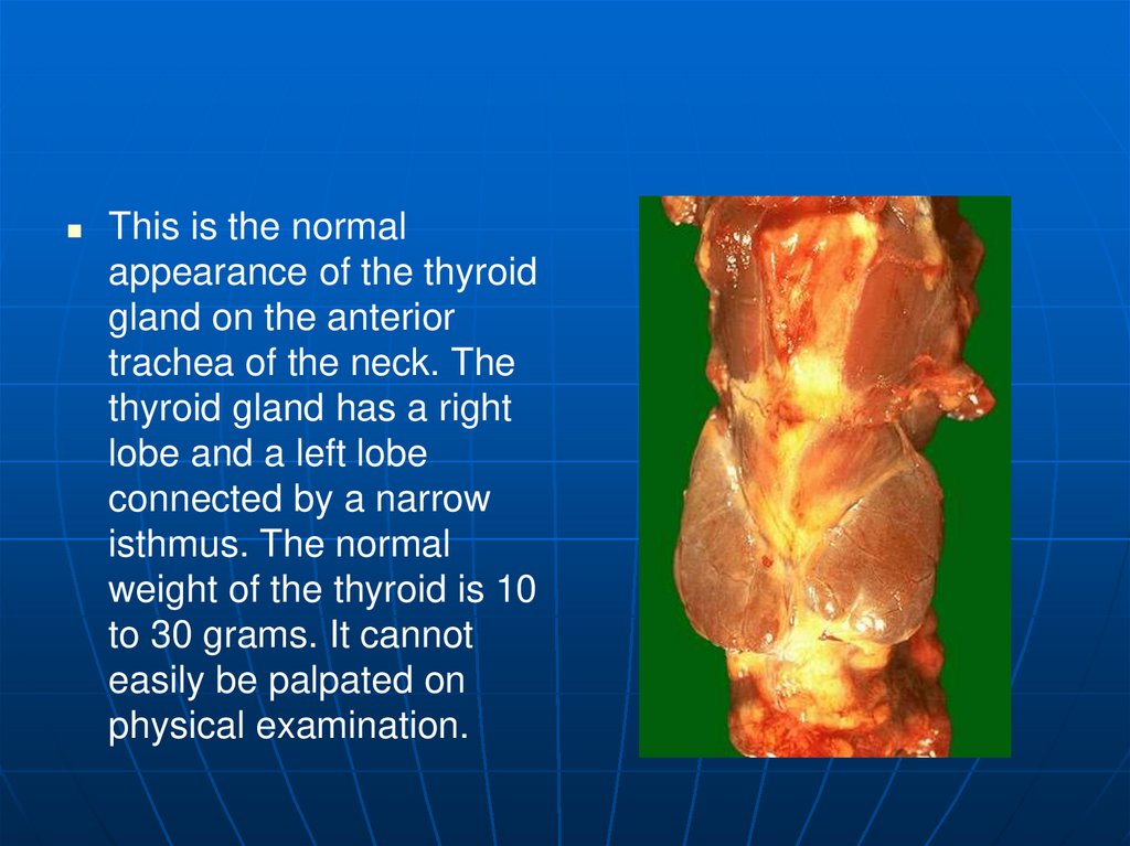
This is the normalappearance of the thyroid
gland on the anterior
trachea of the neck. The
thyroid gland has a right
lobe and a left lobe
connected by a narrow
isthmus. The normal
weight of the thyroid is 10
to 30 grams. It cannot
easily be palpated on
physical examination.
28.
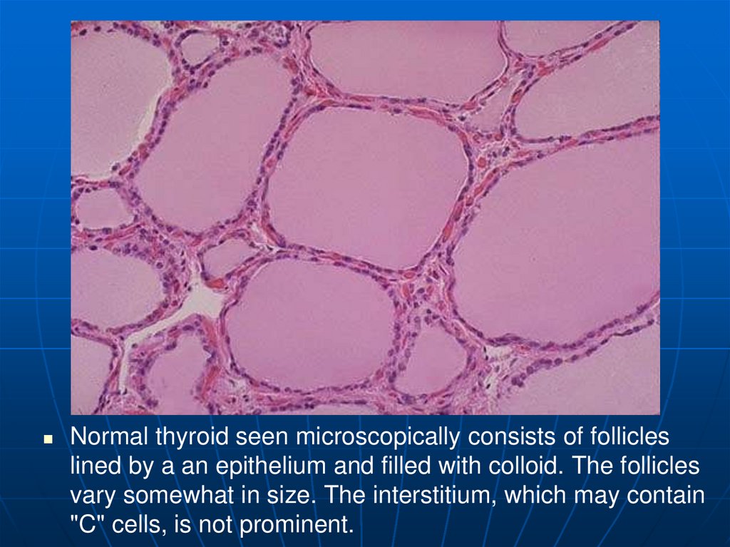
Normal thyroid seen microscopically consists of follicleslined by a an epithelium and filled with colloid. The follicles
vary somewhat in size. The interstitium, which may contain
"C" cells, is not prominent.
29.
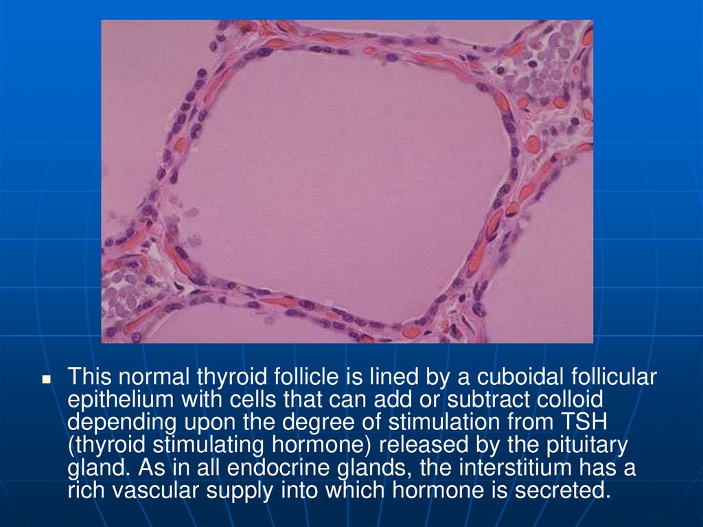
This normal thyroid follicle is lined by a cuboidal follicularepithelium with cells that can add or subtract colloid
depending upon the degree of stimulation from TSH
(thyroid stimulating hormone) released by the pituitary
gland. As in all endocrine glands, the interstitium has a
rich vascular supply into which hormone is secreted.
30. Hyperthyroidism
Clinical findingsHeat intolerance
Tremor
Tachycardia
Hyperactive
Increased body
metabolism and
temperature
• Ocular changes
Main causes
• Graves Disease
• Toxic goiter
• Toxic adenoma
31. Grave’s disease
Grave’s disease is multi-organsystemic autoimmune disorder,
manifested by the triad of basic
features:
hyperthyroidism with diffuse goiter
ophthalmopathy
dermopathy
32. Hyperophthalmia
Grave’s disease• Antibody stimulates
TSH receptors in
extraocular
muscles.
Increased tissue in
orbit causes eye to
protrude.
Won’t go down
Dry conjunctiva
and increased risk
of eye infections.
33.
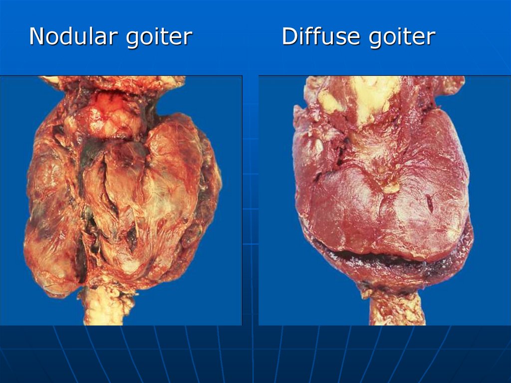
Nodular goiterDiffuse goiter
34. Hyperthyroidism
35.
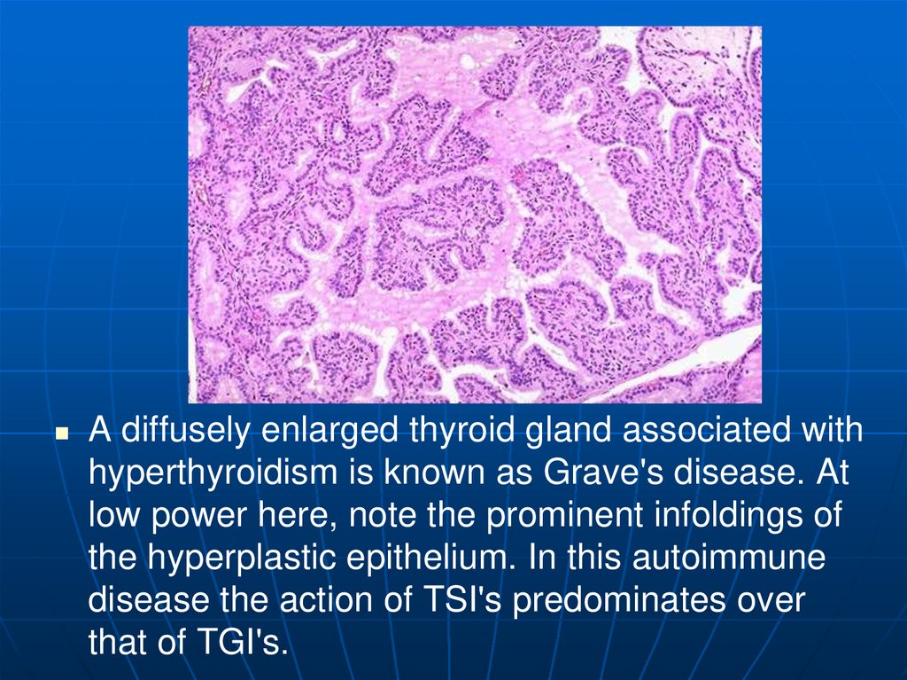
A diffusely enlarged thyroid gland associated withhyperthyroidism is known as Grave's disease. At
low power here, note the prominent infoldings of
the hyperplastic epithelium. In this autoimmune
disease the action of TSI's predominates over
that of TGI's.
36.
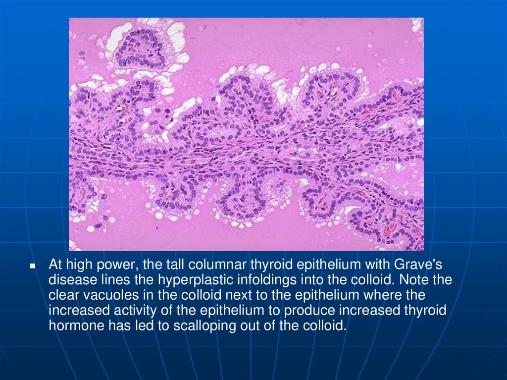
At high power, the tall columnar thyroid epithelium with Grave'sdisease lines the hyperplastic infoldings into the colloid. Note the
clear vacuoles in the colloid next to the epithelium where the
increased activity of the epithelium to produce increased thyroid
hormone has led to scalloping out of the colloid.
37. Tumors and Changes in Size
38. Goiter
NodularUniform increase
Scarring
Cysts
Generally
euthyroid
May cause airway
compression
39. Hashimoto’s Thyroiditis
Many antibodiesT & B cells
Active germinal
centers
Women 5:1
Scarring
In time hypothyroid
Other autoimmune
Arthritis
PA
Lupus
Addison’s
40. Hashimoto’s Thryoiditis
41.
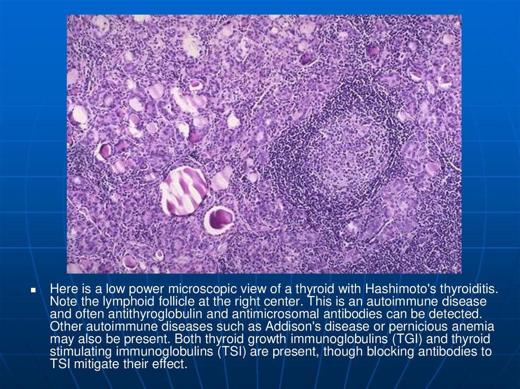
Here is a low power microscopic view of a thyroid with Hashimoto's thyroiditis.Note the lymphoid follicle at the right center. This is an autoimmune disease
and often antithyroglobulin and antimicrosomal antibodies can be detected.
Other autoimmune diseases such as Addison's disease or pernicious anemia
may also be present. Both thyroid growth immunoglobulins (TGI) and thyroid
stimulating immunoglobulins (TSI) are present, though blocking antibodies to
TSI mitigate their effect.
42.
This high power microscopic view of the thyroid withHashimoto's thyroiditis demonstrates the pink Hurthle cells
at the center and right. The lymphoid follicle is at the left.
Hashimoto's thyroiditis initially leads to painless enlargement
of the thyroid, followed by atrophy years later.
43.
This is an example of an immunofluorescence test positivefor anti-microsomal antibody, one of the autoantibodies that
can be seen with autoimmune diseases of the thyroid. Note
the bright green fluorescence in the thyroid epithelial cells,
whereas the colloid in the center of the follicles is dark.
44.
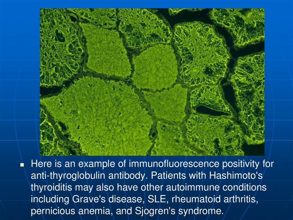
Here is an example of immunofluorescence positivity foranti-thyroglobulin antibody. Patients with Hashimoto's
thyroiditis may also have other autoimmune conditions
including Grave's disease, SLE, rheumatoid arthritis,
pernicious anemia, and Sjogren's syndrome.
45. De Quervain’s Thyroiditis
SubacuteGiant cells
Granuloma
s
Viral?
Painful
neck
46.
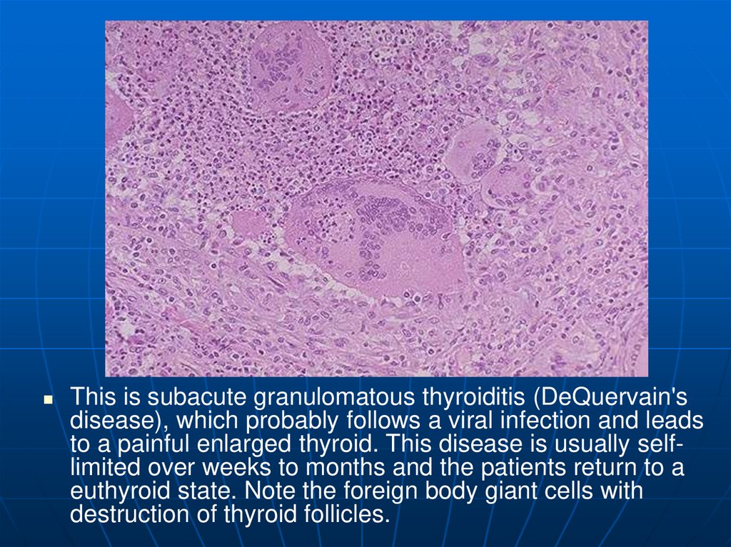
This is subacute granulomatous thyroiditis (DeQuervain'sdisease), which probably follows a viral infection and leads
to a painful enlarged thyroid. This disease is usually selflimited over weeks to months and the patients return to a
euthyroid state. Note the foreign body giant cells with
destruction of thyroid follicles.
47.
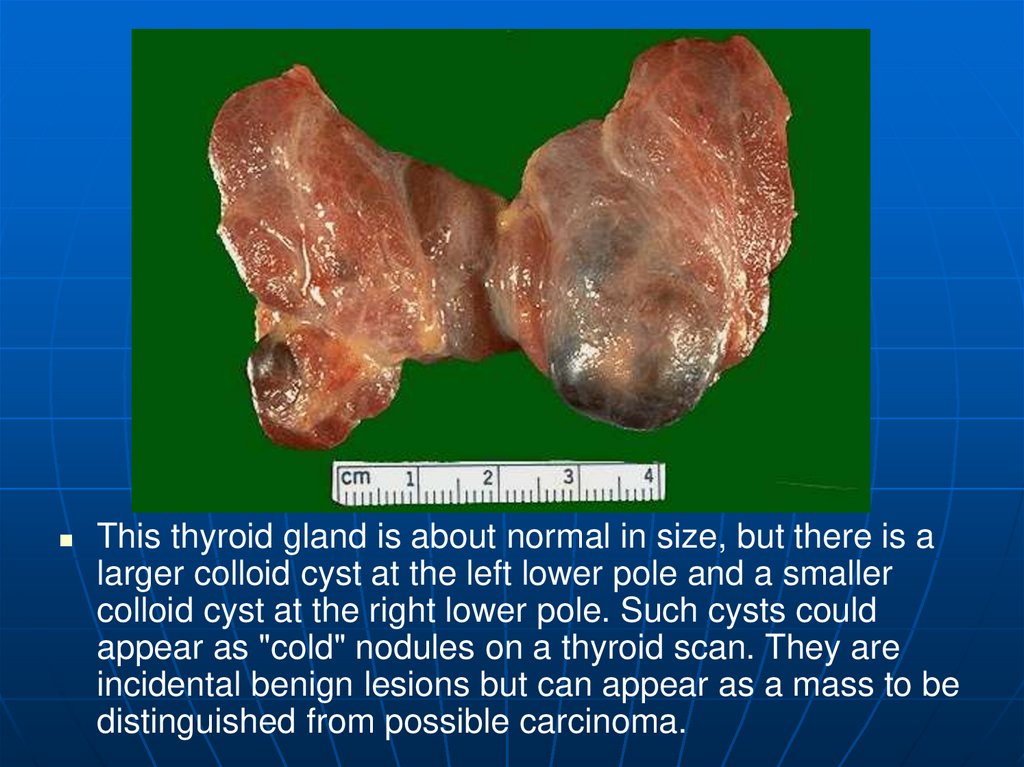
This thyroid gland is about normal in size, but there is alarger colloid cyst at the left lower pole and a smaller
colloid cyst at the right lower pole. Such cysts could
appear as "cold" nodules on a thyroid scan. They are
incidental benign lesions but can appear as a mass to be
distinguished from possible carcinoma.
48.
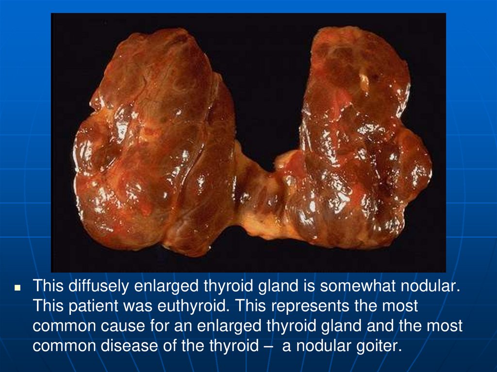
This diffusely enlarged thyroid gland is somewhat nodular.This patient was euthyroid. This represents the most
common cause for an enlarged thyroid gland and the most
common disease of the thyroid – a nodular goiter.
49.
The follicles are irregularly enlarged, with flattened epithelium, consistent withinactivity, in this microscopic appearance at low power of a multinodular
goiter. The earlier phase of a diffuse (non-toxic) goiter leading up to this point
may have resulted from either "endemic" goiter (seen in parts of the world
where dietary deficiency of iodine may occur) or the uncommon "nonendemic"
or sporadic goiter (young adult women are most often affected). Inborn errors
of thyroid hormone biosynthesis leading to goiter are extremely uncommon.
50. Hypothyroidism
GeneticsGland destruction
• Inflammatory
• Surgical removal
• Radiation treatment for hyperthyroidism
Iodine deficiency
• Can’t make T4
Hypothalmic and/or pituitary failure
51. Hypothyroidism
Genetics:Cretinism
Cannot make T4
Growth retarded
Severe mental
retardation
Must recognize
early
52. Hypothyroidism
Clinical• Cold
intolerance
• Bradycardia
• Heart failure
• High lipids
• Lethargic
• Photophobia
• Myxedema
• Skin and hair
changes
53.
This symmetrically smallthyroid gland
demonstrates atrophy.
This patient was
hypothyroid. This is the
end result of Hashimoto's
thyroiditis. Initially, the
thyroid is enlarged and
there may be transient
hyperthyroidism, followed
by a euthyroid state and
then hypothyroidism with
eventual atrophy years
later.
54. Thyroid Adenomas
BenignSolitary
Common
Encapsulated
Generally not
hyperactive
55.
Here is a surgicalexcision of a small mass
from the thyroid gland
that has been cut in half.
A rim of slightly darker
thyroid parenchyma is
seen at the left. The mass
is well-circumscribed.
Grossly it felt firm. By
scintigraphic scan it was
"cold." This is a follicular
adenoma.
56.
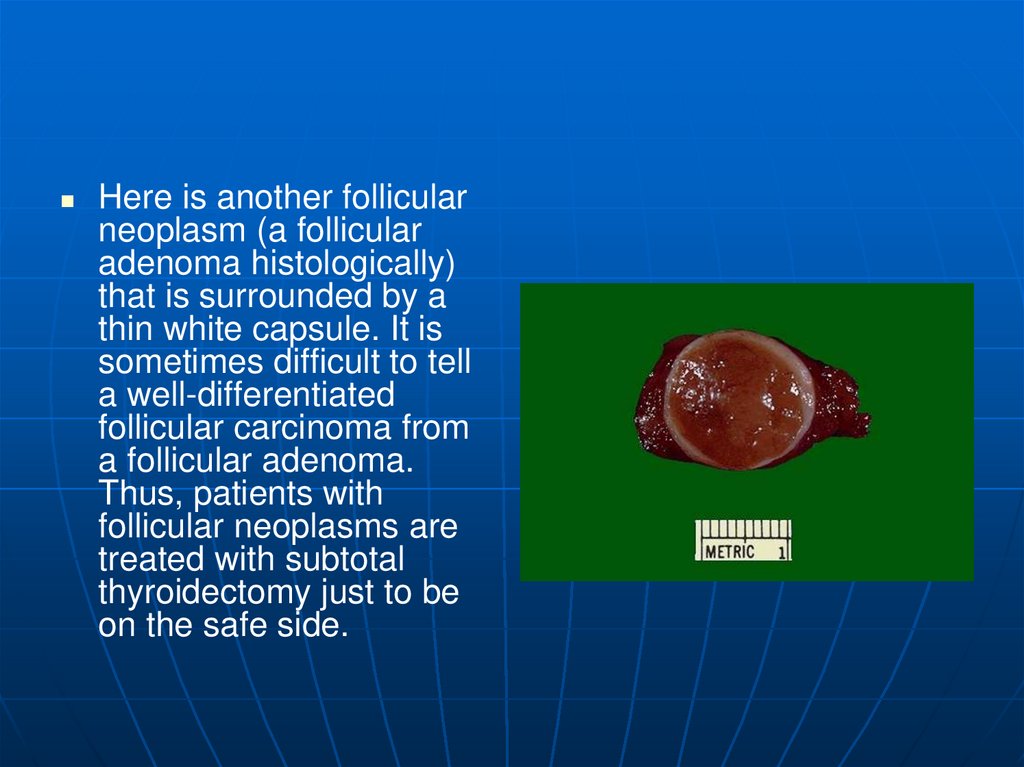
Here is another follicularneoplasm (a follicular
adenoma histologically)
that is surrounded by a
thin white capsule. It is
sometimes difficult to tell
a well-differentiated
follicular carcinoma from
a follicular adenoma.
Thus, patients with
follicular neoplasms are
treated with subtotal
thyroidectomy just to be
on the safe side.
57.
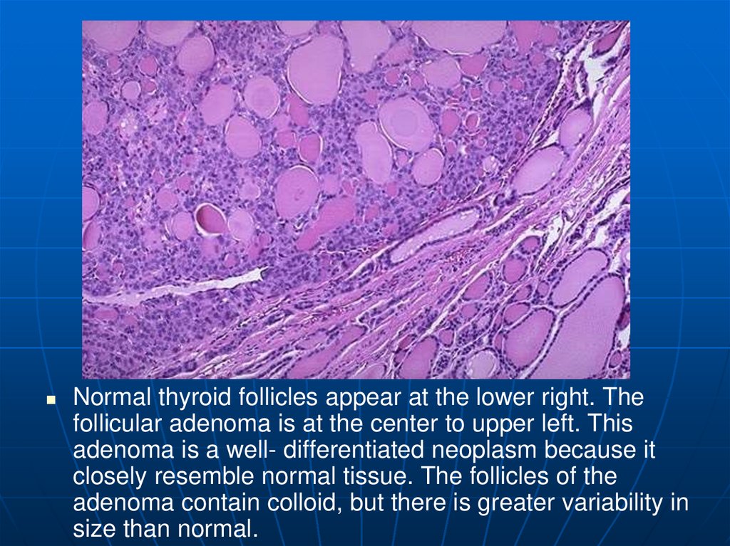
Normal thyroid follicles appear at the lower right. Thefollicular adenoma is at the center to upper left. This
adenoma is a well- differentiated neoplasm because it
closely resemble normal tissue. The follicles of the
adenoma contain colloid, but there is greater variability in
size than normal.
58. Malignancies of Thyroid Origin
Arising from follicular cells• Papillary Carcinoma
• Follicular Carcinoma
• Mixed pattern
Interstitial cells (Calcitonin producing
cells)
Anaplastic, who knows
• Very aggressive tumor
59. Papillary Carcinoma
Papillary groupsMay have
multiple sites
Not actively
producing T4
Readily treated
Spread
Nodes
Lung
Bone
Brain
60. Papillary Carcinoma
61.
Sectioning through a lobe ofexcised thyroid gland reveals
papillary carcinoma. This
neoplasm can be multifocal, as
seen here, because of the
propensity to invade
lymphatics within thyroid, and
lymph node metastases are
common. The larger mass is
cystic and contains papillary
excresences. These tumors
most often arise in middleaged females.
62. Orphan Annie Nuclei
Needleaspirates
Open eyed
nuclei
indicative of
papillary ca
63.
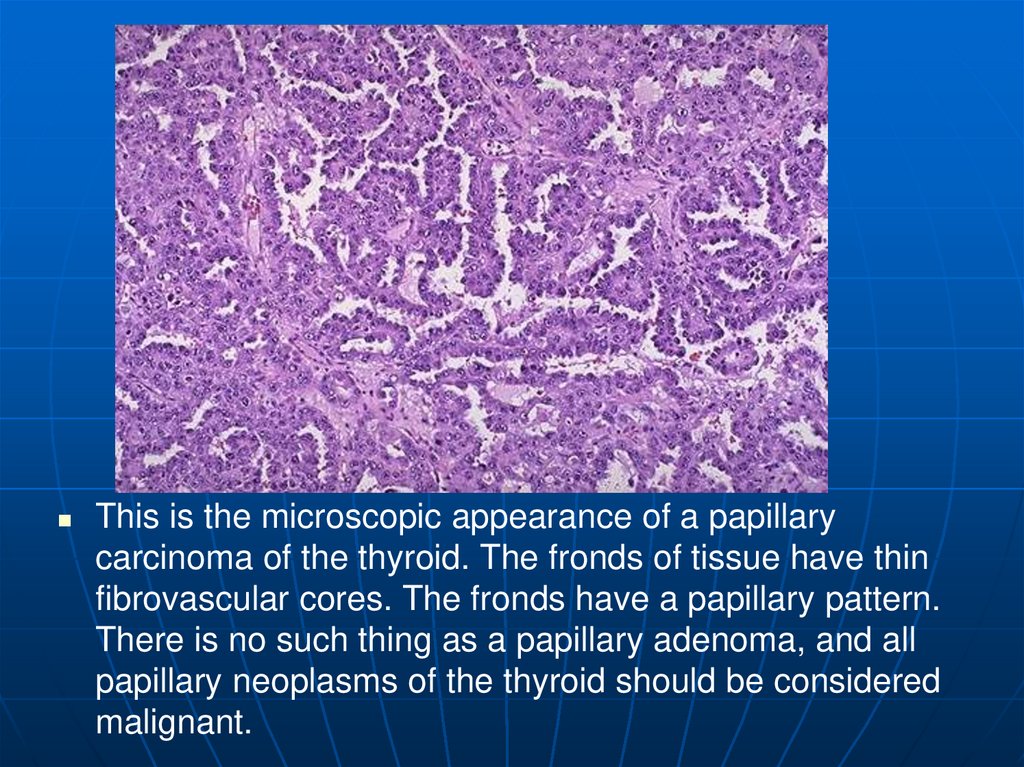
This is the microscopic appearance of a papillarycarcinoma of the thyroid. The fronds of tissue have thin
fibrovascular cores. The fronds have a papillary pattern.
There is no such thing as a papillary adenoma, and all
papillary neoplasms of the thyroid should be considered
malignant.
64.
This is another papillarycarcinoma of thyroid. Note the
small psammoma body in the
center. The cells of the
neoplasm have clear nuclei.
Papillary carcinomas are
indolent tumors that have a
long survival, even with
metastases. The most favorite
site of metastasis is to local
lymph nodes in the neck. In
fact, some papillary
carcinomas may first present
as nodal metastases.
65. C Cell Carcinoma
Interstitial cellsMakes calcitonin
Makes amyloid
• Beta pleated
sheet protein
Often part of a
multiple
endocrine
neoplasia
syndrome
66. C Cell Carcinoma
67.
At the center and to the right is a medullary carcinoma ofthyroid. At the far right is pink hyaline material with the
appearance of amyloid. These neoplasms are derived from
the thyroid "C" cells and, therefore, have neuroendocrine
features such as secretion of calcitonin.
68.
Here the amyloid stroma of the medullary thyroidcarcinoma has been stained with Congo red. Medullary
carcinomas can be sporadic or familial. The familial kind
are associated with multiple endocrine neoplasia
syndrome.
69.
This is the Congo red stained amyloid stroma ofthe medullary carcinoma under polarized light,
which produces a pale greenish appearance.
70.
The anaplastic carcinoma shown here is invadinginto skeletal muscle fibers at the right. This is the
most aggressive thyroid cancer, and luckily the
least common.
71.
There is no resemblance to normal thyroid tissuehence the term "anaplastic" to characterize thisthyroid carcinoma. Note the elongated spindle
cells.
72. Parathyroid
Come from the pharyngeal pouchesMost of us have 4
Make PTH
Mobilizes calcium
Released by low serum calcium
High serum phosphate
73.
Parathyroid hyperplasia isshown here. Three and
one-half glands have
been removed (only half
the gland at the lower left
is present). Parathyroid
hyperplasia is the second
most common form of
primary
hyperparathyroidism, with
parathyroid carcinoma
the least common form.
74.
Here is a normal parathyroid gland. Variable numbers ofsteatocytes are mixed with the parathyroid tissue. There is
a rich vascular supply, as with all endocrine tissues that
secrete their hormonal products directly into the
bloodstream.
75. Hyperparathyroidism
Primary• Parathyroid adenoma 80%
• Hyperplasia 10-15%
• Parathyroid ca <5%
Hypercalcemia
• Stones, bones, abdominal groans and psychic
moans
• Bone wasting
Generalized
Osteoitis fibrosa cystica
76.
In parathyroid hyperplasia, there is little or no adipose tissue, butany or all cell types normally found in parathyroid are present. Note
the pink oxyphil cells here. This is actually "secondary
hyperparathyroidism" with enlarged glands as a consequence of
chronic renal failure with impaired phosphate excretion. The
increased serum phosphate tends to drive serum calcium down,
which in turn drives the parathyroids to secrete more parathormone.
77. Parathyroid Adenoma
78.
Here is a parathyroid adenoma, which is the mostcommon cause for primary hyperparathyroidism. A rim of
normal parathyroid tissue admixed with adipose tissue
cells is seen compressed to the right and lower edge of
the adenoma.
79.
80. Secondary Hyperparathyroidism
Renal failure almost always• Phosphates build up in the blood.
• Cause calcium to drop.
PTH is made
• Phosphate itself can cause release of
PTH
Glands begin to function
autonomously
81.
This is the grossappearance of a
parathyroid carcinoma.
The serum calcium can
be quite high. Note the
large size and irregular
cut surface. These
carcinomas have a
tendency to invade
surrounding tissues in the
neck, complicating their
removal.
82.
This is a parathyroid carcinoma seen at medium power onthe left and higher power on the right. The nests of
neoplastic cells that are not very pleomorphic. Note the
bands of fibrous tissue between the nests. Parathyroid
carcinomas infiltrate surrounding structures in the neck.
83. Hypoparathyroidism
Increased neuromuscular excitability• May lead to tetany
Irritability and possibly even
psychosis
Parkinson-like symptoms
Cataracts
Causes
• Autoimmune destruction
• Accidental removal with thyroid
• Congenital absence
84. Adrenal Gland
Really two glands inone.
• Cortex ->
Salt
Sugar
Sex
• Medulla
Epinephrine
Norepinephrine
85.
Here are normal adrenal glands. Each adultadrenal gland weighs from 4 to 6 grams.
86.
The pair of adrenals in the center are normal. Those at the top comefrom a patient with adrenal atrophy (with either Addison's disease or
long-term corticosteroid therapy). The adrenals at the bottom represent
bilateral cortical hyperplasia. This could be due to a pituitary adenoma
secreting ACTH (Cushing's disease), or Cushing's syndrome from
ectopic ACTH production, or idiopathic adrenal hyperplasia.
87.
These adrenals areblack-red from
extensive
hemorrhage in a
patient with
meningococcemia.
This produces the
WaterhouseFriderichsen
syndrome.
88. Cushing’s Syndrome
Effects of too muchcortisol
Moon face
Central obesity
Buffalo hump
Osteoporosis
Fractures
• Hypertension
• Weakness
89. Cushing’s Disease
Altered feedback regulation at levelof hypothalmus and pituitary
• It only takes a small increase in ACTH
• Loss of cortisol diurnal cycle
Pituitary adenoma
Ectopic ACTH
• Small cell carcinoma of lung
Adrenal tumors autonomously
functioning
90. Cushing’s Disease
91.
This adrenal gland removedsurgically in a patient with
Cushing's syndrome has been
sectioned in half to reveal an
adenoma. Some remaining
atrophic adrenal is seen at the
right. The adenoma is
composed of yellow firm tissue
just like adrenal cortex. This
neoplasm is wellcircumscribed. Histologically, it
is composed of welldifferentiated cells resembling
cortical fasciculata zone. It is
benign.
92.
Microscopically, the adrenal cortical adenoma atthe right resembles normal adrenal fasciculata.
The capsule is at the left. There may be some
cellular pleomorphism.
93.
This high power microscopic appearance of an adrenal corticalcarcinoma demonstrates that the neoplasm closely resembles normal
adrenal cortex. It is difficult to determine malignancy in endocrine
neoplasms based upon cytology alone. Thus, invasion (as seen here
in a vein) and metastases are the most reliable indicators. Luckily,
most endocrine neoplasms are benign adenomas.
94. Hypoadrenalism
Acute loss vs. ChronicPituitary vs. adrenal
Acute
• Waterhouse-Fridericshen
syndrome ->
• Overwhelming infection
with encapsulated
bacteria.
• Leads to vascular
infection.
• Hemorrhagic destruction
of adrenal glands
• Medical crisis
95. Waterhouse-Fridericshen syndrome
96. Waterhouse-Fridericshen syndrome
97.
This is the microscopic appearance of theadrenals with meningococcemia. There is marked
hemorrhagic necrosis with acute adrenal
insufficiency.
98. Addison’s Disease
Slowly developsLoss of adrenal
glands
Lots of ACTH, but
nothing it can do.
Metastatic tumor
TB
Clinical
• Weight loss
• Hypotension
• Hyperpigmentation
99. Adrenal Medulla
PheochromocytomaCatacholamines
Elevated blood pressure
Syncopal episodes
Headaches
Nose bleeds
Anxiety
Maybe an isolated tumor
or part of a multiple
endocrine tumor
syndrome.
100. Pheochromocytoma
101.
This large adrenal neoplasm has been sectioned in half.Note the grey-tan color of the tumor compared to the yellow
cortex stretched around it and a small remnant of remaining
adrenal at the lower right. This patient had episodic
hypertension. This is a tumor arising in the adrenal
medulla--a pheochromocytoma.
102.
There is some residual adrenal cortical tissue atthe lower center right, with the darker cells of
pheochromocytoma seen above and to the left.
103.
By electron microscopy,the neoplastic cells of the
pheochromocytoma
contain neurosecretory
granules. It is these
granules that contain the
catecholamines. The
granules seen here
appear as small black
round objects in the
cytoplasm of the cell. The
cell nucleus is at the
upper left.
104. Diabetes mellitus
105. Diabetes Mellitus
General definition: Chronic disorder of glucosemetabolism with hyperglycemia, triggered by
conditions associated with a relative or absolute
insulin deficiency.
Primary diabetes mellitus: Insulin deficiency due
to islet damage from autoimmune inflammation
(type I) or
— Dysfunction of pancreatic insulin-producing
cells (type II).
106. Diabetes Mellitus
Secondary diabetes mellitus: Insulin deficiencydue to islet damage from pancreatic disease such
as
pancreatitis,
hemochromatosis, or
cystic fibrosis; or
Overproduction of insulin antagonist hormones
such as cortisone and somatotropic hormone
(STH).
107. Diabetes Mellitus Definition
A multisystem disease related to:• Chronic disorder
• Abnormal metabolism of fuels glucose
and fat
• An endocrine disorder causes Abnormal
insulin production
• Impaired insulin utilization
• Both abnormal production and impaired
utilization
107
108. Diabetes Mellitus Definition
Leading cause ofheart disease, stroke,
adult blindness, and
nontraumatic lower
limb amputations
108
109.
Here is a normal pancreatic islet of Langerhanssurrounded by normal exocrine pancreatic acinar
tissue. The islets contain alpha cells secreting
glucagon, beta cells secreting insulin, and delta cells
secreting somatostatin
110.
Immunoperoxidase staining can help identify thenature of the cells present in the islets of Langerhans.
On the right, antibody to insulin has been employed to
identify the beta cells. On the left, antibody to
glucagon identifies the alpha cells.
111.
112.
Type I Diabetes MellitusSynonyms: juvenile-onset diabetes mellitus, insulindependent diabetes mellitus (IDDM).
Autoimmune lymphocytic insulitis in combination with
genetic susceptibility (HLA-DR4 and/or DR3) leads to
formation of autoimmune T-lymphocytes and islet-cell
antibodies.
They destroy the b cells ( A) and leave the glucagonforming cells intact ( B), causing insulin-dependent diabetes
mellitus.
113. Type 1 Diabetes Mellitus
Progressive destruction of pancreaticcells
Autoantibodies cause a reduction of
80% to 90% of normal cell function
before manifestations occur
Causes:
• Genetic predisposition
Related to human leukocyte
antigens (HLAs)
• Exposure to a virus
114.
This is an insulitis of anislet of Langerhans in a
patient who will
eventually develop type I
diabetes mellitus. The
presence of the
lymphocytic infiltrates in
this edematous islet
suggests an autoimmune
mechanism for this
process. The destruction
of the islets leads to an
absolute lack of insulin
that characterizes type I
diabetes mellitus.
115.
A Type I diabetesmellitus: loss of βcells
(IH; insulin) x 200
B Type I diabetes
mellitus: dominance
of α cells
(IH; glucagon) x 200
116. Diabetes Mellitus
Type II Diabetes MellitusSynonyms: adult-onset diabetes mellitus, noninsulindependent diabetes mellitus (NIDDM).
Type IIa is without obesity; type IIb with obesity.
Together with insulin, b cells form amylin (islet
amyloid peptide), which condenses to AE amyloid,
“smothering” the function of the islets. Peripheral
organs and tissues in obese patients also exhibit
insulin resistance due to the protein resistin, secreted
by fat cells, leading to non-insulin-dependent diabetes
mellitus. Immunohistochemical findings reveal normal
counts of insulin-producing cells and glucagonproducing cells.
117. Type 2 Diabetes Mellitus
Accounts for 90% of patientswith diabetes
Usually occurs in people over 40
years of age
80-90% of patients are
overweight
118.
Pancreas continues to producesome endogenous insulin
Insulin produced is either
insufficient or poorly utilized by
the tissues
Insulin resistance
• Body tissues do not respond to
insulin
• Results in hyperglycemia
119.
120.
This islet ofLangerhans
demonstrates pink
hyalinization (with
deposition of amyloid)
in many of the islet
cells. This change is
common in the islets
of patients with type II
diabetes mellitus.
121.
Islet amyloidosis(HE) x 200
Type II diabetes
mellitus: В cells
(IH; insulin) x 200
122. E Type II diabetes mellitus: alpha cells (IH; glucagon) x 200
123. Secondary Diabetes
Results from another medicalcondition or due to the treatment
of a medical condition that
causes abnormal blood glucose
levels
• Cushing syndrome
• Hyperthyroidism
• Parenteral nutrition
124.
125.
126.
Diabetic macroangiopathy follows the pattern ofatherosclerosis .
Complications:
– Coronary sclerosis can lead to myocardial
infarction.
– Cerebral sclerosis can lead to cerebral infarction.
– Popliteal sclerosis can lead to gangrene.
127. Diabetic gangrene
128.
129.
Diabetic microangiopathy: Chronic increased glucoseconcentration leads to glycosylation of proteins, altering the
structure and permeability of the microvascular basement
membranes .
Complications:
– Diabetic retinopathy (a late complication):
Capillary microaneurysms and arteriosclerosis cause
microinfarctions (punctate hemorrhages).
Proliferative retinitis leads to shrinkage of the vitreous body
and retinal detachment.
– Diabetic glomerulosclerosis (Kimmelstiel-Wilson
lesion): Deranged synthesis and breakdown of the glomerular
basement membrane cause thickening of the membrane . This
causes diffuse and, later, nodular deposition of PAS-positive
material in the mesangium and between the glomerular
podocytes and basement membrane, leading to proteinuria
and renal insufficiency.
130.
Diabetic cataract: Osmotic vacuolar degeneration of theepithelium of the lens creates lens opacities.
Diabetic liver: Secondary glycogenosis (glycogen-induced
nuclear defects) occurs in relation to the level of blood
glucose; simultaneous fatty degeneration correlates with
obesity in type IIb diabetes.
Diabetic neuropathy: After approximately 25 years of
diabetes, 50% of patients exhibit axonal and/or myelin
degeneration leading to hyporeflexia and decreased deep
sensation.
Complications: diabetic microangiopathy and diabetic
neuropathy lead to gangrene in the toes.
131. Gestational Diabetes
Develops during pregnancyDetected at 24 to 28 weeks of
gestation
Risk for cesarean delivery,
perinatal death, and neonatal
complications
132. Diabetic retinopathy
133. Diabetic retinopathy Diabetic cataract
134.
135. Diabetes mellitus
136. Diffuse glomerulosclerosis Characterized by diffuse thickening of glomerular capillary basement membranes and increased amount
of mesangialmatrix with mild mesangial cell proliferation. Glomerular
changes always begin in the vascular stalk. The affected
glomeruli eventually develop obliterative diabetic
glomerulosclerosis. These changes are seen in at least
40% of diabetic patients after more than 10 to 20 years.
137. Diabetic microangiopathy, Diabetic neuropathy
138.
An islet cell adenomais seen here,
separated from the
pancreas by a thin
collagenous capsule.
A few normal islets
are seen in the
pancreas at the right
for comparison.
139.
The islet cell adenoma atthe left contrasts with the
normal pancreas with
islets at the right. Some
of these adenomas
function. Those that
produce insulin may lead
to hypoglycemia. Those
that produce gastrin may
lead to multiple gastric
and duodenal ulcerations
(Zollinger-Ellison
syndrome).
140.
This is animmunohistoc
hemical stain
for insulin in
the islet cell
adenoma.
Thus, it is an
insulinoma.
141.
Here is a carcinoid tumorseen on the mucosal
surface at the ileocecal
valve. Note that it is a
small, well-circumscribed
mass that has a yellowish
tint to it. Such neoplasms
are typically benign, even
though they may be
multiple. Most do not
secrete a detectable
hormone.
142.
At low magnification, the smallblue nests of tumor cells in this
carcinoid tumor are grouped
together beneath the mucosa,
but are not encapsulated and
appear to "infiltrate" in the
muscularis, though this is not
strictly invasion. It is rare for a
carcinoid <1 cm to behave in a
malignant fashion, while the
majority >2 cm are malignant.
Most carcinoids are <1 cm.
143.
At high magnification, thesmall nests of tumor cells
in this carcinoid contain
round cells with round
nuclei. Carcinoids can be
found anywhere in the
gastrointestinal tract,
though they are most
common in ileum,
appendix, and colon.
Carcinoids may rarely be
found arising in bronchi of
the lung.
144.
Thisimmunoperoxidase
stain with antibody to
ACTH demonstrates
staining of the cells in
this carcinoid tumor.
This patient had
Cushing's syndrome
due to ectopic ACTH
production from the
carcinoid.
145.
At higher power, theimmunoperoxidase staining
pattern with antibody to ACTH
is shown in this carcinoid
tumor. Carcinoids are capable
of secreting a variety of
hormones. Gastrin secretion
can lead to the ZollingerEllison syndrome (multiple
gastric ulcers). The "carcinoid
syndrome" (quite rare) from
serotonin secretion is typically
a result of a malignant
carcinoid that has
metastasized to the liver.