


 medicine
 biology
Similar presentations:


 and blood, muscle, nervous")


Anatomy & physiology. The tissue level of organization
1.
ANATOMY & PHYSIOLOGYChapter 4 THE TISSUE LEVEL OF ORGANIZATION
PowerPoint Image Slideshow
2. Figure 4.1
FIGURE 4.1Micrograph of Cervical Tissue
This figure is a view of the regular architecture of normal tissue contrasted with the
irregular arrangement of cancerous cells. (credit: “Haymanj”/Wikimedia Commons)
3. MAJOR CHAPTER Objectives
MAJOR CHAPTER OBJECTIVES• Identify the main tissue types and discuss their roles in the human
body
• Identify the four types of tissue membranes and the characteristics
of each that make them functional
• Explain the functions of various epithelial tissues and how their
forms enable their functions
• Explain the functions of various connective tissues and how their
forms enable their functions
• Describe the characteristics of muscle tissue and how these
enable function
• Discuss the characteristics of nervous tissue and how these
enable information processing and control of muscular and
glandular activities
4. 4.1 types of tissues Major section Objectives
4.1 TYPES OF TISSUESMAJOR SECTION OBJECTIVES
• Identify the four main tissue types
• Discuss the functions of each tissue type
• Relate the structure of each tissue type to their function
• Discuss the embryonic origin of tissue
• Identify the three major germ layers
• Identify the main types of tissue membranes
N.B. Any disruption of the structure is a CAUSE
(not only a sign!) of injury or disease.
5. Figure 4.2
FIGURE 4.2Four Types of Tissue: Body
The four types of tissues are exemplified
in nervous tissue, stratified squamous
epithelial tissue, cardiac muscle tissue,
and connective tissue in small intestine.
Clockwise from nervous tissue, LM ×
872, LM × 282, LM × 460, LM × 800.
(Micrographs provided by the Regents of
University of Michigan Medical School ©
2012)
6. Figure 4.3
FIGURE 4.3Embryonic Origin of Tissues and
Major Organs
7. Figure 4.4
FIGURE 4.4Tissue Membranes
The two broad categories of tissue
membranes in the body are (1)
connective tissue membranes, which
include synovial membranes, and (2)
epithelial membranes, which include
mucous membranes, serous
membranes, and the cutaneous
membrane, in other words, the skin.
Careful. p.141: abdominal mesenteries do not line passageways
that lead to the exterior of the body in males – only in females.
8. 4.2 Epithelial tissue Major section Objectives
4.2 EPITHELIAL TISSUEMAJOR SECTION OBJECTIVES
• Explain the structure and function of epithelial tissue
• Distinguish between tight junctions, anchoring junctions, and gap
junctions
• Distinguish between simple epithelia and stratified epithelia, as
well as between squamous, cuboidal, and columnar epithelia
• Describe the structure and function of endocrine and exocrine
glands and their respective secretions
Errors in Review of 4.2, pp. 175, 176: Covering epithelia are strictly
AVASCULAR i.e., they contain NO blood vessels (versus “a few”).
9. Figure 4.5
FIGURE 4.5Types of Cell Junctions
The three basic types of cell-to-cell
junctions are tight junctions, gap
junctions, and anchoring junctions.
Error in text p.143:
“Cells of epithelia are closely
connected and are not
separated by intracellular
intercellular material.”
10. Figure 4.6
FIGURE 4.6Cells of Epithelial Tissue
Simple epithelial tissue is organized as a single layer of cells and stratified epithelial
tissue is formed by several layers of cells.
N.B. Epithelial tissues are classified according to the shape of their surface
cells - as seen in cross section, as well as by their number of cell layers.
11. Figure 4.7
FIGURE 4.7Goblet Cell
(a) In the lining of the small intestine,
columnar epithelium cells are
interspersed with goblet cells.
(b) The arrows in this micrograph point
to the mucous-secreting goblet cells.
LM × 1600. (Micrograph provided by
the Regents of University of Michigan
Medical School © 2012)
12. Figure 4.8a
FIGURE 4.8ASummary of Epithelial Tissue Cells
13. Figure 4.8b
FIGURE 4.8BSummary of Epithelial Tissue Cells
14. Figure 4.9
FIGURE 4.9Types of Exocrine Glands
Exocrine glands are classified by their
structure.
15. Figure 4.10
FIGURE 4.10Modes of Glandular Secretion
(a)
In merocrine secretion, the cell remains intact. – E.g. as seen with goblet cells (mucin), eccrine sweat glands.
(b)
In apocrine secretion, the apical portion of the cell is released, as well. – E.g. as seen with apocrine sweat glands.
(c)
In holocrine secretion, the cell is destroyed as it releases its product and the cell itself becomes part of the secretion.
– E.g. as seen with sebaceous glands producing sebum.
16. Figure 4.11
FIGURE 4.11Sebaceous Glands
These glands secrete oils that lubricate and protect the skin. They are holocrine glands and they are
destroyed after releasing their contents. New glandular cells form to replace the cells that are lost.
LM × 400. (Micrograph provided by the Regents of University of Michigan Medical School © 2012)
17. table 4.1
TABLE 4.1Connective Tissues Examples
18. 4.3 connective tissue supports and protects Major section Objectives
4.3 CONNECTIVE TISSUE SUPPORTS AND PROTECTSMAJOR SECTION OBJECTIVES
• Identify and distinguish between the various sub-types of
connective tissue proper (or fibrous), supportive, and fluid.
• Explain the functions of connective tissues
Add:
Add:
Recognize, name and describe the function(s) of connective
tissue cell types.
Describe the major components of the extracellular matrix of the
various connective tissue subtypes.
19. Figure 4.12
FIGURE 4.12Connective Tissue Proper
Fibroblasts produce this fibrous tissue. Connective tissue proper includes the fixed cells
fibrocytes, adipocytes, and mesenchymal cells. LM × 400. (Micrograph provided by the
Regents of University of Michigan Medical School © 2012)
20. Figure 4.13
FIGURE 4.13Adipose Tissue
This is a loose connective tissue that consists of fat cells with little extracellular matrix.
It stores fat for energy and provides insulation. LM × 800. (Micrograph provided by the
Regents of University of Michigan Medical School © 2012)
21. Figure 4.14
FIGURE 4.14Reticular Tissue
This is a loose connective tissue made up of a network of reticular fibers that provides a
supportive framework for soft organs. LM × 1600. (Micrograph provided by the Regents
of University of Michigan Medical School © 2012)
22. Figure 4.15
FIGURE 4.15Dense Connective Tissue
(a)
Dense regular connective tissue consists of collagenous fibers packed into parallel bundles.
(b)
Dense irregular connective tissue consists of collagenous fibers interwoven into a mesh-like network. From top,
LM × 1000, LM × 200. (Micrographs provided by the Regents of University of Michigan Medical School © 2012)
23. Figure 4.16
FIGURE 4.16Types of Cartilage
Cartilage is a connective tissue
consisting of collagenous fibers
embedded in a firm matrix of chondroitin
sulfates.
(a) Hyaline cartilage provides support
with some flexibility. The example is
from dog tissue.
(b) Fibrocartilage provides some
compressibility and can absorb
pressure.
(c) Elastic cartilage provides firm but
elastic support. From top, LM × 300,
LM × 1200, LM × 1016. (Micrographs
provided by the Regents of University
of Michigan Medical School © 2012)
Error in text p.159:
Your ear lobes do not contain elastic
cartilage although the rest of your
external ear does.
24. Figure 4.17
FIGURE 4.17Blood: A Fluid Connective Tissue
Blood is a fluid connective tissue containing erythrocytes and various types of
leukocytes that circulate in a liquid extracellular matrix. LM × 1600. (Micrograph
provided by the Regents of University of Michigan Medical School © 2012)
25. 4.4 muscle tissue and motion Major section Objectives
4.4 MUSCLE TISSUE AND MOTIONMAJOR SECTION OBJECTIVES
• Identify the three types of muscle tissue
• Compare and contrast the histological appearances and the
functions of each muscle tissue type
• Explain how muscle tissue can enable motion - will be seen later*.
* Error p.163: “The striation is due to the regular alternation of the
contractile proteins actin and myosin, along with the structural
proteins that couple the contractile proteins to connective
tissues.” The protein actin and myosin do NOT contract, they
slide past one another, as we will see later this semester!
26. table 4.2
TABLE 4.2Comparison of Structure and Properties of Muscle Tissue Types
Tissue
Histology
Function
Location
Skeletal
Long cylindrical fiber,
striated, many
peripherally located
nuclei
Voluntary movement, produces
heat, protects organs
Attached to bones and
around entrance points
to body (e.g., mouth,
anus)
Cardiac
Short, branched,
striated, single central
nucleus
Intercalated discs
Contracts to pump blood
Heart
Smooth
Short, spindle-shaped,
no evident striation,
single nucleus in each
fiber
Involuntary movement, moves
food, involuntary control of
respiration, moves secretions,
regulates flow of blood in
arteries by contraction
Walls of major organs
and passageways
27. Figure 4.18
FIGURE 4.18Muscle Tissue
(a) Skeletal muscle cells have prominent
striations and multiple nuclei on their
periphery.
(b) Spindle-shaped smooth muscle
cells have a single nucleus and no
visible striations.
(c) Cardiac muscle cells appear striated
and have a single nucleus. From top,
LM × 1600, LM × 1600, LM × 1600.
(Micrographs provided by the
Regents of University of Michigan
Medical School © 2012)
Add: Cardiac muscle cells
are branched and linked by
intercalated discs rich in GAP
junctions and desmosomes.
28.
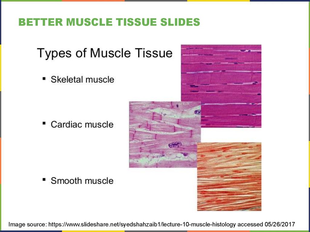
BETTER MUSCLE TISSUE SLIDESImage source: https://www.slideshare.net/syedshahzaib1/lecture-10-muscle-histology accessed 05/26/2017
29. 4.5 nervous tissue for perception, response Major section Objectives
4.5 NERVOUS TISSUE FOR PERCEPTION, RESPONSEMAJOR SECTION OBJECTIVES
• Identify the classes of cells that make up nervous tissue
• Discuss how nervous tissue mediates perception and response
Add:
Recognize, name and describe the function(s) of nervous
tissue cell types – including the distinct glial cell types.
30. Figure 4.19
FIGURE 4.19The Neuron
The cell body of a neuron, also called the soma, contains the nucleus and mitochondria. The
dendrites transfer the nerve impulse to the soma. The axon carries the action potential away
to another excitable cell. LM × 1600. (Micrograph provided by the Regents of University of
Michigan Medical School © 2012)
31. Figure 4.20
FIGURE 4.20Nervous Tissue
Nervous tissue is made up of neurons and neuroglia. The cells of nervous tissue are
specialized to transmit and receive impulses. LM × 872. (Micrograph provided by the
Regents of University of Michigan Medical School © 2012)
32. 4.6 tissue injury and aging Major section Objectives
4.6 TISSUE INJURY AND AGINGMAJOR SECTION OBJECTIVES
• Identify the cardinal signs of inflammation
• List the body’s response to tissue injury
• Explain the process of tissue repair
• Discuss the progressive impact of aging on tissue
• Describe cancerous mutations’ effect on tissue
33. Figure 4.21
FIGURE 4.21Tissue Healing
During wound repair, collagen fibers are laid down randomly by fibroblasts that move
into repair the area.
34. Figure 4.22
FIGURE 4.22Development of Cancer
Note the change in cell size, nucleus
size, and organization in the tissue.
35. disorders & homeostatic imbalances
DISORDERS & HOMEOSTATIC IMBALANCESConnective Tissue: Tendinitis
Tissues and Cancer
Cancer is a generic term for many diseases in which cells escape regulatory signals. Uncontrolled
growth, invasion into adjacent tissues, and colonization of other organs, if not treated early enough,
are its hallmarks. Health suffers when tumors “rob” blood supply from the “normal” organs.
A mutation is defined as a permanent change in the DNA of a cell. Epigenetic modifications,
changes that do not affect the code of the DNA but alter how the DNA is decoded, are also
known to generate abnormal cells. Alterations in the genetic material may be caused by
environmental agents, infectious agents, or errors in the replication of DNA that accumulate with
age. Many mutations do not cause any noticeable change in the functions of a cell. However, if the
modification affects key proteins that have an impact on the cell’s ability to proliferate in an orderly
fashion, the cell starts to divide abnormally.
As changes in cells accumulate, they lose their ability to form regular tissues. A tumor, a
mass of cells displaying abnormal architecture, forms in the tissue. Many tumors are benign,
meaning they do not metastasize nor cause disease.
A tumor becomes malignant, or cancerous, when it breaches the confines of its tissue,
promotes angiogenesis, attracts the growth of capillaries, and metastasizes to other organs.
The specific names of cancers reflect the tissue of origin. Cancers derived from epithelial cells
are referred to as carcinomas. Cancer in myeloid tissue or blood cells form myelomas. Leukemias
are cancers of white blood cells, whereas sarcomas derive from connective tissue.
Cells in tumors differ both in structure and function. Some cells, called cancer stem cells, appear to
be a subtype of cell responsible for uncontrolled growth. Recent research shows that contrary to
what was previously assumed, tumors are not disorganized masses of cells, but have their own
structures.
36. interactive links
INTERACTIVE LINKSView this slideshow http://openstaxcollege.org/l/stemcells about stem cells.
View the University of MichiganWebScope at http://openstaxcollege.org/l/goblet
to find digestive goblet cells (LM).
Watch http://openstaxcollege.org/l/etissues about epithelial tissues’ anatomy.
Watch this animation http://openstaxcollege.org/l/tendonitis to learn about
tendonitis, a painful condition caused by swollen or injured tendons.
View the University of Michigan Webscope at
http://openstaxcollege.org/l/cardiovascular to see a blood smear (LM).
Visit this link http://openstaxcollege.org/l/10quiz to test your connective tissue
knowledge with this 10-question quiz.
Watch this video http://openstaxcollege.org/l/musctissue about muscle tissue.
Follow this link http://openstaxcollege.org/l/nobel about nervous tissue.
Watch this video http://openstaxcollege.org/l/healinghand of a hand healing.
Watch this video http://openstaxcollege.org/l/tumor to learn more about tumors.
37. key terms & CHAPTER Review
KEY TERMS & CHAPTER REVIEWThis PowerPoint presentation is copyright 2011-2015, Rice University.
All Rights Reserved.
Last modified: 05/2017 / Dr. F. Jolicoeur