


 biology
Similar presentations:

 and blood, muscle, nervous")


Tissues. The histophysiology of the epithelial tissue
1.
Tissues.The histophysiology of
the Epithelial tissue.
2.
The plan of the lecture1. Tissue.
2. The basic types of tissues.
3. The common characteristics of epithelia.
Histogenesis of the epithelia.
4. The epithelial reactivity and the regeneration.
5. The general characteristics of glands.
6. The morphology of the secretory cycle.
3.
The tissue – is the morphological or morphofisiological system.Tissue is the team of the same differentiated cells (F. Shter, 1917).
Tissue is the number of connecting cells, which are modified for
the realization of the function (V.P. Karpov, 1917).
“Although some cells in the body are essentially
migratory and therefore to some extent independent
entities, most exist in aggregations which carry out similar
or closely related functions, and which behave in a
coordinated manner. Such groups are termed tissues”.
4.
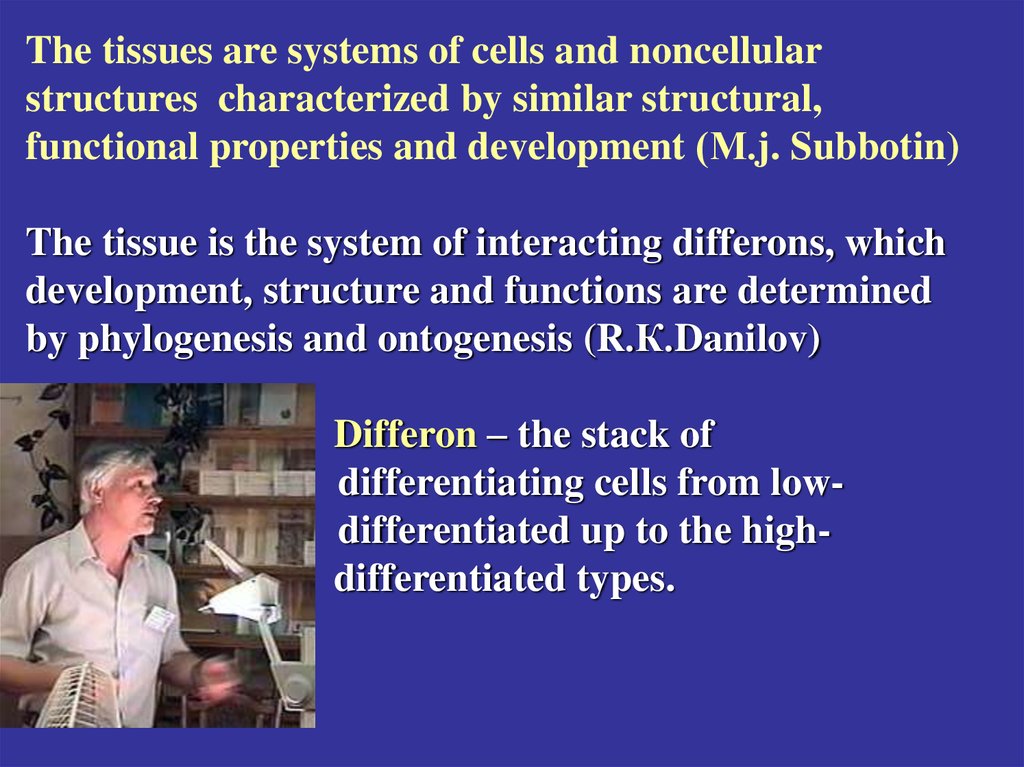
The tissues are systems of cells and noncellularstructures characterized by similar structural,
functional properties and development (М.j. Subbotin)
The tissue is the system of interacting differons, which
development, structure and functions are determined
by phylogenesis and ontogenesis (R.К.Danilov)
Differon – the stack of
differentiating cells from lowdifferentiated up to the highdifferentiated types.
.
5.
Differentiation – the morphofunctionalexchange of the same organized cells.
The main result of the differentiation is the pool
of the active functioning cells.
6.
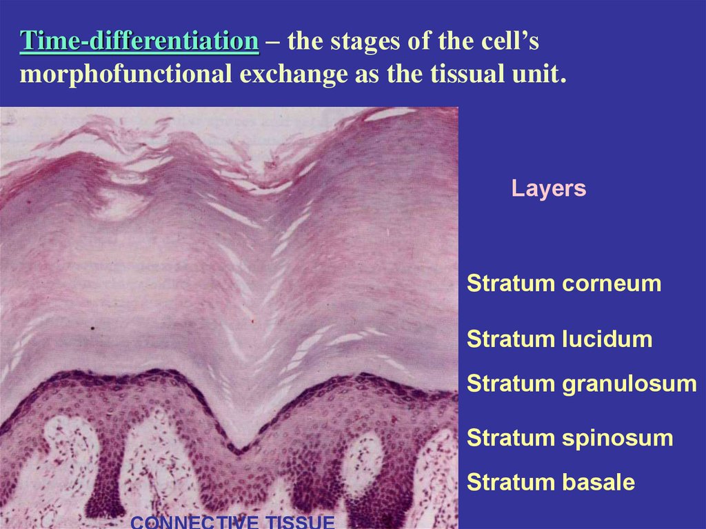
Time-differentiation – the stages of the cell’smorphofunctional exchange as the tissual unit.
Layers
Stratum corneum
Stratum lucidum
Stratum granulosum
Stratum spinosum
Stratum basale
CONNECTIVE TISSUE
7.
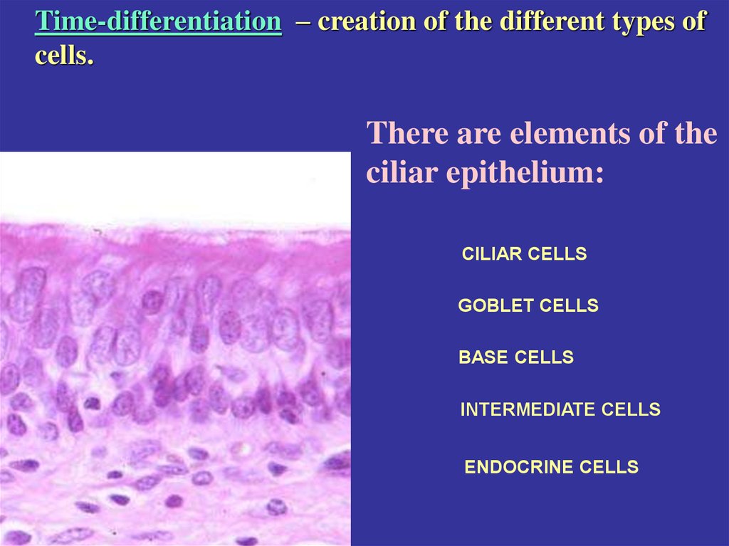
Time-differentiation – creation of the different types ofcells.
There are elements of the
ciliar epithelium:
CILIAR CELLS
GOBLET CELLS
BASE CELLS
INTERMEDIATE CELLS
ENDOCRINE CELLS
8.
Biochemical-differentiation – creation of the cellsproducing specific proteins.
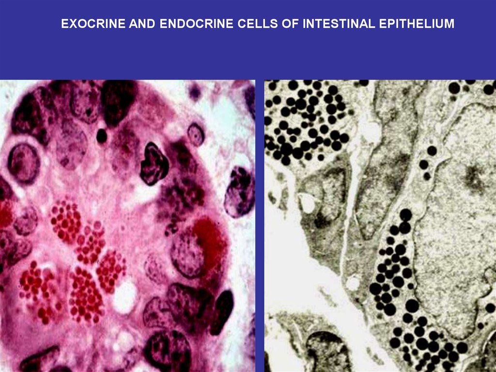
EXOCRINE AND ENDOCRINE CELLS OF THE INTESTINAL EPITHELIUM
9.
First of all start to differentiate the steam cells source thedifferon.
Steam cell’s characteristics:
1. They self-support the cell’s pool.
2. Mitosis.
3. An ability to start differentiation for some daughters cells
after division of the mother cell.
The differentiation is supervised by the nerve, endocrine and the
immune systems.
10.
Regeneration – the capability of the tissue to recover itselfafter violation. There are known different mechanisms of the
regeneration at the different tissues.
Intracellular regeneration – organell’s recovering. Most
typical for the nerve tissue, myocardium, salivary glands. The
reason – there are no steam cell at that tissues.
Cell regeneration – possible by mitosis of the steam cells. Most
typical for epithelium and muscular tissue.
Histotypical regeneration – an exchange of the parenchymal
cells by the stromal one.
11.
Physiological regeneration – the recovering of the cell’spopulation after the death of the some cells.
Reparation – the recovering of the cell’s population or the cell’s
structure after the violation.
12.
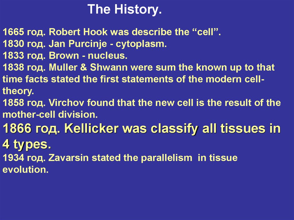
The History.1665 год. Robert Hook was describe the “cell”.
1830 год. Jan Purcinje - cytoplasm.
1833 год. Brown - nucleus.
1838 год. Muller & Shwann were sum the known up to that
time facts stated the first statements of the modern celltheory.
1858 год. Virchov found that the new cell is the result of the
mother-cell division.
1866 год. Kellicker was classify all tissues in
4 types.
1934 год. Zavarsin stated the parallelism in tissue
evolution.
13.
THE BASIC TYPES OF TISSUESEPITHELIAL
CONNECTIVE (SUPPORT) AND BLOOD
MUSCLE
NERVOUS
14. THE COMMON CHARACTERISTICS OF EPITHELIA
• COVER SURFACES OR LINE CAVITIES• FORM CONTINUOUS LAYERS
• INDIVIDUAL CELLS ARE TIGHTLY JONED BY JUNCTIONS
• REST ON BASEMENT MEMBRANES WITH UNDERLYING
CONNECTIVE TISSUE
• AVASCULAR (NO BLOOD VESSELS)
• SURFACE AND CYTOPLASM ARE SPECIALIZED INTO THE
APICAL AND BASAL PARTS
• ARE RENEWING TISSUES (POSESS STEM CELLS)
15.
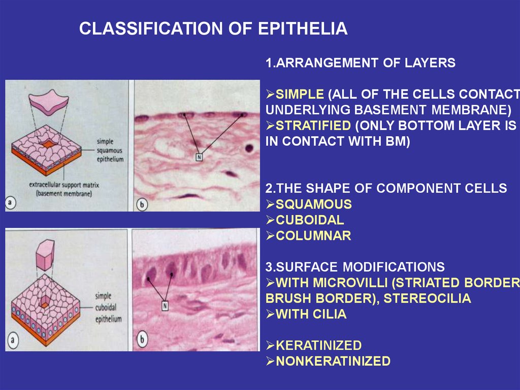
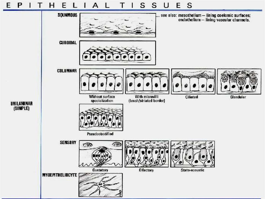
CLASSIFICATION OF EPITHELIA1.ARRANGEMENT OF LAYERS
SIMPLE (ALL OF THE CELLS CONTACT
UNDERLYING BASEMENT MEMBRANE)
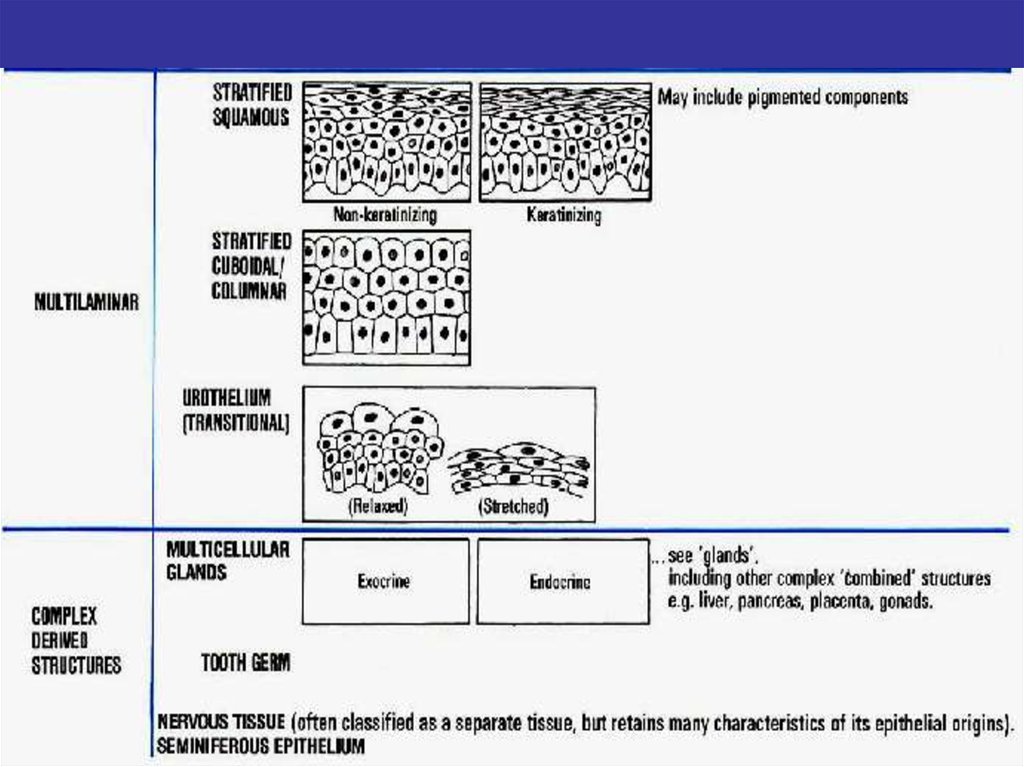
STRATIFIED (ONLY BOTTOM LAYER IS
IN CONTACT WITH BM)
2.THE SHAPE OF COMPONENT CELLS
SQUAMOUS
CUBOIDAL
COLUMNAR
3.SURFACE MODIFICATIONS
WITH MICROVILLI (STRIATED BORDER,
BRUSH BORDER), STEREOCILIA
WITH CILIA
KERATINIZED
NONKERATINIZED
16. THE LOCATION OF THE MAJOR TYPES OF EPITHELIA
SIMPLESQUAMOUS
•BLOOD VESSELS
•SEROUS MEMBRANES
•Henle’s loops OF KINDEY
SIMPLE CUBOIDAL
•KIDNEY TUBULES
•SMALL DUCTS OF GLANDS
SIMPLE
COLUMNAR
•STOMACH
•GALL BLADDER & BILE DUCTS
•INTESTINAL MUCOSA
WITH MICROVILLI
PSEUDOSTRITIFIED •RESPIRATORY PASSAGES
(CILIATED)
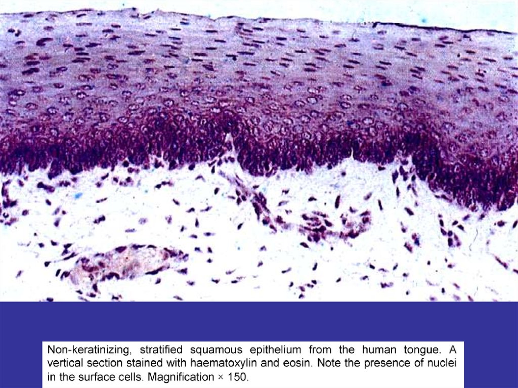
STRATIFIED
NONKERATINIZED
•ESOPHAGUS
•ANTERIOR CORNEAL SURFACE
•PART OF ORAL CAVITY
STRATIFIED
KERATINIZED
•SKIN
•PART OF ORAL CAVITY
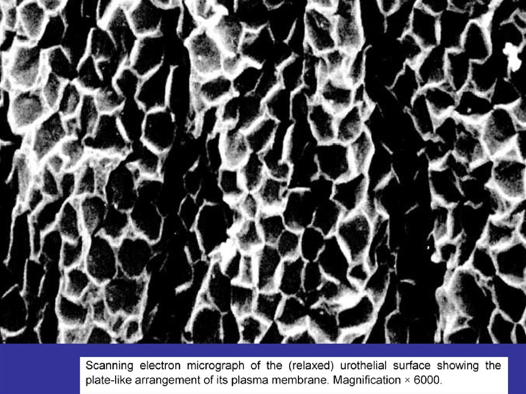
TRANSITIONAL
•URINARY PASSAGES
17.
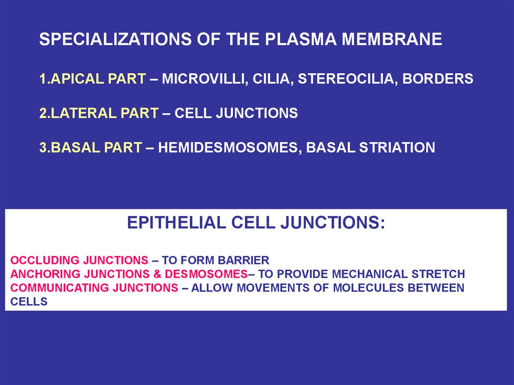
SPECIALIZATIONS OF THE PLASMA MEMBRANE1.APICAL PART – MICROVILLI, CILIA, STEREOCILIA, BORDERS
2.LATERAL PART – CELL JUNCTIONS
3.BASAL PART – HEMIDESMOSOMES, BASAL STRIATION
EPITHELIAL CELL JUNCTIONS:
OCCLUDING JUNCTIONS – TO FORM BARRIER
ANCHORING JUNCTIONS & DESMOSOMES– TO PROVIDE MECHANICAL STRETCH
COMMUNICATING JUNCTIONS – ALLOW MOVEMENTS OF MOLECULES BETWEEN
CELLS
18.
19.
20.
21.
22.
23.
24.
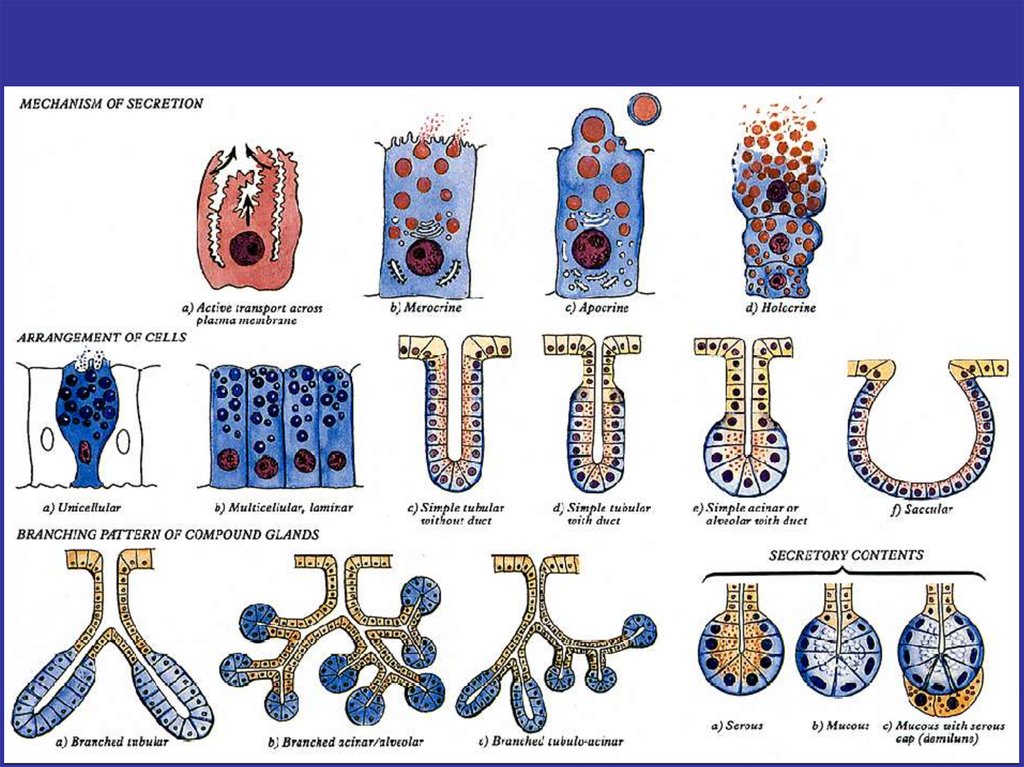
25. SECRETORY EPITHELIA AND GLANDS
EXOCRINE GLANDS• ENDOCRINE
SIMPLE
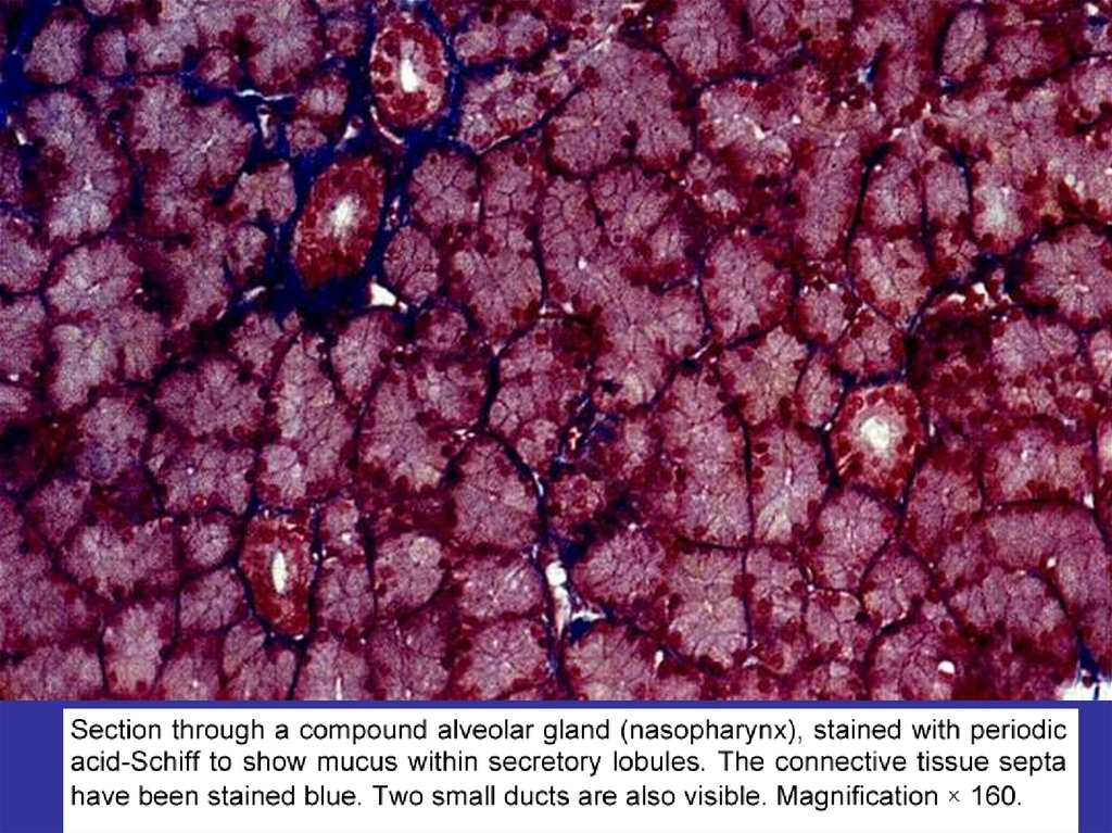
COMPOUND
• EXOCRINE
ALVEOLAR
TUBULAR
MIXED
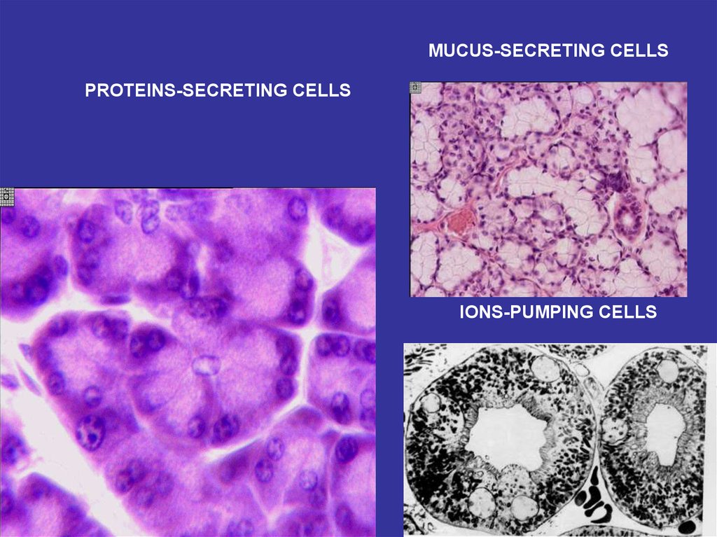
PROTEINS SECRETING
BRANCHED
NONBRANCHED
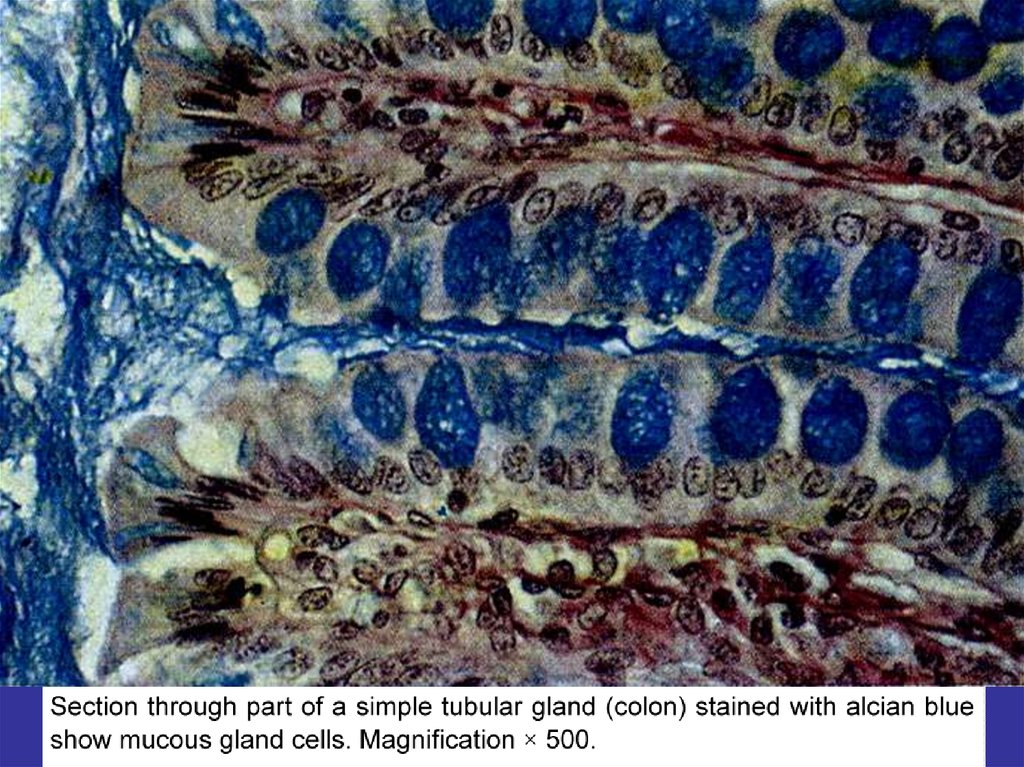
MUCUS SECRETING
LIPIDS (STEROIDS)
SECRETING
IONS-PUMPING
MECHANISMS OF SECRETION
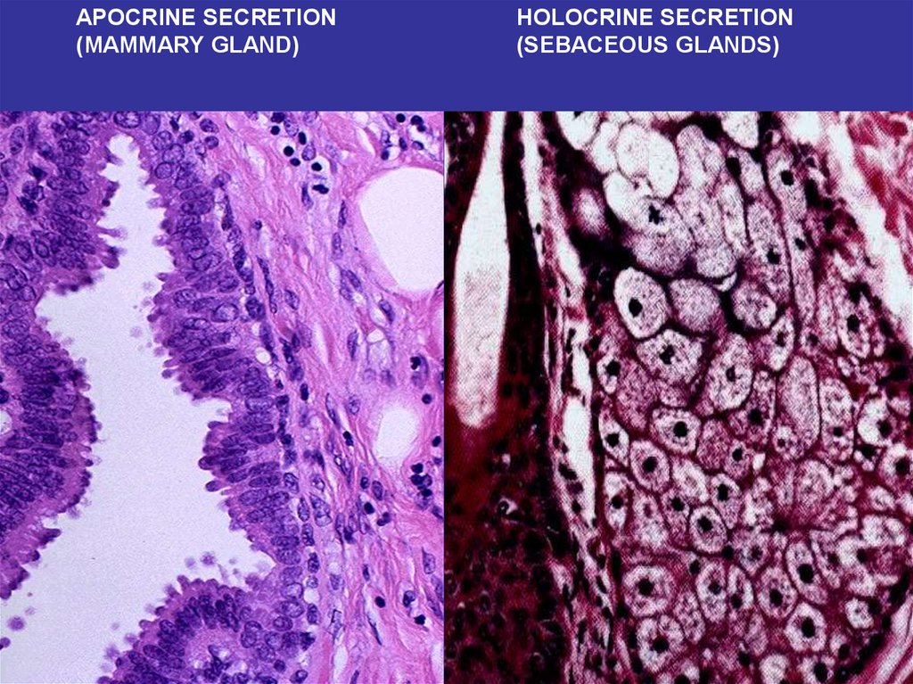
MEROCRINE
APOCRINE
HOLOCRINE
26.
27.
EXOCRINE AND ENDOCRINE CELLS OF INTESTINAL EPITHELIUM28.
MUCUS-SECRETING CELLSPROTEINS-SECRETING CELLS
IONS-PUMPING CELLS
29.
30.
31.
APOCRINE SECRETION(MAMMARY GLAND)
HOLOCRINE SECRETION
(SEBACEOUS GLANDS)