


 biology
Similar presentations:


 and blood, muscle, nervous")
Features of examination of a dental patient
1.
Department of Dentistry of General PracticeFeatures of the development, structure and physiology of the
organs of the maxillofacial region.
Features of examination of a dental patient.
Associate professor of the Department of Dentistry of General Practice
Dombrovskaya Julia Andreevna
Sankt-Petersburg
2023
1
2.
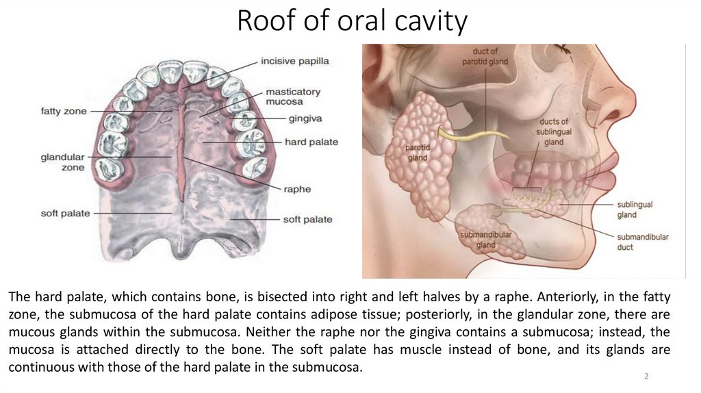
Roof of oral cavityThe hard palate, which contains bone, is bisected into right and left halves by a raphe. Anteriorly, in the fatty
zone, the submucosa of the hard palate contains adipose tissue; posteriorly, in the glandular zone, there are
mucous glands within the submucosa. Neither the raphe nor the gingiva contains a submucosa; instead, the
mucosa is attached directly to the bone. The soft palate has muscle instead of bone, and its glands are
continuous with those of the hard palate in the submucosa.
2
3.
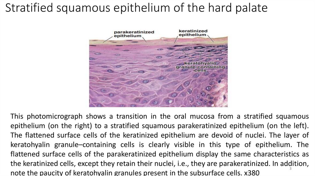
Stratified squamous epithelium of the hard palateThis photomicrograph shows a transition in the oral mucosa from a stratified squamous
epithelium (on the right) to a stratified squamous parakeratinized epithelium (on the left).
The flattened surface cells of the keratinized epithelium are devoid of nuclei. The layer of
keratohyalin granule–containing cells is clearly visible in this type of epithelium. The
flattened surface cells of the parakeratinized epithelium display the same characteristics as
the keratinized cells, except they retain their nuclei, i.e., they are parakeratinized. In addition,
3
note the paucity of keratohyalin granules present in the subsurface cells. х380
4.
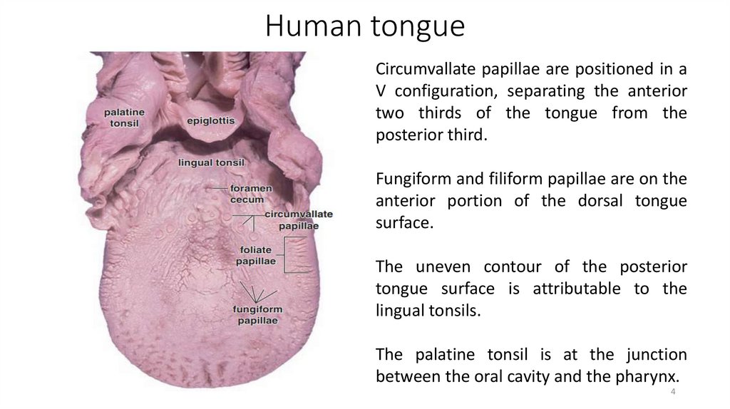
Human tongueCircumvallate papillae are positioned in a
V configuration, separating the anterior
two thirds of the tongue from the
posterior third.
Fungiform and filiform papillae are on the
anterior portion of the dorsal tongue
surface.
The uneven contour of the posterior
tongue surface is attributable to the
lingual tonsils.
The palatine tonsil is at the junction
between the oral cavity and the pharynx.
4
5.
Lingual papillae1. Structurally, the filiform papillae are posteriorly bent conical projections
of the epithelium. These papillae do not possess taste buds and are
composed of stratified squamous keratinized epithelium.
2. Fungiform papillae are slightly rounded, elevated structures situated
among the filiform papillae. A highly vascularized connective tissue core
forms the center of the fungiform papilla and projects into the base of the
surface epithelium. Because of the deep penetration of connective tissue
into the epithelium (arrows), combined with a very thin keratinized surface,
the fungiform papillae appear as small red dots when the dorsal surface of
the tongue is examined by gross inspection.
3. In a section, foliate papillae can be distinguished from fungiform
papillae because they appear in rows separated by deep clefts (arrows). The
foliate papillae are covered by stratified squamous nonkeratinized
epithelium containing numerous taste buds on their lateral surfaces.
4. Circumvallate papillae are covered by stratified squamous epithelium
that may be slightly keratinized. Each circumvallate papilla is surrounded
by a trench or cleft. Numerous taste buds are on the lateral walls of the
papillae. The deep trench surrounding the circumvallate papillae and the
presence of taste buds on the sides rather than on the free surface
are
5
features that distinguish circumvallate from fungiform papillae.
6.
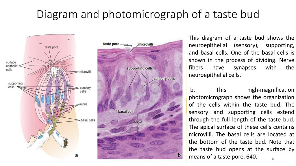
Diagram and photomicrograph of a taste budThis diagram of a taste bud shows the
neuroepithelial (sensory), supporting,
and basal cells. One of the basal cells is
shown in the process of dividing. Nerve
fibers
have
synapses
with
the
neuroepithelial cells.
b.
This
high-magnification
photomicrograph shows the organization
of the cells within the taste bud. The
sensory and supporting cells extend
through the full length of the taste bud.
The apical surface of these cells contains
microvilli. The basal cells are located at
the bottom of the taste bud. Note that
the taste bud opens at the surface by
means of a taste pore. 640.
6
7.
Diagram of taste receptors and their signaling mechanisma.
b.
c.
This diagram shows the signaling mechanism of bitter,
sweet and umami receptors in the neuroepithelial cells.
These cells selectively express only one class of receptor
proteins; for simplicity all three taste receptors are
depicted in the apical cell membrane. See text for details.
PLC – phospholipase C, IP2–inositol-1,4-diphosphate, IP3
– inositol 1,4,5-trisphosphate (IP3).
Signaling mechanism in sour sensation is generated by H
protons that primary blocks K channels. The H protons
enter the cell via amiloride-sensitive Na channels and
through tastespecific H channels (PKD1L3 and PKD2L1)
exclusively expressed in cells involved in sour taste
transduction.
Salty sensation derives from Na ions that enter the
neuroepithelial cells through the amiloride-sensitive Na
channels. Intracellular Na causes a depolarization of
membrane and activation of additional voltage-sensitive
Na and Ca2 channels. Calcium mediated release of
neurotransmitters from synaptic vesicles results in
7
stimulating gustatory nerve fiber.
8.
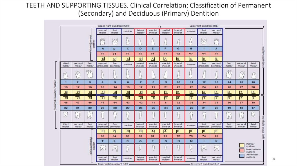
TEETH AND SUPPORTING TISSUES. Clinical Correlation: Classification of Permanent(Secondary) and Deciduous (Primary) Dentition
8
9.
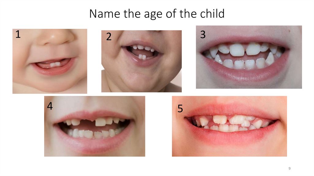
Name the age of the child1
3
2
4
5
9
10.
Teeth consist of several layers of specialized tissues10
11.
Structure of young enamel.a. This electron micrograph shows enamel rods cut obliquely. Arrows indicate the
boundaries between adjacent rods. х14,700.
b. Parts of two adjacent rods are seen at higher magnification. Arrows mark the boundary
between the two rods.
The dark needlelike objects are young hydroxyapatite crystals; the substance between the hydroxyapatitecrystals is
the organic matrix of the developing enamel. As the enamel matures, the hydroxyapatite crystals grow, and the bulk
of the organic matrix is removed. х60,000.
11
12.
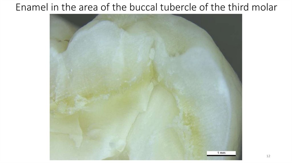
Enamel in the area of the buccal tubercle of the third molar12
13.
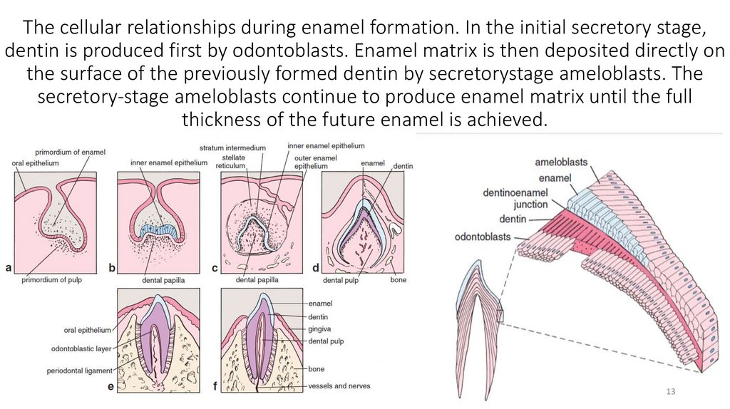
The cellular relationships during enamel formation. In the initial secretory stage,dentin is produced first by odontoblasts. Enamel matrix is then deposited directly on
the surface of the previously formed dentin by secretorystage ameloblasts. The
secretory-stage ameloblasts continue to produce enamel matrix until the full
thickness of the future enamel is achieved.
13
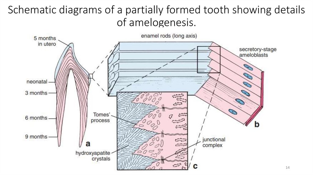
14.
Schematic diagrams of a partially formed tooth showing detailsof amelogenesis.
14
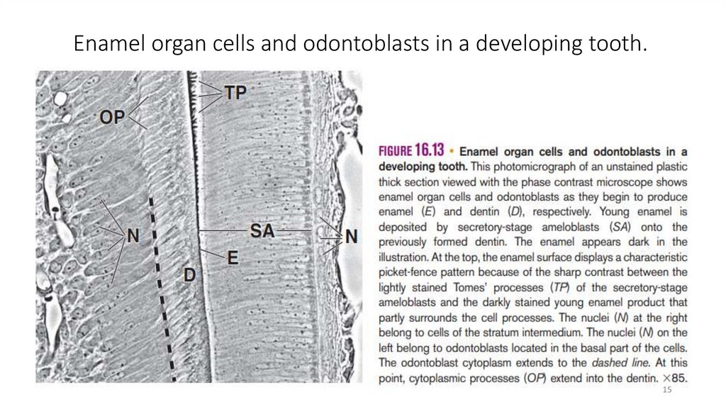
15.
Enamel organ cells and odontoblasts in a developing tooth.15
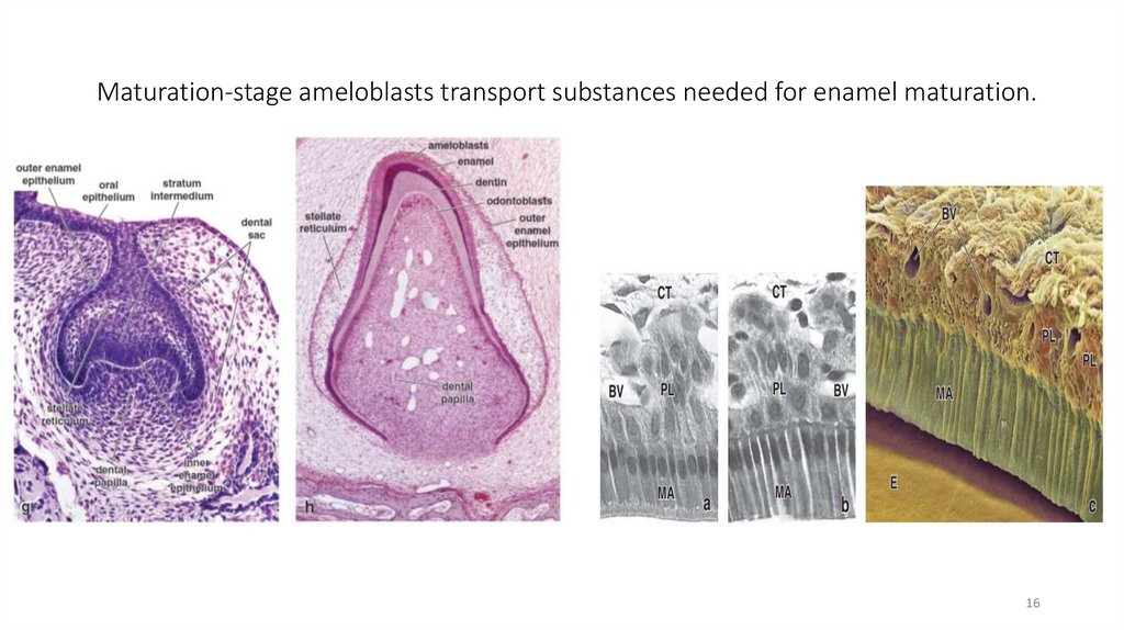
16.
Maturation-stage ameloblasts transport substances needed for enamel maturation.16
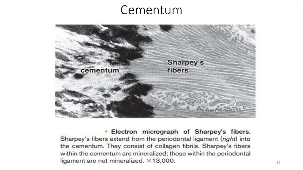
17.
Cementum17
18.
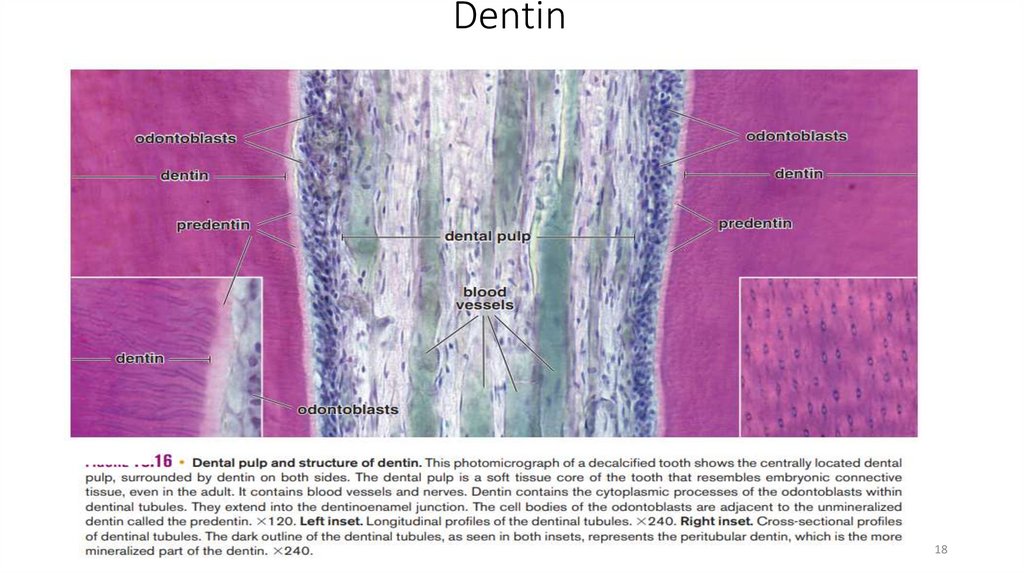
Dentin18
19.
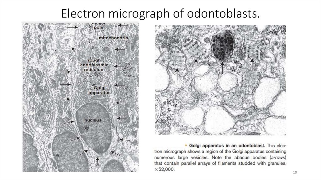
Electron micrograph of odontoblasts.19
20.
Dental Pulp and Central Pulp Cavity (Pulp Chamber)20
21.
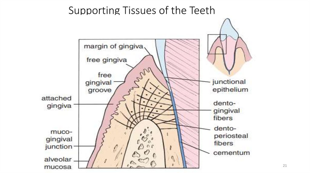
Supporting Tissues of the Teeth21
22.
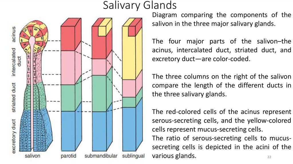
Salivary GlandsDiagram comparing the components of the
salivon in the three major salivary glands.
The four major parts of the salivon–the
acinus, intercalated duct, striated duct, and
excretory duct—are color-coded.
The three columns on the right of the salivon
compare the length of the different ducts in
the three salivary glands.
The red-colored cells of the acinus represent
serous-secreting cells, and the yellow-colored
cells represent mucus-secreting cells.
The ratio of serous-secreting cells to mucussecreting cells is depicted in the acini of the
various glands.
22
23.
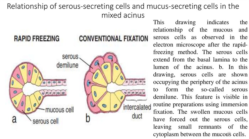
Relationship of serous-secreting cells and mucus-secreting cells in themixed acinus
This
drawing
indicates
the
relationship of the mucous and
serous cells as observed in the
electron microscope after the rapidfreezing method. The serous cells
extend from the basal lamina to the
lumen of the acinus. b. In this
drawing, serous cells are shown
occupying the periphery of the acinus
to form the so-called serous
demilune. This feature is visible in
routine preparations using immersion
fixation. The swollen mucous cells
have forced out the serous cells,
leaving small remnants of the
23
cytoplasm between the mucous
cells.
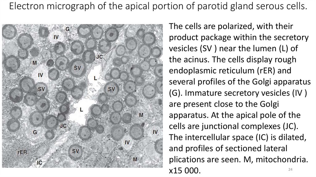
24.
Electron micrograph of the apical portion of parotid gland serous cells.The cells are polarized, with their
product package within the secretory
vesicles (SV ) near the lumen (L) of
the acinus. The cells display rough
endoplasmic reticulum (rER) and
several profiles of the Golgi apparatus
(G). Immature secretory vesicles (IV )
are present close to the Golgi
apparatus. At the apical pole of the
cells are junctional complexes (JC).
The intercellular space (IC) is dilated,
and profiles of sectioned lateral
plications are seen. M, mitochondria.
24
x15 000.
25.
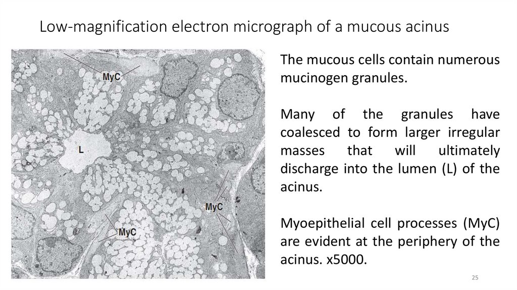
Low-magnification electron micrograph of a mucous acinusThe mucous cells contain numerous
mucinogen granules.
Many of the granules have
coalesced to form larger irregular
masses that will ultimately
discharge into the lumen (L) of the
acinus.
Myoepithelial cell processes (MyC)
are evident at the periphery of the
acinus. x5000.
25
26.
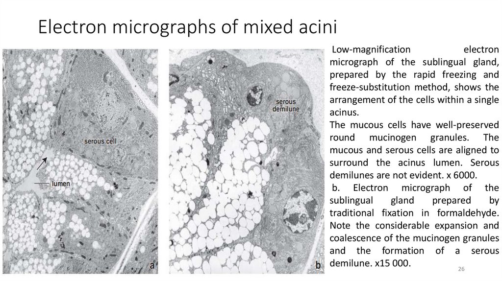
Electron micrographs of mixed aciniLow-magnification
electron
micrograph of the sublingual gland,
prepared by the rapid freezing and
freeze-substitution method, shows the
arrangement of the cells within a single
acinus.
The mucous cells have well-preserved
round mucinogen granules. The
mucous and serous cells are aligned to
surround the acinus lumen. Serous
demilunes are not evident. x 6000.
b. Electron micrograph of the
sublingual
gland
prepared
by
traditional fixation in formaldehyde.
Note the considerable expansion and
coalescence of the mucinogen granules
and the formation of a serous
demilune. x15 000.
26
27.
Major Salivary Glands. Parotid Gland. The parotid glands are completelyserous.
The parotid gland in the
human is composed
entirely of serous acini and
their ducts. Typically,
adipose cells are also
distributed throughout the
gland. The lower portion of
the figure reveals an
excretory duct within a
connective tissue septum.
x120. Inset.
Higher magnification of the
27
serous acinar cells. x320
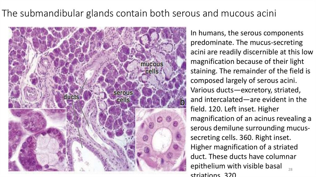
28.
The submandibular glands contain both serous and mucous aciniIn humans, the serous components
predominate. The mucus-secreting
acini are readily discernible at this low
magnification because of their light
staining. The remainder of the field is
composed largely of serous acini.
Various ducts—excretory, striated,
and intercalated—are evident in the
field. 120. Left inset. Higher
magnification of an acinus revealing a
serous demilune surrounding mucussecreting cells. 360. Right inset.
Higher magnification of a striated
duct. These ducts have columnar
epithelium with visible basal 28
29.
Saliva29
30.
The sublingual gland also contains both serous and mucous elementsHere, the mucous acini predominate. The
mucous acini are conspicuous because of
their light staining. Critical examination of
the mucous acini at this relatively low
magnification reveals that they are not
spherical structures but, rather, elongate or
tubular structures with branching
outpockets. Thus, the acinus is rather large,
and much of it is usually not seen within the
plane of a single section. The ducts of the
sublingual gland that are observed with the
greatest frequency in a section are the
interlobular ducts. 120. Inset. The serous
component of the gland is composed largely
of demilunes (asterisks), artifacts of
30
conventional fixation. 320.
31.
Features of examination of a dental patient.31