Similar presentations:
Muscle Tissue
1. Muscle Tissue
2. Muscle Tissue
• Muscles tissue distributed almost everywhere• Some functions of muscular tissue
–
–
–
–
Propels food we eat along gastrointestinal tract
Expels waste we produce
Changes amount of air that enters the lung
Pumps the blood to body tissues
3. Muscle Tissue
• Three types of muscle tissue:– Skeletal muscle, cardiac muscle, smooth muscle
– Composes 40-50% of weight of the adult
• 700 skeletal muscles in the muscular system
4. 3 Types of Muscle Tissue
CardiacSkeletal
Smooth
5. Introduction to Skeletal Muscle: Functions of Skeletal Muscle
• Functions of Skeletal Muscle–
–
–
–
Body movement
Maintenance of posture
Protection and support
Storage and movement of materials
• sphincters,
– Heat production
• shiver when cold to generate heat
6. Introduction to Skeletal Muscle: Functions of Skeletal Muscle
What are the five major functions ofskeletal muscle?
Body movement, maintenance of posture,
protection and support, storage and movement
of material, and heat production.
7. Introduction to Skeletal Muscle: Characteristics Skeletal Muscle Tissue
• Characteristics– Excitability
• responsive to nervous system stimulation
• neurons secreting neurotransmitters that bind to muscle cells
– Conductivity
• electrical change traveling along plasma membrane
• initiated in response to neurotransmitter binding
– Contractility
• contractile proteins within muscle cells
• slide past each other
• tension used to pull on bones of skeleton
8. Introduction to Skeletal Muscle: Characteristics Skeletal Muscle Tissue
• Characteristics (continued)– Elasticity
• due to protein fibers acting like compressed coils
• when contraction ended, tension in proteins released
• muscle returns to original length
– Extensibility
• lengthening of a muscle cell
• e.g., extension of the triceps brachii when flex elbow joint
9. Anatomy of Skeletal Muscle: Gross Anatomy
Skeletal muscle
–
–
–
–
–
Composed of thousands of muscle cells
Typically as long as the entire muscle
Often referred to as muscle fibers
Organized into bundles, termed fascicles
Muscle composed of fibers, connective tissue, blood vessels,
nerves
10. Skeletal Muscle High Magnification
NucleiA band
I band
11. Anatomy of Skeletal Muscle: Gross Anatomy
• Connective tissue components–
–
Three concentric layers of connective tissue:
epimysium, perimysium, endomysium
Provide
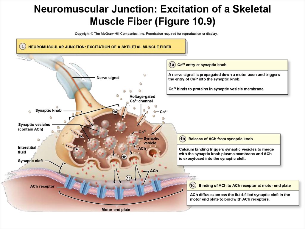
protection
sites for blood vessel and nerve distribution
means of attachment to skeleton or other structures
12. Anatomy of Skeletal Muscle: Gross Anatomy
• Connective tissue components (continued)–
–
–
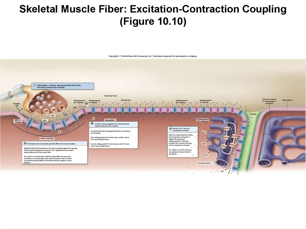
Epimysium
layer of dense irregular connective tissue
surrounds whole skeletal muscle
Perimysium
dense irregular tissue surrounding the fascicles
contains extensive blood vessels and nerves supplying fibers
Endomysium
innermost connective tissue layer
delicate areolar connective tissue
surrounds and electrically insulates each muscle fiber
contains reticular protein fibers
–
help bind together neighboring muscle fibers
13. Connective Tissue and Fascicles
EpimysiumFascicle
Perimysium
Endomysium
Epimysium + Perimysium + Endomysium = Tendon
14. Anatomy of Skeletal Muscle: Gross Anatomy
• Connective tissue components (continued)–
–
Tendon
cordlike structure composed of dense regular connective
tissue
formed by the three connective tissue layers
attach the muscle to bone, skin or another muscle
Aponeurosis
thin, flattened sheet of dense irregular tissue
formed from the three connective tissue layers
15. Tendon and Aponeurosis of Palmaris Longus muscle
16. Anatomy of Skeletal Muscle: Gross Anatomy
• Connective tissue components (continued)–
Deep fascia
additional sheet of dense irregular connective tissue
external to the epimysium
separates individual muscles
binds together muscles with similar functions
contains nerves, blood vessels, and lymph vessels
fills spaces between muscles
17. Superficial and Deep Fasciae
DeepSuperficial
10-17
18. Anatomy of Skeletal Muscle: Gross Anatomy
Connective tissue components (continued)–
Superficial fascia
superficial to deep fascia
composed of areolar and adipose connective tissue
separates muscles from skin
19. Skin
20. Superficial Fascia
21. Deep Fascia
22. Superficial Muscles
23. Deeper Muscles
24. Even Deeper Muscles
25. Yet Even Deeper Muscles
26. Soft Tissue and Bone
27. Bone
28. Anatomy of Skeletal Muscle: Gross Anatomy
• Blood vessels and nerves–
Skeletal muscles vascularized by extensive blood vessels
–
Deliver oxygen and nutrients, removing waste products
Innervated by motor neurons
–
Axons
extend through connective layers
almost make contact with individual muscle fiber
junction termed the neuromuscular junction
–
Skeletal muscle termed voluntary muscle
because fibers consciously controlled by nervous system
29. Structural Organization of Skeletal Muscle (Figure 10.1)
Copyright © The McGraw-Hill Companies, Inc. Permission required for reproduction or display.Tendon
Structural
Organization
of Skeletal
Muscle
(Figure
10.1)
Deep fascia
Epimysium
Skeletal muscle
Artery
Vein
Nerve
Perimysium
Fascicle
Endomysium
Muscle fiber
30. Anatomy of Skeletal Muscle: Gross Anatomy
What are the locations of theendomysium, perimysium, and
epimysium?
The endomysium is the layer of
connective tissue surrounding the whole
skeletal muscle and providing protection.
The perimysium surrounds the muscle
fascicles and contains extensive blood
vessels and nerves.
The endomysium is the innermost layer
surrounding and electrically insulating
muscle fibers.
31. Anatomy of Skeletal Muscle: Microscopic Anatomy
Sarcoplasma
–
–
–
Cytoplasm of muscle fibers (cells comprising muscle)
Contains typical cellular structures
e.g., Golgi apparatus, ribosomes, vesicles
Has specialized cellular structure
32. Anatomy of Skeletal Muscle: Microscopic Anatomy
• Multinucleated cell–
–
–
Elongated cells extending length of muscle
Myoblasts
embryonic cells which fuse
form single skeletal muscle fibers during development
each contributing a nucleus to total nuclei
Thus fibers multinucleated cells
33. Anatomy of Skeletal Muscle: Microscopic Anatomy
• Multinucleatedcell (continued)
–
Satellite cells
myoblasts
remaining,
unfused, in
adult skeletal
tissue
may be
stimulated to
differentiate if
tissue injured
(Figure 10.2)
Copyright © The McGraw-Hill Companies, Inc. Permission required for reproduction or display.
Myoblasts
Muscle fiber
Myoblasts fuse
to form a skeletal
muscle fiber.
Satellite cell
Muscle fiber
Satellite cell
Nuclei
34. Anatomy of Skeletal Muscle: Microscopic Anatomy
• Sarcolemma and T-tubules–
–
Plasma membrane of a skeletal muscle fiber
sarcolemma
Invaginations of the sarcolemma
T-tubules, or transverse tubules
35. Anatomy of Skeletal Muscle: Microscopic Anatomy
Sarcolemma and T-tubules (continued)–
Na+/ K+ pumps along sarcolemma and T-tubules
create concentration gradients for Na+ and K+
three Na+ pumped out while two K+ pumped in
resting membrane potential maintained by pumps
– inside of cell relatively negative in comparison to
outside
– responsible for excitability of skeletal muscle fibers
36. Anatomy of Skeletal Muscle: Microscopic Anatomy
• Sarcolemma and T-tubules (continued)–
Voltage-gated Na+ channels and voltage-gated K+ channels
also present
necessary for propagation of electrical change along
sarcolemma
37. Anatomy of Skeletal Muscle: Microscopic Anatomy
• Sarcoplasmic reticulum–
–
–
–
Internal membrane complex
Similar to smooth endoplasmic reticulum
Surround bundles of contractile proteins
Terminal cisternae
blind sacs of sarcoplasmic reticulum
serve as reservoirs for calcium ions
combine in twos with central T-tubule to form triads
38. Structure and Organization of a Skeletal Muscle Fiber: Sarcolemma and T-Tubules (Figure 10.3 b)
Copyright © The McGraw-Hill Companies, Inc. Permission required for reproduction or display.Interstitial fluid
– K+
+
+ + +
+ + +
–
– – –
– – –
+
+
– –
– –
+
+
+
+
+
– –
– –
+
+
+
–
in
– Na+
–
2K+
+
Sarcolemma
Sarcoplasm
(b) Sarcolemma and T-tubules
Voltage-gated
K+ channel
–
3 Na+ out
Voltage-gated
Na+ channel
Na+/K+
pump
+
T-tubule
39.
• Sarcoplasmic reticulum(continued)
– Ca2+ pumps embedded in
sarcoplasmic reticulum
• move Ca2+ into
sarcoplasmic reticulum
• stored bound to
specialized proteins,
calmodulin and
calsequestrin
– Voltage-gated Ca2+ channels
• open to release Ca2+ from
sarcoplasmic reticulum into
sarcoplasm
• causes muscle contraction
Copyright © The McGraw-Hill Companies, Inc. Permission required for reproduction or display.
SR membrane
Ca2+
Ca2+ pump
Voltage-gated
Ca2+ channel
Calmodulin
Calsequestrin
Sarcoplasm
Terminal cisterna
(c) Sarcoplasmic reticulum
(Figure 10.3c)
40. Anatomy of Skeletal Muscle: Microscopic Anatomy
• Muscle fibers and myofibrils–
–
Myofibrils
long cylindrical structures
extend length of muscle fiber
compose 80% of volume of muscle fiber
each fiber with hundreds to thousands
Myofilaments
bundles of protein filaments
takes many to extend length of myofibril
two types: thick and thin
41. Structure and Organization of a Skeletal Muscle Fiber (Figure 10.3 a)
Copyright © The McGraw-Hill Companies, Inc. Permission required for reproduction or display.Muscle
Fascicle
Muscle fiber
Triad
Sarcoplasmic
reticulum
T-tubule
Terminal
cisternae
Sarcolemma
Nucleus
Myofibrils
Sarcomere
Myofilaments
Nucleus
Openings into
T-tubules
Sarcoplasm
Nucleus
(a) Skeletal muscle fiber
Mitochondrion
42. Anatomy of Skeletal Muscle: Microscopic Anatomy
Muscle fibers and myofibrils (continued)• Thick filaments
–
Assembled from bundles of protein molecules, myosin
each myosin protein with two intertwined strands
each strand with a globular head and elongated tail
tails pointing toward center of thick filaments
heads pointing toward edges of thick filaments
head with a binding site for actin (thin filaments)
head with site where ATP attaches and is split
43. Anatomy of Skeletal Muscle: Microscopic Anatomy
Muscle fibers and myofibrils (continued)• Thin filaments
–
–
–
–
–
Primarily composed of two strands of protein, actin
Two strands twisted around each other
Many small spherical molecules, globular actin
Connected to form a fibrous strand, filamentous actin
Globular actin with myosin binding site
where myosin head attaches during contraction
44. Anatomy of Skeletal Muscle: Microscopic Anatomy
Muscle fibers and myofibrils• Thin filaments (continued)
–
–
Tropomyosin
twisted “stringlike” protein
cover small bands of the actin strands
covers myosin binding sites in a noncontracting muscle
Troponin
globular protein attached to tropomyosin
binding site for Ca2+
together form troponin-tropomyosin complex
45. Molecular Structure of Thick and Thin Filaments (Figure 10.4)
Copyright © The McGraw-Hill Companies, Inc. Permission required for reproduction or display.Muscle fiber
Myofibril
Myofilaments
Myosin molecule
Heads
Actin binding site
ATP and ATPase binding site
Tail
Myosin heads
(a) Thick filament
Troponin
Tropomyosin
G-actin
(b) Thin filament
F-actin
Myosin binding site
Ca2+ binding site
46. Anatomy of Skeletal Muscle: Microscopic Anatomy
• Organization of a sarcomere–
–
–
–
Myofilaments arranged in repeating units, sarcomeres
Number varies with length of myofibril
Composed of overlapping thick and thin filaments
Delineated at both ends by Z discs
specialized proteins perpendicular to myofilaments
anchors for thin filaments
47. Anatomy of Skeletal Muscle: Microscopic Anatomy
Organization of a sarcomere• Overlapping filaments (continued)
–
–
–
Form alternating patterns of light and dark regions
Appears striated under a microscope
due to size and density differences between thick and thin
filaments
Each thin filament with three thick filaments
form triangle at its periphery
48. Skeletal Muscle (striations)
Skeletal musclefiber
Nuclei
A band
I band
49. Sarcomere
MyofibrilSarcomere
Sarcomere
Z disc
H band
A band
I band
M line
50. Structure of a Sarcomere (Figure 10.5 a)
Copyright © The McGraw-Hill Companies, Inc. Permission required for reproduction or display.Muscle fiber
Sarcomeres
I band
A band
I band
Myofibril
Z disc
H zone
M line
Sarcomere
(a)
Z disc
Myofilaments
51. Anatomy of Skeletal Muscle: Microscopic Anatomy
Organization of a sarcomere (continued)• Overlapping filaments
–
I bands
region containing only thin filaments
extend from both directions of Z disc
bisected by Z disc
appear light under a microscope
disappear at maximal muscle contraction
52. Anatomy of Skeletal Muscle: Microscopic Anatomy
Organization of a sarcomere• Overlapping filaments (continued)
–
A band
central region of sarcomere
contains entire thick filament
contains partially overlapping thin filaments
appears dark under a microscope
53. Anatomy of Skeletal Muscle: Microscopic Anatomy
Organization of a sarcomere• Overlapping filaments (continued)
–
–
H zone
central portion of A band
thick filaments only present; no thin filament overlap
disappears during maximal muscle contraction
M line
protein meshwork structure at center of H zone
attachment site for thick filaments
54. Structure of a Sarcomere (Figure 10.5 b)
Copyright © The McGraw-Hill Companies, Inc. Permission required for reproduction or display.Sarcomere
Z disc
Connectin
Z disc
Thick filament
Thin filament
M line
Thin filament
H zone
I band
(b)
A band
I band
55. Structure of a Sarcomere (Figure 10.5 c)
Copyright © The McGraw-Hill Companies, Inc. Permission required for reproduction or display.Transverse
sectional plane
M line
Thick filaments
and accessory
proteins
(c)
H zone
Thick filaments
A band
Thick filaments
Thin filaments
I band
Thin filaments
Connectin
Z disc
Thin filaments
Connectin
and accessory
proteins
56. Anatomy of Skeletal Muscle: Microscopic Anatomy
Organization of a sarcomere• Other structural and functional proteins
–
Connectin
protein extending from Z discs to M line
extends through core of each thick filament
stabilizes the position of thick filaments
springlike to produce passive tension during contraction
during relaxation, passive tension released
57. Anatomy of Skeletal Muscle: Microscopic Anatomy
Organization of a sarcomere• Other structural and functional proteins (continued)
–
–
Nebulin
actin-binding protein
part of I band of the sarcomere
plays possible role in creating orderly structure of sarcomere
Dystrophin
anchors myofibrils adjacent to sarcolemma to sarcolemma
proteins
links internal myofilament proteins to external proteins
abnormal structure or amounts of proteins in muscular
dystrophy
58. Anatomy of Skeletal Muscle: Microscopic Anatomy
• Mitochondria and other structuresassociated with energy production
–
–
–
–
Muscle with high ATP requirement
Abundant mitochondria for aerobic cellular respiration
Glycogen stores for immediate fuel molecule
Creatinine phosphate
molecule unique to muscle tissue
provides fibers means of supplying ATP anaerobically
59. Anatomy of Skeletal Muscle: Microscopic Anatomy
• Mitochondria and other structuresassociated with energy production
(continued)
–
Myoglobin
molecule unique to muscle tissue
reddish globular protein similar to hemoglobin
binds oxygen when muscle at rest
releases it during muscular contraction
provides additional oxygen to enhance aerobic cellular
respiration
60. Anatomy of Skeletal Muscle: Microscopic Anatomy
What are the primary components ofthick and thin filaments?
Thick filaments are composed of myosin
protein.
Thin filaments are composed primarily of actin
protein. Tropomyosin and tropin are associated
regulatory proteins.
61. Anatomy of Skeletal Muscle: Microscopic Anatomy
In which band are there thick filamentsonly, with no thin filament overlap?
H zone
62. Anatomy of Skeletal Muscle: Innervation of Skeletal Muscle Fibers
• Motor unit–
Motor neuron nerve cells
transmit nerve signals from brain or spinal cord
have axons that branch
individually innervate numerous skeletal muscle fibers
single motor neuron + fibers it controls = motor unit
63. Anatomy of Skeletal Muscle: Innervation of Skeletal Muscle Fibers
• Motor unit (continued)–
Varied number of fibers a neuron innervates
small motor units less than five muscle fibers
large motor units with several thousand
inverse relationship between size of motor unit and degree
of control
– e.g., small motor units innervating eye
– need greater control
– e.g., large motor units innervating lower limbs
– need less precise control
64. Anatomy of Skeletal Muscle: Innervation of Skeletal Muscle Fibers
• Motor unit (continued)–
–
Fibers dispersed throughout most of a muscle
Stimulation producing weak contraction over a wide area
65. Anatomy of Skeletal Muscle: Innervation of Skeletal Muscle Fibers
• Neuromuscular junctions–
–
–
Location where motor neuron innervates muscle
Usually mid-region of muscle fiber
Has synaptic knob, motor end plate, synaptic cleft
66. Neuromuscular Junction High Magnification
Skeletal muscle fiberAxon of motor nerve
Motor end plate
67. Anatomy of Skeletal Muscle: Innervation of Skeletal Muscle Fibers
Neuromuscular junctions (continued)• Synaptic knob
–
–
–
–
The expanded tip of the axon
Axon enlarged and flattened in this region
Houses synaptic vesicles, small membrane sacs
filled with neurotransmitter, acetylcholine (ACh)
Has Ca2+ pumps embedded in plasma membrane
establish calcium gradient, with more outside the neuron
68. Neuromuscular Junction TEM: High Magnification
Synaptic vesicles ofPrimary synaptic cleft
Secondary synaptic
cleft (junctional folds)
synaptic terminal
Mitochondria of
synaptic terminal
69. Anatomy of Skeletal Muscle: Innervation of Skeletal Muscle Fibers
Neuromuscular junctions• Synaptic knob (continued)
–
–
–
Has voltage-gated Ca2+ channels in membrane
Ca2+ flowing down concentration gradient if opened
Vesicles normally repelled from membrane of synaptic knob
because both normally negatively charged
70. Anatomy of Skeletal Muscle: Innervation of Skeletal Muscle Fibers
Neuromuscular junctions• Motor end plate
–
–
–
Specialized region of sarcolemma
Has numerous folds
increase surface area covered by knob
Has vast numbers of ACh receptors
plasma membrane protein channels
opened by binding of ACh
allow Na+ entry and K+ exit
71. Anatomy of Skeletal Muscle: Innervation of Skeletal Muscle Fibers
Neuromuscular junctions (continued)• Synaptic cleft
–
–
–
Narrow fluid-filled space
Separates synaptic knob and motor end plate
Acetylcholinesterase residing here
enzyme that breaks down ACh molecules
after their release into synaptic cleft
72. Structure and Organization of a Neuromuscular Junction (Figure 10.7a)
Copyright © The McGraw-Hill Companies, Inc. Permission required for reproduction or display.Neuromuscular
junction
Structure and
Organization of a
Neuromuscular
Junction
(Figure
10.7a)
Synaptic knob
Nerve signal
Synaptic
cleft
Endomysium
Sarcolemma
(a)
Motor end
plate
Myofilaments
Myofibril
73. Structure and Organization of a Neuromuscular Junction (Figure 10.7b)
Copyright © The McGraw-Hill Companies, Inc. Permission required for reproduction or display.Ca2+ pump
Interstitial fluid
Ca2+
Voltage-gated
Ca2+ channels
Structure and
Organization of
a
Neuromuscular
Junction
(Figure
10.7b)
Synaptic knob
Vesicle
with ACh
Synaptic
cleft
ACh
Sarcolemma
Sarcoplasm
–Na+
Ach receptor
K+
Junction fold
Motor end plate
(b)
74. Anatomy of Skeletal Muscle: Innervation of Skeletal Muscle Fibers
What is a motor unit, and why does itvary in size?
A motor unit is a single motor neuron and the
muscle fibers it controls.
There is an inverse relationship between size
and degree of control. Muscles needing greater
power but less control have bigger motor units.
75. Physiology of Skeletal Muscle Contraction
• During muscle contraction–
–
–
–
–
Protein filaments within sarcomeres interact
Sarcomeres shorten
Tension is exerted on portion of skeleton where muscle attached
Contracting fiber decreases in length
Movement occurs
76. Overview of Events in Skeletal Muscle Contraction (Figure 10.8)
Copyright © The McGraw-Hill Companies, Inc. Permission required for reproduction or display.1 NEUROMUSCULAR JUNCTION: EXCITATION OF A SKELETAL MUSCLE FIBER
Release of neurotransmitter acetycholine (ACh) from synaptic vesicles and
subsequent binding of Ach to Ach receptors.
2
Neuromuscular
junction
Synaptic vesicle (contains ACh)
Action potential
1
Muscle
fiber
T-tubule
ACh
Ach receptor
Terminal
cisterna
of SR
Sarcoplasmic
reticulum
SARCOLEMMA, T-TUBULES, AND SARCOPLASMIC
RETICULUM: EXCITATION-CONTRACTION COUPLING
ACh binding triggers propagation of an action potential
along the sarcolemma and T-tubules to the sarcoplasmic
reticulum, which is stimulated to release Ca2+.
2
Ca2+
Sarcolemma
Sarcomere
Ca2+
3
Thin filament
Ca2+
3
SARCOMERE: CROSSBRIDGE CYCLING
Ca2+ binding to troponin triggers sliding of thin
filaments past thick filaments of sarcomeres;
sarcomeres shorten, causing muscle contraction.
Thick filament
77. Skeletal Muscle Contraction—Neuromuscular Junction: Excitation of a Skeletal Muscle Fiber
• First physiological event– Muscular fiber excitation by motor neuron
– Occurs at neuromuscular junction
– Results in release of ACh and subsequent binding of ACh
receptors
78. Skeletal Muscle Contraction—Neuromuscular Junction: Excitation of a Skeletal Muscle Fiber
• Calcium entry at synaptic knob– Nerve signal propagated down motor axon
– Triggers opening of voltage-gated Ca2+ channels
– Movement of calcium down concentration gradient
• from interstitial fluid into synaptic knob
– Binding of calcium with proteins on synaptic vesicles
79. Skeletal Muscle Contraction—Neuromuscular Junction: Excitation of a Skeletal Muscle Fiber
• Release of ACh from synaptic knob– Merging of synaptic vesicles with synaptic knob membrane
• triggered by binding of Ca2+
– Exocytosis of ACh into synaptic cleft
– About 300 vesicles per nerve signal
80. Skeletal Muscle Contraction—Neuromuscular Junction: Excitation of a Skeletal Muscle Fiber
• Binding of ACh at motor end plate– Diffusion of ACh across synaptic cleft
– Binds with ACh receptors within motor end plate
– Causes excitation of muscle fiber
81. Neuromuscular Junction: Excitation of a Skeletal Muscle Fiber (Figure 10.9)
Copyright © The McGraw-Hill Companies, Inc. Permission required for reproduction or display.1
NEUROMUSCULAR JUNCTION: EXCITATION OF A SKELETAL MUSCLE FIBER
1a Ca2+ entry at synaptic knob
A nerve signal is propagated down a motor axon and triggers
the entry of Ca2+ into the synaptic knob.
Nerve signal
Ca2+ binds to proteins in synaptic vesicle membrane.
Voltage-gated
Ca2+ channel
Synaptic knob
Ca2+
1a
Synaptic vesicles
(contain ACh)
Ca2+
Synaptic
vesicle
ACh
Interstitial
fluid
1b
Synaptic cleft
1b Release of ACh from synaptic knob
Calcium binding triggers synaptic vesicles to merge
with the synaptic knob plasma membrane and ACh
is exocytosed into the synaptic cleft.
ACh
1c
1c Binding of ACh to ACh receptor at motor end plate
ACh receptor
ACh diffuses across the fluid-filled synaptic cleft in the
motor end plate to bind with ACh receptors.
Motor end plate
82. Skeletal Muscle Contraction—Neuromuscular Junction: Excitation of a Skeletal Muscle Fiber
What triggers the binding of synapticvesicles to the synaptic knob
membrane to cause exocytosis of ACh?
Nerve signal triggers the entry of calcium into
the synaptic knob. Calcium binding to synaptic
vesicles triggers the exocytosis of ACh.
83. Skeletal Muscle Contraction—Neuromuscular Junction: Excitation of a Skeletal Muscle Fiber
Clinical View: Myasthenia Gravis–
–
–
–
–
–
–
Autoimmune disease, primarily in women
Antibodies binding ACh receptors in neuromuscular junctions
Receptors removed from muscle fiber by endocytosis
Results in decreased muscle stimulation
Rapid fatigue and muscle weakness
Eye and facial muscles often involved first
May be followed by swallowing problems, limb weakness
84. Skeletal Muscle Contraction—Sarcolemma, T-Tubules, Sarcoplasmic Reticulum: Excitation-Contraction Coupling
• Second physiological event– Excitation-contraction coupling
– Links skeletal muscle stimulation to events of contraction
– Consists of three events:
• development of end-plate potential at motor end plate
• initiation and propagation of action potential along
sarcolemma
• release of Ca2+ from sarcoplasmic reticulum
85. Skeletal Muscle Contraction—Sarcolemma, T-Tubules, Sarcoplasmic Reticulum: Excitation-Contraction Coupling
• Development of an end-plate potential at themotor end plate
–
–
–
–
Binding of ACh to ACh receptors on motor end plate
Receptors stimulated to open
Allows Na+ to rapidly diffuse into muscle fiber
Allows K+ to slowly diffuse out
86. Skeletal Muscle Contraction—Sarcolemma, T-Tubules, Sarcoplasmic Reticulum: Excitation-Contraction Coupling
• Development of an end-plate potential at themotor end plate (continued)
– Net gain of positive charge inside fiber
– Reverses electrical charge difference at motor end plate
• reverse termed an end plate potential (EPP)
• transient, localized at motor end plate
– Can be stimulated again almost immediately
87. Skeletal Muscle Contraction—Sarcolemma, T-Tubules, Sarcoplasmic Reticulum: Excitation-Contraction Coupling
• Initiation and propagation of action potentialalong the sarcolemma and T-tubules
– Action potential triggered by EPP
• first, inside of sarcolemma becoming relatively positive
– due to influx of Na+ from voltage-gated channels
– termed depolarization
• then, inside of sarcolemma returning to resting potential
– due to outflux of K+ from voltage-gated channels
– termed repolarization
88. Skeletal Muscle Contraction—Sarcolemma, T-Tubules, Sarcoplasmic Reticulum: Excitation-Contraction Coupling
• Initiation and propagation of action potentialalong the sarcolemma and T-tubules
(continued)
– Action potential propagated along sarcolemma and T-tubules
• inflow of Na+ at initial portion of sarcolemma
• causes adjacent regions to experience electrical changes
• initiate voltage-gated Na+ channels in this region to open
• action potential propagated down the sarcolemma and ttubules
– Refractory period
• time between depolarization and repolarization
• muscle unable to be restimulated
89. Skeletal Muscle Contraction—Sarcolemma, T-Tubules, Sarcoplasmic Reticulum: Excitation-Contraction Coupling
• Release of calcium from the sarcoplasmicreticulum
– Opening of voltage-gated Ca2+ channels
• found in terminal cisternae of sarcoplasmic reticulum
• triggered by action potential
– Diffusion of Ca2+ out of cisternae
– Diffusion of Ca2+ into sarcoplasm
– Now interacts with thick and thin filaments
90. Skeletal Muscle Fiber: Excitation-Contraction Coupling (Figure 10.10)
Copyright © The McGraw-Hill Companies, Inc. Permission required for reproduction or display.2
SARCOLEMMA, T-TUBULES, AND SARCOPLASMIC RETICULUM:
EXCITATION-CONTRACTION COUPLING
Interstitial fluid
+
+
+
+
– –
– –
– –
– –
– –
– –
– –
– –
– –
– –
+
Release of Ca2+ from the
sarcoplasmic reticulum
Ca2+
When the action potential reaches
the sarcoplasmic reticulum, it
triggers the opening of
voltage-gated Ca2+ channels
located in the terminal cisternae
of the sarcoplasmic reticulum
Ca2+ diffuses out of the cisternae
sarcoplasmic reticulum into the
sarcoplasm.
2c
Ca2+
Ca2+
Terminal cisterna
Ca2+
+
– –
– –
– –
– –
– –
– –
–
– – –
Second, voltage-gated K+ channels open, and K+ moves
out to cause repolarization.
+
+
– – –
The result is a reversal in the electrical charge difference across the
membrane of a muscle fiber at the motor end plate, which is called
an end-plate potential (EPP). (The inside which was negative is now
positive.)
First, voltage-gated Na+ channels open, and Na+ moves
in to cause depolarization.
+
+
Development of an end-plate potential (EPP) at the motor end plate
Binding of ACh to ACh receptors in the motor end plate triggers the opening
of these chemically gated ion channels. Na+ rapidly diffuses into and K+
slowly diffuses out of the muscle fiber.
An action potential is propagated along the sarcolemma
and T-tubules.
+
+
2a
+
+
Motor end plate
+
+
Sarcoplasm
Initiation and propagation of an action potential
along sarcolemma and T-tubules
T-tubule
Voltage-gated
Ca2+ channels
2c
K+
+
+
+
+
+
+
+
+
+
+
+
+
2b
+
+
ACh
+
+
ACh
receptor
+
+
K+
+
+
2a
Na+
+
+
+
+
+
EPP
Na+
Terminal cisterna
of sarcoplasmic
reticulum
Voltage-gated
K+ channel
Voltage-gated
Na+ channel
– –
Synaptic
cleft
– –
Sarcolemma
Voltage-gated
Voltage-gated
Na+ channel 2b K+ channel
Sarcolemma
91. Skeletal Muscle Contraction—Sarcolemma, T-Tubules, Sarcoplasmic Reticulum: Excitation-Contraction Coupling
What two events are linked in thephysiologic process call excitationcontraction coupling?
The events of skeletal muscle stimulation at the
neuromuscular junction are coupled to the
events of contraction caused by sliding
myofilaments.




 biology


