


 biology
Similar presentations:


Biology of the Cell
1. Biology of the Cell
2. Understanding the Cell
• All body processes dependent upon cells fortheir activities
• Cells known as “the functional units of the
body”
• Knowledge of cell structure and function
crucial for understanding anatomy and
physiology
3. Introduction to Cells: How Cells Are Studied
• Cells– Studied through the discipline of cytology
– Discovered after the invention of microscopes
– Measured in micrometers (1/10,000 cm)
• Microscopy
– The use of a microscope to view small-scale structures
– Accomplished through staining techniques to provide contrast
4. Microscopy
5. TEM vs. SEM
6. Figure 4.1
Copyright © The McGraw-Hill Companies, Inc. Permission required for reproduction or display.Cilia
(a) Light microscopy
Cilia
SEM 3000x
LM 720x
TEM 50,000x
Cilia
(b) Transmission electron microscopy
(c) Scanning electron microscopy
a: © The McGraw-Hill Companies, Inc./Al Telser, photographer; b: © VVG/SPL/Photo Researchers, Inc.
c: © Eye of Science/Photo Researchers, Inc.
7. Introduction to Cells: Cell Size and Shape
• Cells vary greatly in size and shape–
–
–
–
E.g., an erythrocyte between 7-8 nm
E.g., an oocyte of 120 nm
Most microscopic
Shapes spherical, cubelike, columnlike, cylindrical, disc-shaped, or
irregular
8. Figure 4.2
Copyright © The McGraw-Hill Companies, Inc. Permission required for reproduction or display.Size
10 m
Human height
1m
Some muscle and
nerve cells
0.1 m
Unaided eye
Ostrich egg
1 cm
1 mm
Human
oocyte
10 µm
Most plant and animal cells
(average ~ 30 µm)
Red blood cell
Most bacteria
1µm
Mitochondrion
100 nm
Viruses
Ribosomes
10 nm
Large macromolecules (proteins)
1 nm
Small molecules (amino acids)
0.1 nm
Atom
Electron microscope
100 µm
Light microscope
Figure 4.2
9. Figure 4.3
Copyright © The McGraw-Hill Companies, Inc. Permission required for reproduction or display.Figure 4.3
Irregular: Nerve cells
Biconcave disc: Red blood cells
Cube-shaped: Kidney tubule cells
Column-shaped: Intestinal
lining cells
Spherical: Cartilage cells
Cylindrical: Skeletal muscle cells
10. Introduction to Cells: Common Features and General Functions
Overview of Cellular Components• Plasma membrane
– Forms the outer limiting barrier
– Separates internal contents of cell from external environment
– Cilia, flagellum, microvilli
• modified extension of plasma membrane
11. Plasma Membrane
12. Introduction to Cells: Common Features and General Functions
Overview of Cellular Components (continued)• Nucleus
–
–
–
–
Largest structure in the cell
Enclosed by a nuclear envelope
Contains the genetic material, DNA
Inner fluid called nucleoplasm
• Cytoplasm
– Cellular contents between plasma membrane and the nucleus
– Includes cytosol, organelles, and inclusions
13. Nucleus
14. Cytoplasm
CytoplasmMitochondria
Nucleus
Peroxisomes
Vesicles
15. Introduction to Cells: Common Features and General Functions
Cytoplasmic Components• Cytosol (intracellular fluid)
– Viscous fluid of the cytoplasm
– High water content
– Contains dissolved macromolecules and ions
16. Introduction to Cells: Common Features and General Functions
Cytoplasmic Components (continued)• Organelles
–
–
–
–
Organized structures within cells
“Little organs”
Unique shape and function
Membrane-bound organelles
• enclosed by a membrane
• separates contents from the cytosol
• e.g., endoplasmic reticulum, Golgi apparatus, lysosomes, peroxisomes,
mitochondria
17. Introduction to Cells: Common Features and General Functions
Cytoplasmic Components• Organelles (continued)
– Non-membrane-bound organelles
• not enclosed within a membrane
• generally composed of protein
• e.g., ribosomes, cytoskeleton, centrosome, proteasomes
• Inclusions
– Large diverse group of molecules
• not bound by membrane
• not considered organelles
• e.g., pigments, glycogen, triglycerides
18. Figure 4.4
Copyright © The McGraw-Hill Companies, Inc. Permission required for reproduction or display.Membrane-bound organelles
Rough endoplasmic reticulum
Smooth endoplasmic reticulum
Mitochondrion
Golgi apparatus
Nucleus
Peroxisome
Nuclear membrane
Nucleoplasm
Lysosome
Nucleolus
Non-membranebound organelles
Cytoplasm
Ribosomes
Free
ribosomes
Fixed
ribosomes
Plasma membrane
Centrosome
Proteasome
Modifications of
plasma membrane
Cytoskeleton
Microvilli
Cilia
Flagellum
Cytosol
(intracellular fluid)
Inclusions
Vesicle
19. The Structure of a Cell
20. Introduction to Cells: Common Features and General Functions
General Cell Functions• Performed by most cells
– Maintain integrity and shape of cell
• dependent on plasma membrane and internal contents
– Obtain nutrients and form chemical building blocks
• harvest energy for survival
– Dispose of wastes
• avoid accumulation disrupting cellular activities
21. Introduction to Cells: Common Features and General Functions
General Cell Functions (continued)• Performed by some cells
– Cell division
• make more cells of the same type
• help maintain the tissue by providing new cells
22. Plasma Membrane
Inner leafletOuter leaflet
Extracellular matrix
Plasma membranes
Cytoplasm
23.
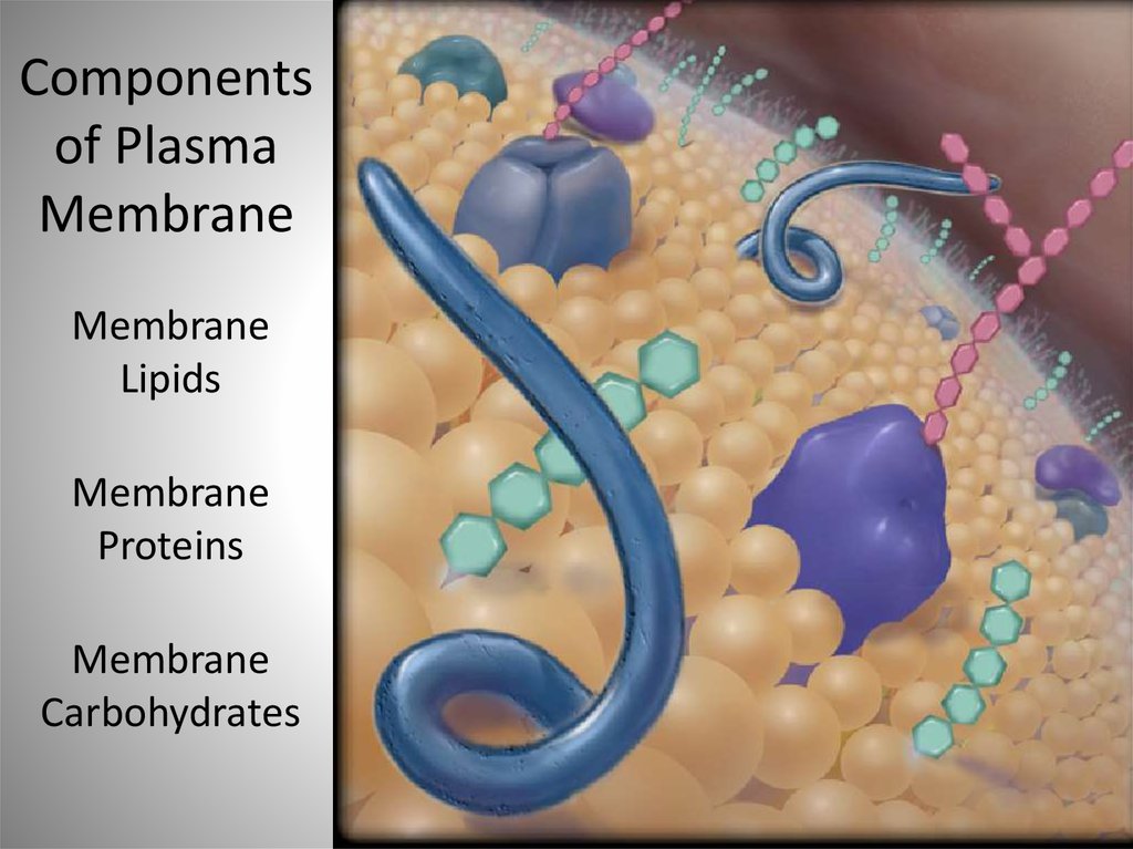
Componentsof Plasma
Membrane
Membrane
Lipids
Membrane
Proteins
Membrane
Carbohydrates
24. Plasma Membrane
25. Chemical Structure of the Plasma Membrane: Lipid Components
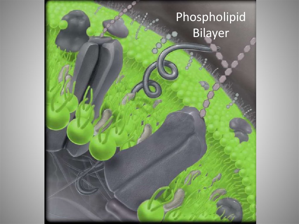
• Phospholipids–
–
–
–
–
–
–
Most membrane lipids of this type
Polar “head” and two hydrophobic “tails”
Form two parallel sheets of molecules
Lie tail to tail with tails forming internal area membrane
Head directed outward
Structure termed phospholipid bilayer
Ensures cytosol and fluid surrounding cells remain separate
• surrounding fluid termed interstitial fluid
26.
PhospholipidBilayer
27.
Fatty Acid TailsPolar Heads
Phospholipid Molecules
28.
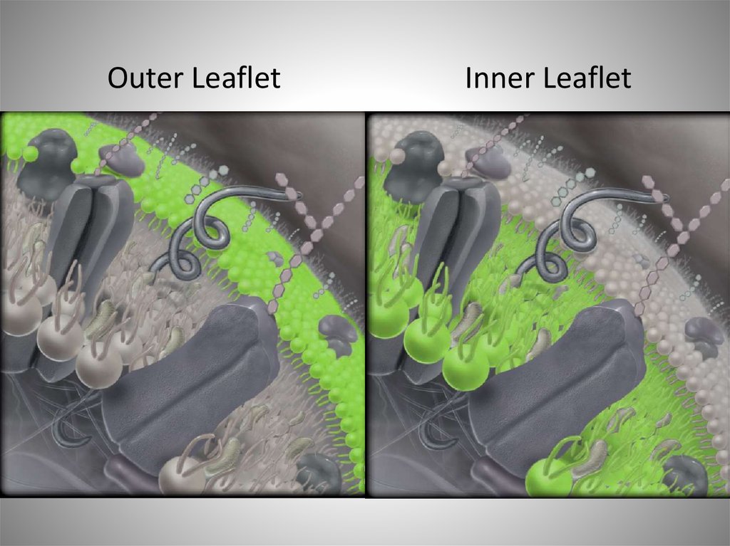
Outer LeafletInner Leaflet
29. Chemical Structure of the Plasma Membrane: Lipid Components
• Cholesterol– Scattered within phospholipid bilayer
– Strengthens the membrane
– Stabilizes the membrane against temperature extremes
• Glycolipids
– Lipids with attached carbohydrate groups
– Located on outer phospholipid region only
– Helps to form the glycocalyx
• the “coating of sugar” on cell’s surface
30. Figure 4.5
Copyright © The McGraw-Hill Companies, Inc. Permission required for reproduction or display.Interstitial fluid
Phospholipid
Glycolipid
Carbohydrate
Polar head of
phospholipid
molecule
Phospholipid
bilayer
Glycoprotein
Nonpolar tails
of phospholipid
molecule
Protein
Cholesterol
Integral protein
Peripheral protein
Filaments of
cytoskeleton
Cytosol
Functions of Plasma Membrane
Cytosol
1. Physical barrier: Establishes a flexible boundary, protects cellular contents, and supports
cell structure. Phospholipid bilayer separates substances inside and outside the cell
2. Selective permeability: Regulates entry and exit of ions, nutrients, and waste molecules
through the membrane
3. Electrochemical gradients: Establishes and maintains an electrical charge difference
across the plasma membrane
4. Communication: Contains receptors that recognize and respond to molecular signals
(a) Plasma membrane
Phospholipid
bilayer
Phospholipid
bilayer
Cytosol
(b) Phospholipid bilayer
b: © Don W. Fawcett/Photo Researchers, Inc.
31. Membrane Lipid Cholesterol
32. Membrane Lipid Glycolipid
33. Membrane Carbohydrates Glycocalyx
34. Chemical Structure of the Plasma Membrane: Membrane Proteins
• Membrane proteins–
–
–
–
Compose half of plasma membrane by weight
Can “float” and move about fluid bilayer
Most of a membrane’s functions determined by resident proteins
Classified as integral or peripheral proteins
35. Membrane Protein
36.
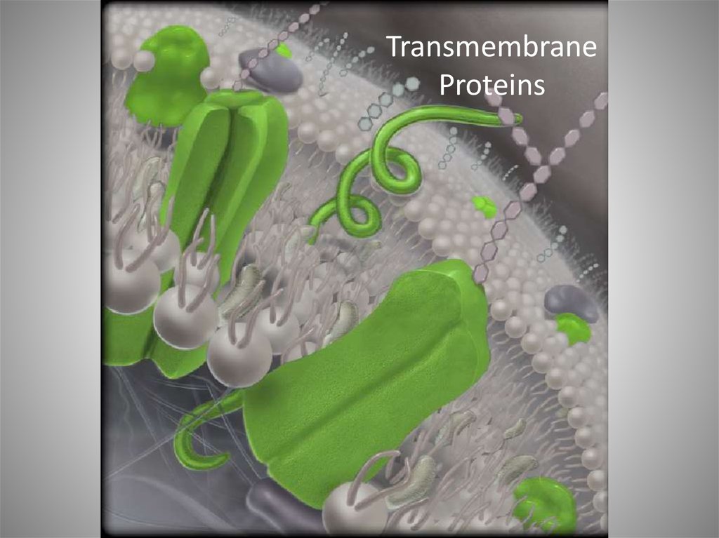
TransmembraneProteins
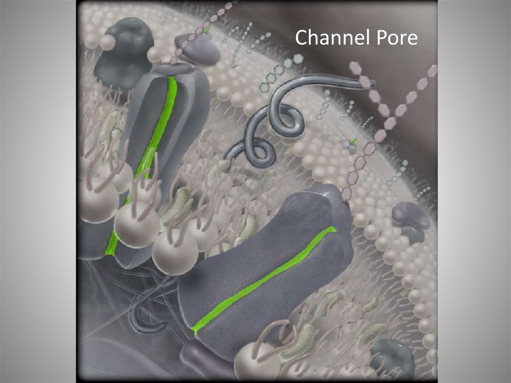
37. Chemical Structure of the Plasma Membrane: Membrane Proteins
• Integral proteins–
–
–
–
Embedded within and extend across lipid bilayer
Hydrophobic regions interacting with hydrophobic interior
Hydrophilic regions interacting with hydrophilic regions
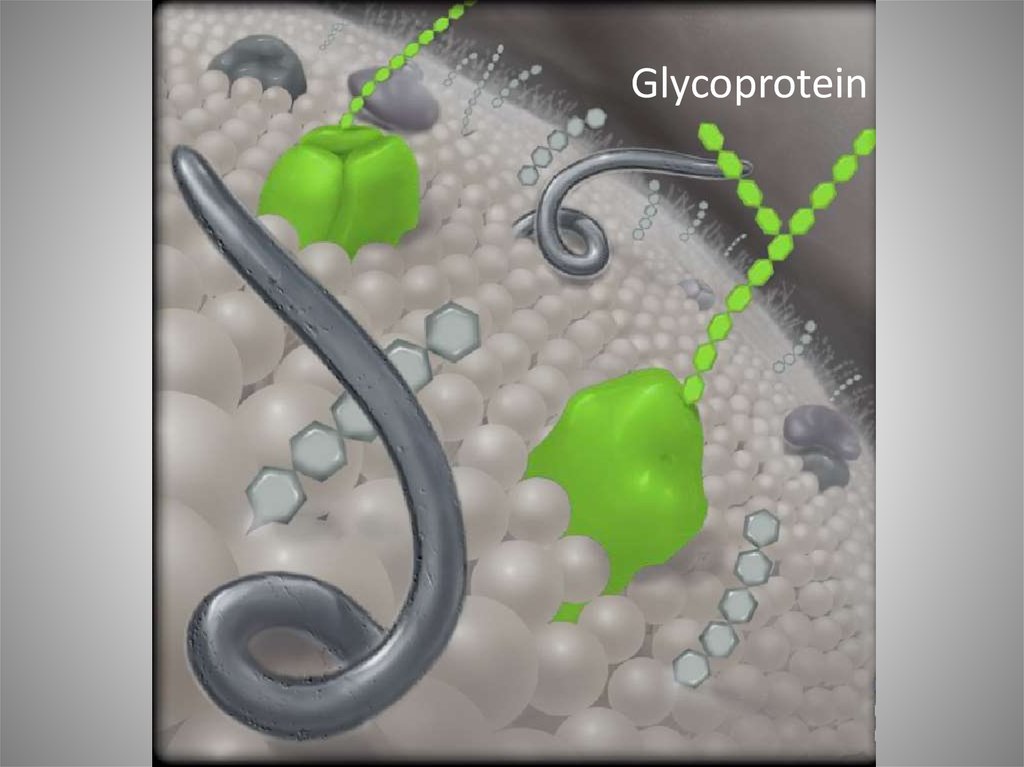
Often glycoproteins with carbohydrate portion
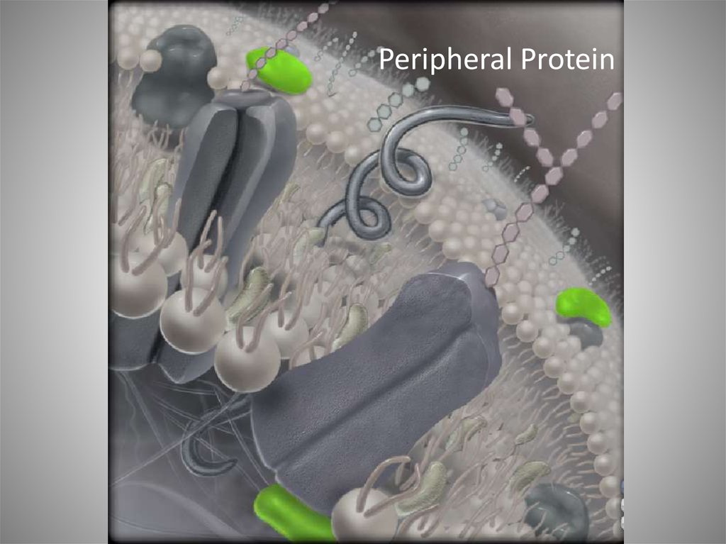
• Peripheral proteins
– Not embedded in lipid bilayer
– Attach loosely to surfaces of the membrane
38.
Channel Pore39.
Peripheral Protein40.
Glycoprotein41. Chemical Structure of the Plasma Membrane: Membrane Proteins
• Often categorized functionally– Transport proteins
• regulate movement of substances across membrane
• e.g., channels, carriers, and pumps
– Cell surface receptors
• bind ligand molecules released from a specific cell
• bind receptors on another cell
• e.g., neurotransmitters and hormones
42. Chemical Structure of the Plasma Membrane: Membrane Proteins
• Often categorized functionally (continued)– Identity markers
• communicate to other cells
• e.g., immune system cells distinguishing healthy cells from foreign cells
– Enzymes
• catalyze chemical reactions
43. Chemical Structure of the Plasma Membrane: Membrane Proteins
• Often categorized functionally (continued)– Anchoring sites
• Secure cytoskeleton to plasma membrane
– Cell-adhesion proteins
• Perform cell to cell attachments
44. Figure 4.6
Copyright © The McGraw-Hill Companies, Inc. Permission required for reproduction or display.Ligand
Interstitial
fluid
Substrate
Product
Interstitial
fluid
Cytosol
Cytoskeleton protein
Transport protein
Receptor
Identity marker
Enzyme
Anchoring site
Cell-adhesion protein
45. Membrane Transport
• One important function of plasma membrane– Regulating movement of materials into and out of a cell
requires substances from interstitial fluid
requires waste elimination into interstitial fluid
occurs through processes of membrane transport
can be categorized as passive or active transport
46. Membrane Transport
• Passive processes of membrane transport– Do not require energy
– Depend on substances moving down concentration gradient
• move from where there is more of a substance to where there is less
– Two types:
• diffusion
• osmosis
47. Membrane Transport
• Active processes of membrane transport– Require energy
– E.g., movement of a substance up its concentration gradient
• termed active transport
– E.g., release of a membrane-bound vesicle
• termed vesicular transport
48. Membrane Transport— Passive Processes: Diffusion
• Environmental conditions affecting rate of diffusion– “Steepness” of concentration gradient
• measure of the difference in concentration between two areas
• steeper gradient with a faster rate of diffusion
– Temperature
• reflects kinetic energy and random movement
• higher movement with higher temperature
• results in faster rate of diffusion
49. Figure 4.7
Copyright © The McGraw-Hill Companies, Inc. Permission required for reproduction or display.50. Membrane Transport— Passive Processes: Diffusion
Cellular Diffusion• Simple diffusion
–
–
–
–
–
–
Molecules passing between phospholipid molecules
Solutes small and nonpolar
Include respiratory gases (O2 and CO2), some fatty acids, ethanol, urea
Cannot be regulated by plasma membrane
Movement dependent on concentration gradient alone
Continue to move as long as gradient exists
51. Figure 4.8
Copyright © The McGraw-Hill Companies, Inc. Permission required for reproduction or display.Small nonpolar solutes move down
their concentration gradients.
Interstitial
fluid
Oxygen
Cytosol
Carbon dioxide
52. Membrane Transport— Passive Processes: Diffusion
Cellular Diffusion (continued)• Facilitated diffusion
– Transport process for small charged or polar solutes
– Require assistance from plasma membrane proteins
– Two types of facilitated diffusion
• channel-mediated diffusion
• carrier-mediated diffusion
– Maximum rate of transport determined by number of channels and
carriers
• higher rate with greater number of transport proteins
53. Membrane Transport— Passive Processes: Diffusion
Cellular Diffusion (continued)• Facilitated diffusion
– Transport process for small charged or polar solutes
– Require assistance from plasma membrane proteins
– Two types of facilitated diffusion
• channel-mediated diffusion
• carrier-mediated diffusion
– Maximum rate of transport determined by number of channels and
carriers
• higher rate with greater number of transport proteins
54. Membrane Transport— Passive Processes: Diffusion
Cellular Diffusion (continued)• Channel-mediated diffusion
– Movement of small ions through water-filled protein channels
– Channels specific for one ion type
– Leak channels
• continuously open
– Gated channel
• usually closed
• open in response to stimulus
55. Figure 4.9a
Copyright © The McGraw-Hill Companies, Inc. Permission required for reproduction or display.Ions move down their concentration
gradient through water-filled channels.
Na+
Interstitial
fluid
Cytosol
(a) Channel-mediated diffusion
Na+
leak channel
K+ leak
channel
K+
56. Membrane Transport— Passive Processes: Diffusion
Cellular Diffusion (continued)• Na+ channels
– Na+ leak channels
• allow Na+ to pass through continuously
– Chemically gated Na+ channels
• allow Na+ to move through in response to a particular chemical
57. Membrane Transport— Passive Processes: Diffusion
Cellular Diffusion (continued)• Carrier-mediated diffusion
–
–
–
–
–
–
Small, polar molecules assisted across membrane by carrier protein
Transport substances such as glucose
Binding of substance causing change in carrier protein shape
Releases substances on other side of membrane
Move substances down their gradient
Carrier transporting only one substance termed a uniporter
58. Figure 4.9b
Copyright © The McGraw-Hill Companies, Inc. Permission required for reproduction or display.Carrier proteins change shape to transport
molecules across the plasmamembrane.
Glucose
Interstitial
fluid
Cytosol
Glucose carrier protein
(b) Carrier-mediated diffusion
59. Membrane Transport— Passive Processes: Osmosis
• Osmosis– Passive movement of water through selectively permeable membrane
• membrane allowing passage of water
• membrane preventing passage of most solutes
– Occurs in response to differences in water concentration
• different concentrations on either side of a membrane
60. Membrane Transport— Passive Processes: Osmosis
Plasma Membrane: A Selectively Permeable Membrane• Two ways water crosses membrane
– “Slip between” molecules of phospholipid bilayer
– Moves through integral protein water channels
• termed aquaporins
61. Membrane Transport— Passive Processes: Osmosis
Plasma Membrane: A Selectively Permeable Membrane(continued)
• Two types of solutes
– Permeable solutes
• pass through bilayer
• small and nonpolar solutes
• e.g., oxygen, carbon dioxide
– Nonpermeable solutes
• prevented from passing through bilayer
• charged, polar, or large solutes
• e.g., ions, glucose, proteins
62. Membrane Transport— Passive Processes: Osmosis
Concentration Gradients Across the Plasma Membrane• Differences in solute concentration across membrane
– May exist between cytosol and interstitial fluid
– Also cause water concentrations to exist
– Greater concentration of solutes with lower concentration of water
63. Membrane Transport— Passive Processes: Osmosis
Movement of Water Into or Out of a Cell by Osmosis• Net movement of water by osmosis
–
–
–
–
–
–
Dependent on concentration gradient between cytosol and solution
Moves down its gradient
E.g., moves from solution of 1% solutes to solution containing 3% solutes
Moves until equilibrium is reached
Equal concentration of water inside and outside cell
Moves toward solution with lower water concentration
64. Figure 4.10
Copyright © The McGraw-Hill Companies, Inc. Permission required for reproduction or display.Figure 4.10
Plasma membrane
Cytosol
Interstitial
fluid
Protein
Aquaporin
Water
molecule
Permeable to water
Ca2+
Cl-–
K+
Impermeable
to most solutes
(charged, polar, large)
Na+
Glucose
Lower water
concentration
(higher solute
concentration)
Concentration
gradient
Higher water
concentration
(lower solute
concentration)
65. Membrane Transport— Passive Processes: Osmosis
Osmotic Pressure– Pressure exerted by movement of water across semipermeable
membrane
– Due to difference in solution concentration
– Steeper gradient, more water moved by osmosis
– Steeper gradient, greater osmotic pressure
66. Membrane Transport— Passive Processes: Osmosis
Osmotic Pressure (continued)• Figure 4.11
–
–
–
–
Semipermeable membrane allowing for passage of water only
Side A with more solutes initially
Water moving from side B to side A by osmosis
Continues until fluids equal in concentration
67. Figure 4.11
Copyright © The McGraw-Hill Companies, Inc. Permission required for reproduction or display.Side A
Side B
Side A
Side B
Non-permeable solutes (glucose, Na+, protein)
Water molecules
Higher solute Semipermeable
concentration,
membrane
lower water
concentration
Lower solute
concentration,
higher water
concentration
Initial setup: Side A contains proportionately
more solute and less water.
Semipermeable
membrane
Final setup: Water moved by osmosis from side
B down the water gradient to side A until the
concentrations of side A and side B are equal.
68. Membrane Transport— Passive Processes: Osmosis
Osmotic Pressure (continued)• Can be measured indirectly
– Could put stopper on side A in figure 4.11b
– Could exert force to return fluid to original level
– Would create hydrostatic pressure within the tube
• the pressure exerted by a fluid on wall of its container
– Osmotic pressure equal to hydrostatic pressure applied
• = total pressure needed to return fluid to original level
69. Membrane Transport— Passive Processes: Osmosis
Osmosis and Tonicity• Cell gains or loses water with osmosis
– Accompanying change in cell volume and osmotic pressure
– Tonicity
• ability of a solution to change the volume or pressure of the cell by osmosis
70. Membrane Transport— Passive Processes: Osmosis
Osmosis and Tonicity (continued)• Isotonic solution
–
–
–
–
Both cytosol and solution with same relative concentration of solutes
E.g., physiological saline with a concentration of 0.9% NaCl
Isotonic to erythrocytes
No net movement of water
71. Membrane Transport— Passive Processes: Osmosis
Osmosis and Tonicity (continued)• Hypotonic solution
– Solution with a lower concentration of solutes than cytosol
– E.g., erythrocytes in pure water
– Water moving down concentration gradient
• from outside the cell to inside
– Increased volume and pressure of cell
– May cause cell lysis (rupture)
• hemolysis, term for ruptured red blood cells
72. Membrane Transport— Passive Processes: Osmosis
Osmosis and Tonicity (continued)• Hypertonic solution
–
–
–
–
–
–
Solution with a higher concentration of solutes than cytosol
E.g., erythrocytes in 3% NaCl pure water
Water moves down concentration gradient
Moves from inside the cell to outside
Decreased volume and pressure of cell
May cause cell to shrink
• termed crenation
73. Figure 4.12
Copyright © The McGraw-Hill Companies, Inc. Permission required for reproduction or display.Isotonic solution
Hypotonic solution
Interstitial fluid is less
concentrated than cytosol.
Interstitial fluid is the same
concentration as cytosol.
Erythrocyte
Water
leaves
cell.
Erythrocyte
Erythrocyte
SEM 11,550x
SEM 9030x
SEM 6900x
Interstitial fluid is more
concentrated than cytosol.
Water
enters
cell.
No net
movement
of water.
Normal erythrocytes
(a)
Hypertonic solution
Erythrocytes nearing hemolysis
(b)
Erythrocytes undergoing crenation
(c)
a: © Dennis Kunkel Microscopy, Inc./Phototake; b: © Dennis Kunkel Microscopy, Inc./Phototake; c: © Dennis Kunkel Microscopy, Inc./Phototake
74. Membrane Transport: Active Processes
Active Transport– Opposes the movement of solutes by diffusion
– Solutes moved against a concentration gradient
– Maintains gradient between cell and interstitial fluid
75. Membrane Transport: Active Processes
Active Transport (continued)• Primary active transport
–
–
–
–
–
Uses energy directly from breakdown of ATP
Phosphate group added to transport protein
Results in a change in protein’s shape
Results in movement of solute across membrane
Addition of phosphate to protein termed phosphorylation
76. Membrane Transport: Active Processes
Figure 4.13• Ion pumps
– Active transport proteins
that move ions across
membrane
– Help cell maintain
internal concentration of
ions
– E.g., Ca2+ pumps in
plasma membrane of
erythrocytes
• prevent cell rigidity
from accumulated
calcium
Copyright © The McGraw-Hill Companies, Inc. Permission required for reproduction or display.
Erythrocyte
ADP
+Pi
ATP
Ca2+
pump
Ca2+
Cytosol
Interstitial
fluid
77. Membrane Transport: Active Processes
Active Transport (continued)• Sodium-potassium pump
–
–
–
–
–
–
Special kind of ion pump, an exchange pump
Moves one ion into cell against gradient
Moves another ion out of cell against gradient
Three Na+ pumped out for two K+ pumped in
Maintains steep membrane gradient
Requires ATP
78. Membrane Transport: Active Processes
Active Transport• Sodium-potassium pump (continued)
– Maintains an electrochemical gradient
• electrical charge difference across plasma membrane
• due to unequal distribution of positive and negative substances across
membrane
• voltage differences termed membrane potential
• at rest, termed resting membrane potential
79. Figure 4.14
Copyright © The McGraw-Hill Companies, Inc. Permission required for reproduction or display.Interstitial
fluid (IF)
Cytosol
Phospholipid bilayer
Figure 4.14
ATP
binding
site
K+
ATP
Na+
Transport
protein
1 Three sodium ions (Na+) and ATP bind to sites on the
cytoplasmic surface of the Na+/ K+ pump.
IF
Cytosol
IF
K+
Na+/K+
Pump
K+
Cytosol
Breakdown of ATP
(releases energy)
ADP
Na+
P
Transport protein
resumes original
shape
Transport protein changes
shape (requires energy
from ATP breakdown)
4 This transport protein reverts back to its original
shape, resulting in the release of the K+ ions into
the cytosol. The Na+/ K+ pump is now ready to
begin the process again.
IF
Cytosol
K+
Na+
P
3 Two K+ ions from the interstitial fluid then bind to
sites on the outer cellular surface of the Na+/ K+
pump. At the same time, the Pi produced earlier
by ATP hydrolysis is released into the cytosol.
2 ATP is split into ADP and Pi, resulting in both the
binding of the Pi to the pump and release of energy
that causes the Na+/ K+ pump to change
conformation (shape) and release the Na+ ions into
the interstitial fluid.
80. Membrane Transport: Active Processes
Active Transport (continued)• Secondary active transport
–
–
–
–
–
Moves substance against concentration gradient
Uses energy provided by movement of second substance down gradient
Kinetic energy providing “power” to pump other substance
Na+ moving down concentration gradient
Ultimately dependent on Na+/K+ pumps for energy
81. Membrane Transport: Active Processes
Active Transport• Secondary active transport (continued)
– Two substances moved in same direction
• proteins termed symporters
• process symport secondary active transport
• e.g., glucose transported up its gradient into cell
– Na+ and glucose moved in same direction
82. Membrane Transport: Active Processes
Active Transport• Secondary active transport (continued)
– Two substances moved in opposite directions
• proteins termed antiporters
• process termed antiport secondary active transport
• e.g., H+ transported up its gradient out of cell
– Na+ and H+ moved in opposite directions
83. Figure 4.15
Copyright © The McGraw-Hill Companies, Inc. Permission required for reproduction or display.Glucose is transported
up its gradient into cell.
Na+ diffuses down
its gradient into cell.
Interstitial fluid
Symporter
Antiporter
Cytosol
H+ is transported up
its gradient out of cell.
(a) Symporter: Substances
move in the same direction.
(b) Antiporter: Substances
move in opposite directions.
84. Membrane Transport: Active Processes
Vesicular Transport– Requires vesicles
• membrane-bounded sac filled with materials
– Requires energy to transport vesicles
– Exocytosis
• vesicle fuses with membrane
• releases substances outside the cell
– Endocytosis
• vesicle encloses material outside cell
• fuses with membrane to release inside cell
85. Membrane Transport: Active Processes
Vesicular Transport (continued)• Exocytosis
–
–
–
–
How large substances are secreted from cell
Macromolecules too large to be moved across membrane
Material packed within intracellular transport vehicles
Vesicle and plasma membrane fusion
• requires ATP
– Contents released to outside of cell
– E.g., release of neurotransmitters from nerve cells
86. Figure 4.16
Copyright © The McGraw-Hill Companies, Inc. Permission required for reproduction or display.Cytosol
Interstitial fluid
Secretory
vesicle
Plasma
membrane
Figure 4.16
Vesicle membrane
1 Vesicle nears plasma membrane
Membrane
proteins
2 Fusion of vesicle membrane with plasma membrane
Plasma
membrane
opens
3 Plasma membrane opens to outside of cell
4
Release of vesicle components into the interstitial
fluid and integration of vesicle membrane
components into the plasma membrane
87. Membrane Transport: Active Processes
Vesicular Transport (continued)• Endocytosis
–
–
–
–
Cellular uptake of large substances from external environment
Used for the uptake of materials for digestion
Used for retrieval of membrane from exocytosis
Used for regulating membrane protein composition
• to alter cellular processes
– Three types:
• phagocytosis, pinocytosis, and receptor-mediated endocytosis
88. Membrane Transport: Active Processes
Vesicular Transport (continued)• Steps of endocytosis
– Substances within interstitial fluid packaged into a vesicle
– Vesicle formed at cell surface
– Inward fold of membrane to form pocket
• termed invagination
– Deepens and pinches off when layer fuses
• requires energy
– Intracellular vesicle with material formerly outside cell
89. Membrane Transport: Active Processes
Vesicular Transport (continued)• Phagocytosis
–
–
–
–
Occurs when cell engulfs large particle external to cell
Forms large extensions termed pseudopodia
Surround particle, enclosing it in membrane sac
Fuses with lysosome
• contents digested here
– Only in a few cell types
• E.g., white blood cells engulfing microbes
90. Figure 4.17a
Copyright © The McGraw-Hill Companies, Inc. Permission required for reproduction or display.Figure 4.17a
Pseudopodia
Particle
Invagination
Interstitial
fluid
Plasma
membrane
Newly
formed
vesicle
(a) Phagocytosis
Cytosol
91. Membrane Transport: Active Processes
Vesicular Transport (continued)• Pinocytosis
–
–
–
–
–
Internalization of droplets of interstitial fluid
Multiple, small vesicles formed
All dissolved solutes taken into cell
Performed by most cells
E.g., cells of capillary wall
92. Figure 4.17b
Copyright © The McGraw-Hill Companies, Inc. Permission required for reproduction or display.Figure 4.17b
Plasma
membrane
Interstitial
fluid
Cytosol
Vesicle
(b) Pinocytosis
93. Membrane Transport: Active Processes
Vesicular Transport (continued)• Receptor-mediated endocytosis
–
–
–
–
Movement of specific molecules from interstitial environment into a cell
Requires binding to a receptor
Enables cell to obtain bulk quantities of substances
E.g., transport of cholesterol from blood to a cell
• cholesterol in blood in structures termed low-density lipoproteins
• LDLs internalized by this process
94. Membrane Transport: Active Processes
Vesicular Transport (continued)• Steps of receptor-mediated endocytosis
–
–
–
–
–
–
–
Molecule binding to protein receptors in membrane
Form ligand-receptor complex
Accumulate at special regions containing clathrin protein
Fold inward to form clathrin-coated pit
Form clathrin-coated vesicle
Moves into cytosol
Fusion of lipid bilayers requiring ATP
95. Membrane Transport: Active Processes
Vesicular Transport (continued)• Steps of receptor-mediated endocytosis
–
–
–
–
–
–
–
Molecule binding to protein receptors in membrane
Form ligand-receptor complex
Accumulate at special regions containing clathrin protein
Fold inward to form clathrin-coated pit
Form clathrin-coated vesicle
Moves into cytosol
Fusion of lipid bilayers requiring ATP
96. Figure 4.17c
Copyright © The McGraw-Hill Companies, Inc. Permission required for reproduction or display.Receptors
Plasma
membrane
Clathrincoated pit
(c) Receptor-mediated endocytosis
Interstitial
fluid
Cytosol
Clathrincoated
vesicle
97. Figure 4.18a
Copyright © The McGraw-Hill Companies, Inc. Permission required for reproduction or display.Figure 4.18a
Do not require expenditure of cellular energy; substance moves
into or out of a cell down its concentration gradient.
(a) Passive Processes
DIFFUSION: Movement of a solute from an area of higher concentration to an area of lower concentration.
Simple Diffusion: Small and nonpolar substances move between phospholipid molecules
of the plasma membrane.
Carbon dioxide
Oxygen
Interstitial fluid
Cytosol
Facilitated Diffusion: Small, charged, or polar substances move assisted by a transport protein (channel or carrier).
Na+
Channel-Mediated: Ion (e.g., Na+)
movement is facilitated by channels
across the plasma membrane.
Channel
Carrier
Glucose
Carrier-Mediated: Small polar molecule
movement (e.g., glucose) is facilitated by
protein carriers across the plasma membrane.
Cytosol
Interstitial fluid
OSMOSIS: Movement of water across a selectively permeable membrane from an area of higher water concentration
to an area of lower water concentration.
Cytosol
Interstitial fluid
Solute
Aquaporin
Water
Plasma membrane
98. Figure 4.18b
Copyright © The McGraw-Hill Companies, Inc. Permission required for reproduction or display.Figure 4.18b
(b) Active Processes
Require expenditure of cellular energy; substance moved up its
concentration gradient or involves a vesicle.
ACTIVE TRANSPORT: Movement of a substance up its concentration gradient via a protein pump.
Primary Active Transport: Pumps are powered directly by splitting an ATP molecule.
Transport protein changes
shape (requires energy
from ATP breakdown)
ADP + P
Na+
Note: The two ion species
are not simultaneously
attached to the pump
ATP
K+
Cytosol
Interstitial fluid
Secondary Active Transport: Pumps are powered by energy harnessed as a second substance
(usually Na+) moves through a channel down a concentration gradient.
Cytosol
Interstitial
fluid
Glucose
Na+
Antiport: Two
substances are moved
in opposite directions.
Symport: Two
substances are moved
in the same direction
H+
VESICULAR TRANSPORT: Movement of a substance across the plasma membrane via a vesicle.
Exocytosis: Movement of a
substance out of
a cell via a vesicle.
Endocytosis: Movement of a substance into a cell via a vesicle. The three types of endocytosis
include phagocytosis, pinocytosis, and receptor-mediated endocytosis.
Vesicles
Receptor-Mediated Endocytosis:
Movement of a specific substance
into a cell following the binding of
the substance to a receptor.
Vesicle
Receptors
Plasma
membrane
opens
Cytosol
Particle
Interstitial
fluid
Phagocytosis:
Movement of
large substances
into a cell.
Pinocytosis: Movement
of fluid into a cell.
Pseudopodia
99. Membrane Transport: Active Processes
Clinical View: Familial Hypercholesteremia–
–
–
–
–
–
Inherited genetic disorder
Defects in LDL receptor or proteins of LDLs
Interfere with normal receptor-mediated endocytosis of cholesterol
Results in greatly elevated cholesterol
Causes atherosclerosis
Greatly increased risk of heart attack