


 biology
Similar presentations:


Membrane Structure and Function
1. Chapter 7
Membrane Structure andFunction
PowerPoint® Lecture Presentations for
Biology
Eighth Edition
Neil Campbell and Jane Reece
Lectures by Chris Romero, updated by Erin Barley with contributions from Joan Sharp
Copyright © 2008 Pearson Education, Inc., publishing as Pearson Benjamin Cummings
2. Overview: Life at the Edge
• The plasma membrane is the boundary thatseparates the living cell from its surroundings
• The plasma membrane exhibits selective
permeability, allowing some substances to
cross it more easily than others
Copyright © 2008 Pearson Education, Inc., publishing as Pearson Benjamin Cummings
3.
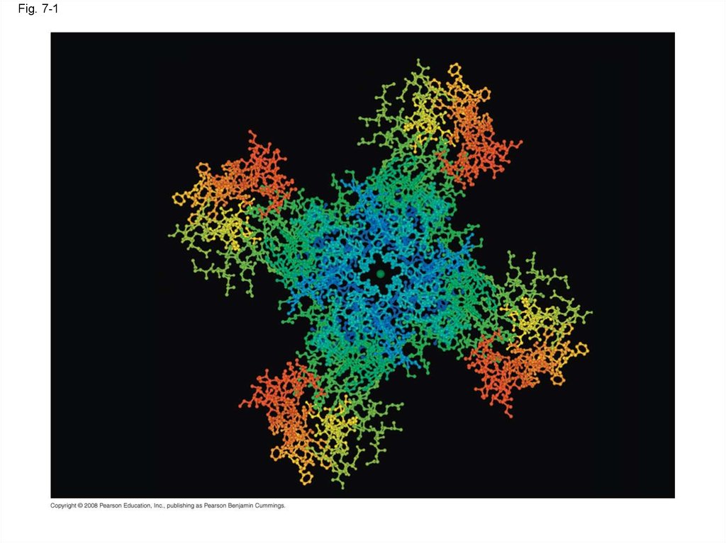
Fig. 7-14. Concept 7.1: Cellular membranes are fluid mosaics of lipids and proteins
• Phospholipids are the most abundant lipid inthe plasma membrane
• Phospholipids are amphipathic molecules,
containing hydrophobic and hydrophilic regions
• The fluid mosaic model states that a
membrane is a fluid structure with a “mosaic” of
various proteins embedded in it
Copyright © 2008 Pearson Education, Inc., publishing as Pearson Benjamin Cummings
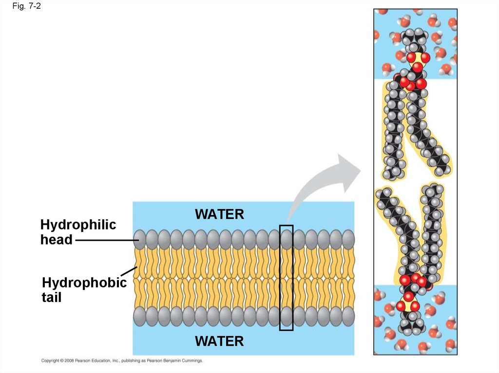
5. Membrane Models: Scientific Inquiry
• Membranes have been chemically analyzedand found to be made of proteins and lipids
• Scientists studying the plasma membrane
reasoned that it must be a phospholipid bilayer
Copyright © 2008 Pearson Education, Inc., publishing as Pearson Benjamin Cummings
6.
Fig. 7-2Hydrophilic
head
WATER
Hydrophobic
tail
WATER
7.
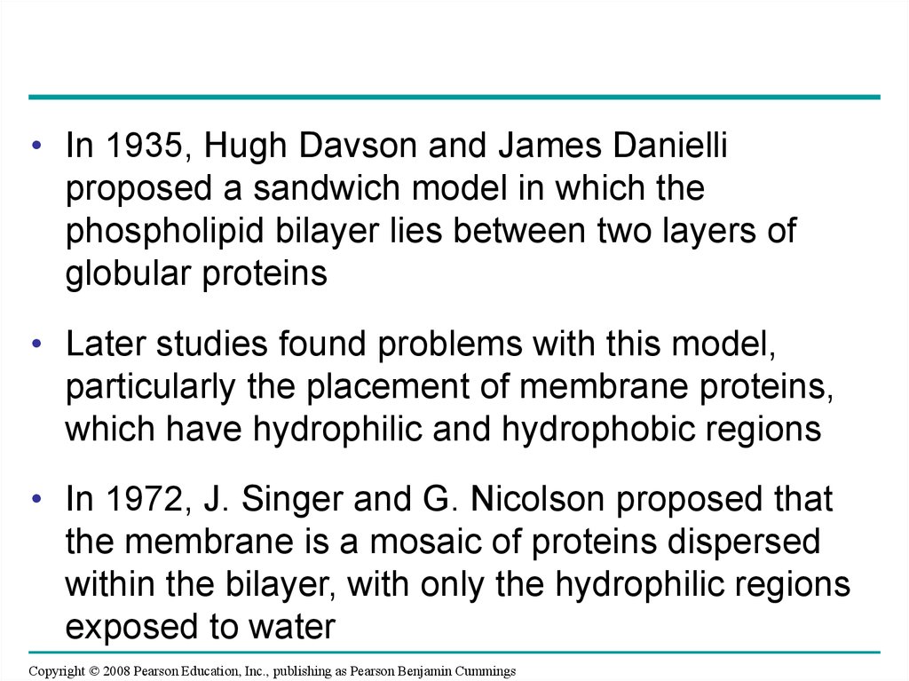
• In 1935, Hugh Davson and James Danielliproposed a sandwich model in which the
phospholipid bilayer lies between two layers of
globular proteins
• Later studies found problems with this model,
particularly the placement of membrane proteins,
which have hydrophilic and hydrophobic regions
• In 1972, J. Singer and G. Nicolson proposed that
the membrane is a mosaic of proteins dispersed
within the bilayer, with only the hydrophilic regions
exposed to water
Copyright © 2008 Pearson Education, Inc., publishing as Pearson Benjamin Cummings
8.
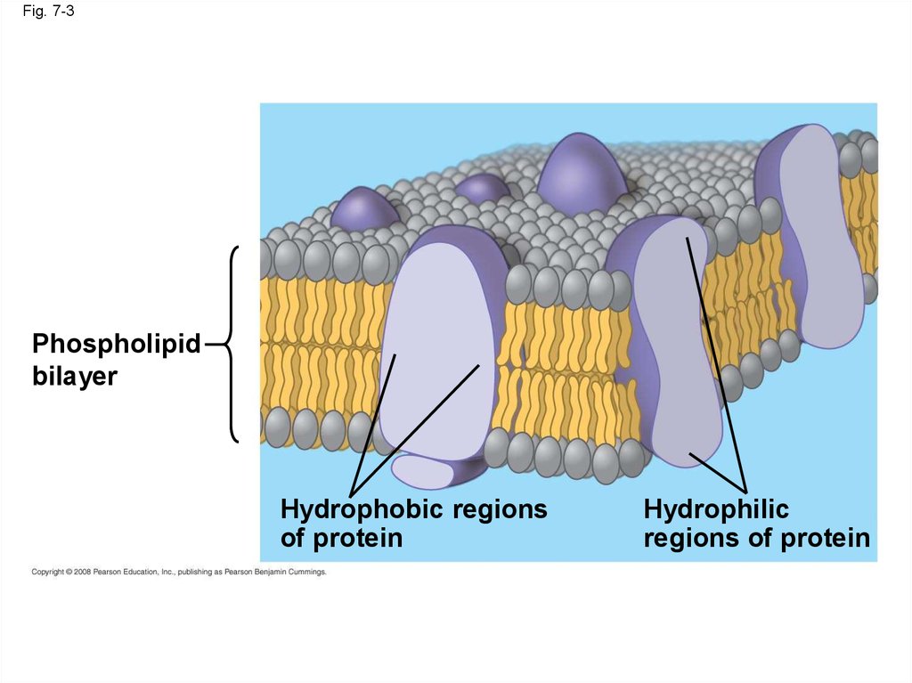
Fig. 7-3Phospholipid
bilayer
Hydrophobic regions
of protein
Hydrophilic
regions of protein
9.
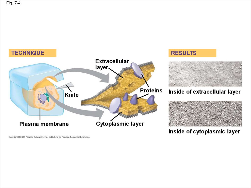
• Freeze-fracture studies of the plasmamembrane supported the fluid mosaic model
• Freeze-fracture is a specialized preparation
technique that splits a membrane along the
middle of the phospholipid bilayer
Copyright © 2008 Pearson Education, Inc., publishing as Pearson Benjamin Cummings
10.
Fig. 7-4TECHNIQUE
RESULTS
Extracellular
layer
Knife
Plasma membrane
Proteins
Inside of extracellular layer
Cytoplasmic layer
Inside of cytoplasmic layer
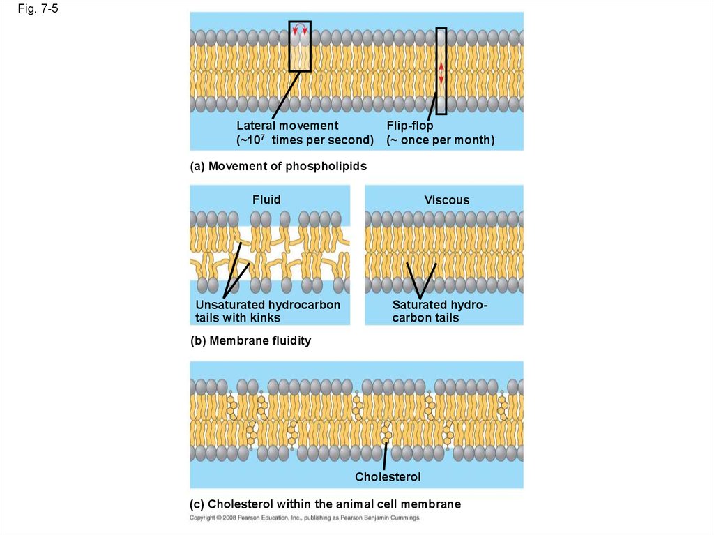
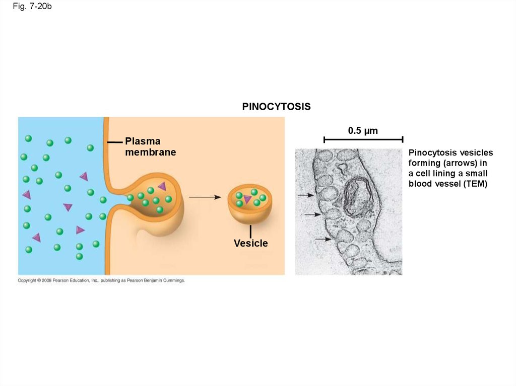
11. The Fluidity of Membranes
• Phospholipids in the plasma membrane canmove within the bilayer
• Most of the lipids, and some proteins, drift
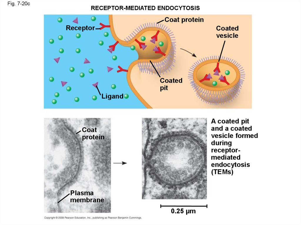
laterally
• Rarely does a molecule flip-flop transversely
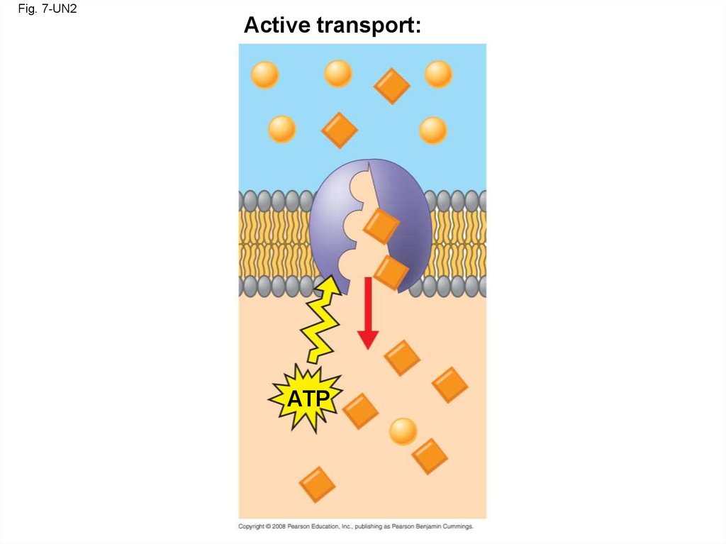
across the membrane
Copyright © 2008 Pearson Education, Inc., publishing as Pearson Benjamin Cummings
12.
Fig. 7-5Lateral movement
(~107 times per second)
Flip-flop
(~ once per month)
(a) Movement of phospholipids
Fluid
Unsaturated hydrocarbon
tails with kinks
Viscous
Saturated hydrocarbon tails
(b) Membrane fluidity
Cholesterol
(c) Cholesterol within the animal cell membrane
13.
Fig. 7-5aLateral movement
( 107 times per second)
(a) Movement of phospholipids
Flip-flop
( once per month)
14.
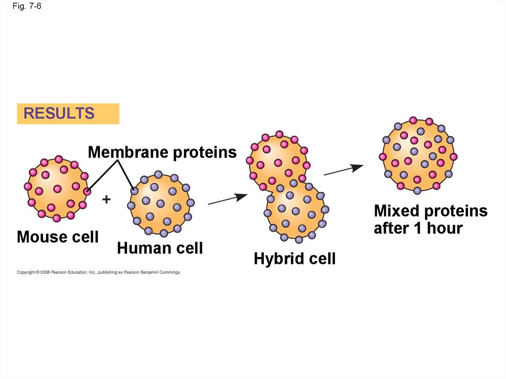
Fig. 7-6RESULTS
Membrane proteins
Mouse cell
Mixed proteins
after 1 hour
Human cell
Hybrid cell
15.
• As temperatures cool, membranes switch froma fluid state to a solid state
• The temperature at which a membrane
solidifies depends on the types of lipids
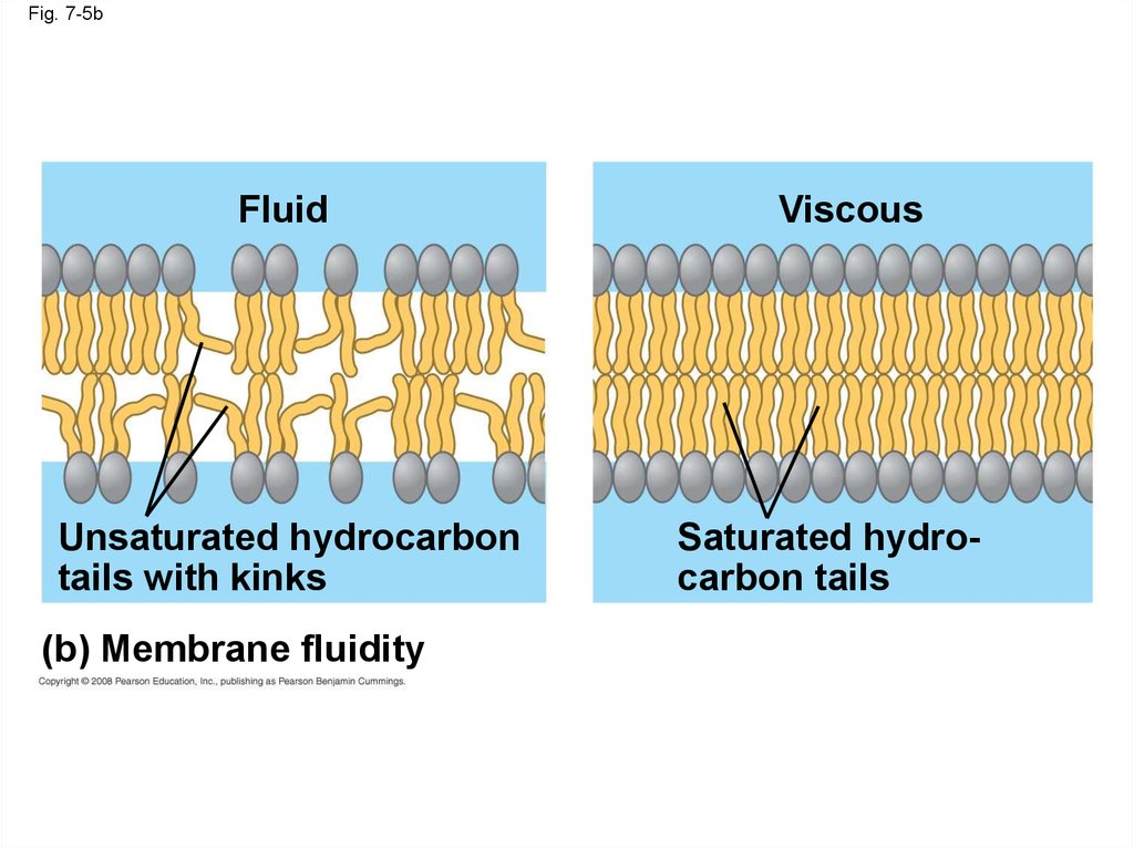
• Membranes rich in unsaturated fatty acids are
more fluid that those rich in saturated fatty
acids
• Membranes must be fluid to work properly;
they are usually about as fluid as salad oil
Copyright © 2008 Pearson Education, Inc., publishing as Pearson Benjamin Cummings
16.
Fig. 7-5bFluid
Unsaturated hydrocarbon
tails with kinks
(b) Membrane fluidity
Viscous
Saturated hydrocarbon tails
17.
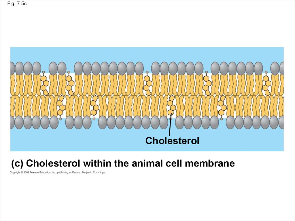
• The steroid cholesterol has different effects onmembrane fluidity at different temperatures
• At warm temperatures (such as 37°C),
cholesterol restrains movement of
phospholipids
• At cool temperatures, it maintains fluidity by
preventing tight packing
Copyright © 2008 Pearson Education, Inc., publishing as Pearson Benjamin Cummings
18.
Fig. 7-5cCholesterol
(c) Cholesterol within the animal cell membrane
19. Membrane Proteins and Their Functions
• A membrane is a collage of different proteinsembedded in the fluid matrix of the lipid bilayer
• Proteins determine most of the membrane’s
specific functions
Copyright © 2008 Pearson Education, Inc., publishing as Pearson Benjamin Cummings
20.
Fig. 7-7Fibers of
extracellular
matrix (ECM)
Glycoprotein
Carbohydrate
Glycolipid
EXTRACELLULAR
SIDE OF
MEMBRANE
Cholesterol
Microfilaments
of cytoskeleton
Peripheral
proteins
Integral
protein
CYTOPLASMIC SIDE
OF MEMBRANE
21.
• Peripheral proteins are bound to the surfaceof the membrane
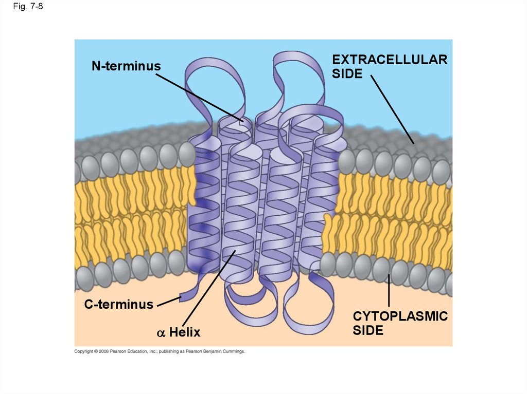
• Integral proteins penetrate the hydrophobic
core
• Integral proteins that span the membrane are
called transmembrane proteins
• The hydrophobic regions of an integral protein
consist of one or more stretches of nonpolar
amino acids, often coiled into alpha helices
Copyright © 2008 Pearson Education, Inc., publishing as Pearson Benjamin Cummings
22.
Fig. 7-8N-terminus
C-terminus
Helix
EXTRACELLULAR
SIDE
CYTOPLASMIC
SIDE
23.
• Six major functions of membrane proteins:– Transport
– Enzymatic activity
– Signal transduction
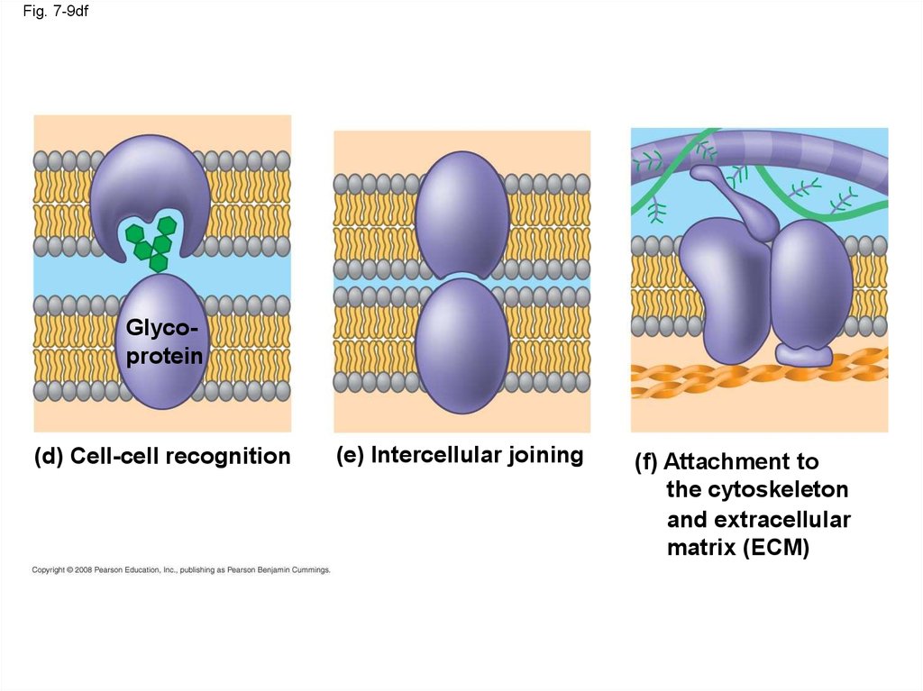
– Cell-cell recognition
– Intercellular joining
– Attachment to the cytoskeleton and
extracellular matrix (ECM)
Copyright © 2008 Pearson Education, Inc., publishing as Pearson Benjamin Cummings
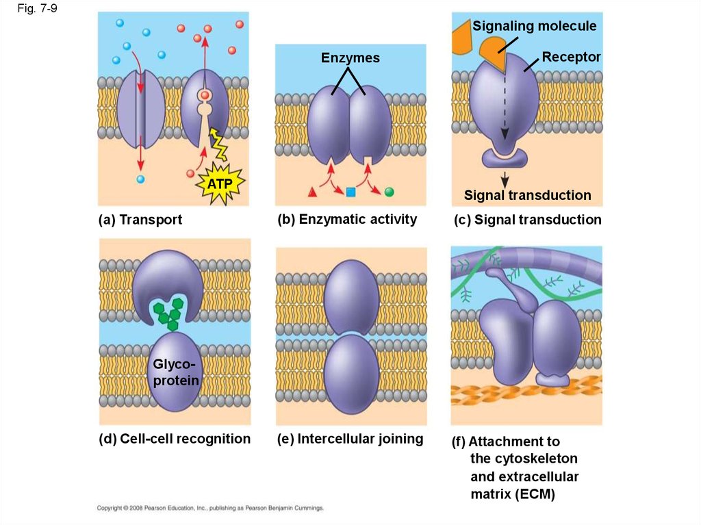
24.
Fig. 7-9Signaling molecule
Enzymes
ATP
(a) Transport
Receptor
Signal transduction
(b) Enzymatic activity
(c) Signal transduction
(e) Intercellular joining
(f) Attachment to
the cytoskeleton
and extracellular
matrix (ECM)
Glycoprotein
(d) Cell-cell recognition
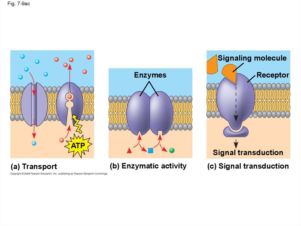
25.
Fig. 7-9acSignaling molecule
Enzymes
ATP
(a) Transport
Receptor
Signal transduction
(b) Enzymatic activity
(c) Signal transduction
26.
Fig. 7-9dfGlycoprotein
(d) Cell-cell recognition
(e) Intercellular joining
(f) Attachment to
the cytoskeleton
and extracellular
matrix (ECM)
27. The Role of Membrane Carbohydrates in Cell-Cell Recognition
• Cells recognize each other by binding tosurface molecules, often carbohydrates, on the
plasma membrane
• Membrane carbohydrates may be covalently
bonded to lipids (forming glycolipids) or more
commonly to proteins (forming glycoproteins)
• Carbohydrates on the external side of the
plasma membrane vary among species,
individuals, and even cell types in an individual
Copyright © 2008 Pearson Education, Inc., publishing as Pearson Benjamin Cummings
28. Synthesis and Sidedness of Membranes
• Membranes have distinct inside and outsidefaces
• The asymmetrical distribution of proteins,
lipids, and associated carbohydrates in the
plasma membrane is determined when the
membrane is built by the ER and Golgi
apparatus
Copyright © 2008 Pearson Education, Inc., publishing as Pearson Benjamin Cummings
29.
Fig. 7-10ER
1
Transmembrane
glycoproteins
Secretory
protein
Glycolipid
Golgi
2
apparatus
Vesicle
3
4
Secreted
protein
Plasma membrane:
Cytoplasmic face
Extracellular face
Transmembrane
glycoprotein
Membrane glycolipid
30. Concept 7.2: Membrane structure results in selective permeability
• A cell must exchange materials with itssurroundings, a process controlled by the
plasma membrane
• Plasma membranes are selectively permeable,
regulating the cell’s molecular traffic
Copyright © 2008 Pearson Education, Inc., publishing as Pearson Benjamin Cummings
31. The Permeability of the Lipid Bilayer
• Hydrophobic (nonpolar) molecules, such ashydrocarbons, can dissolve in the lipid bilayer
and pass through the membrane rapidly
• Polar molecules, such as sugars, do not cross
the membrane easily
Copyright © 2008 Pearson Education, Inc., publishing as Pearson Benjamin Cummings
32. Transport Proteins
• Transport proteins allow passage ofhydrophilic substances across the membrane
• Some transport proteins, called channel
proteins, have a hydrophilic channel that
certain molecules or ions can use as a tunnel
• Channel proteins called aquaporins facilitate
the passage of water
Copyright © 2008 Pearson Education, Inc., publishing as Pearson Benjamin Cummings
33.
• Other transport proteins, called carrier proteins,bind to molecules and change shape to shuttle
them across the membrane
• A transport protein is specific for the substance
it moves
Copyright © 2008 Pearson Education, Inc., publishing as Pearson Benjamin Cummings
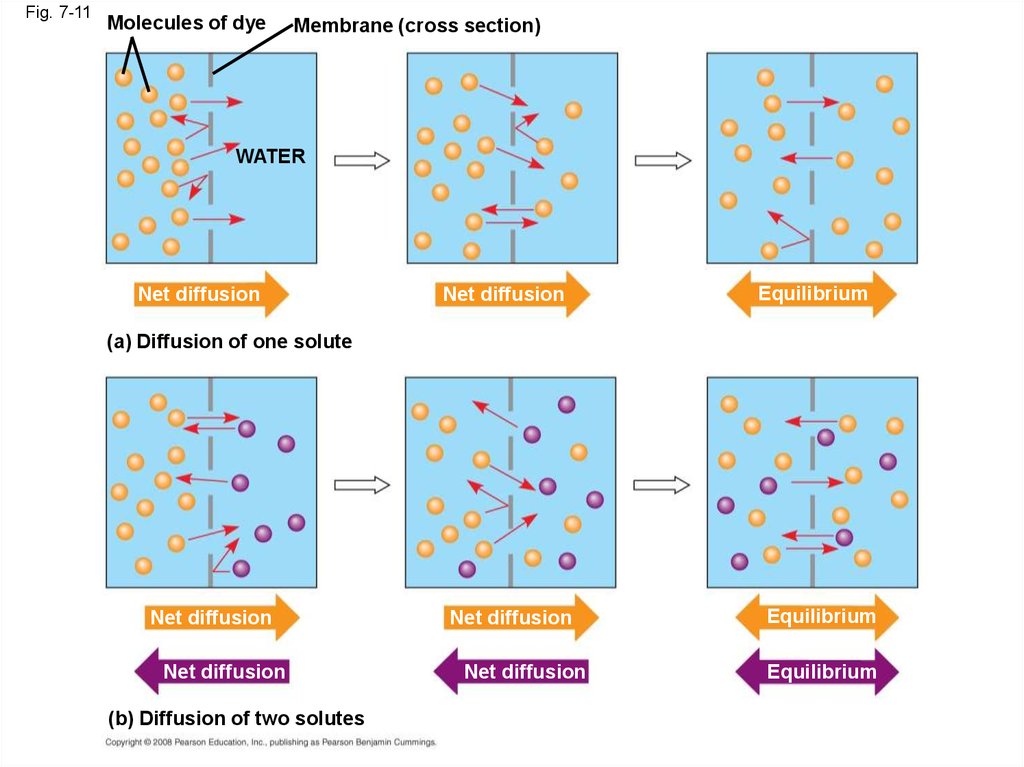
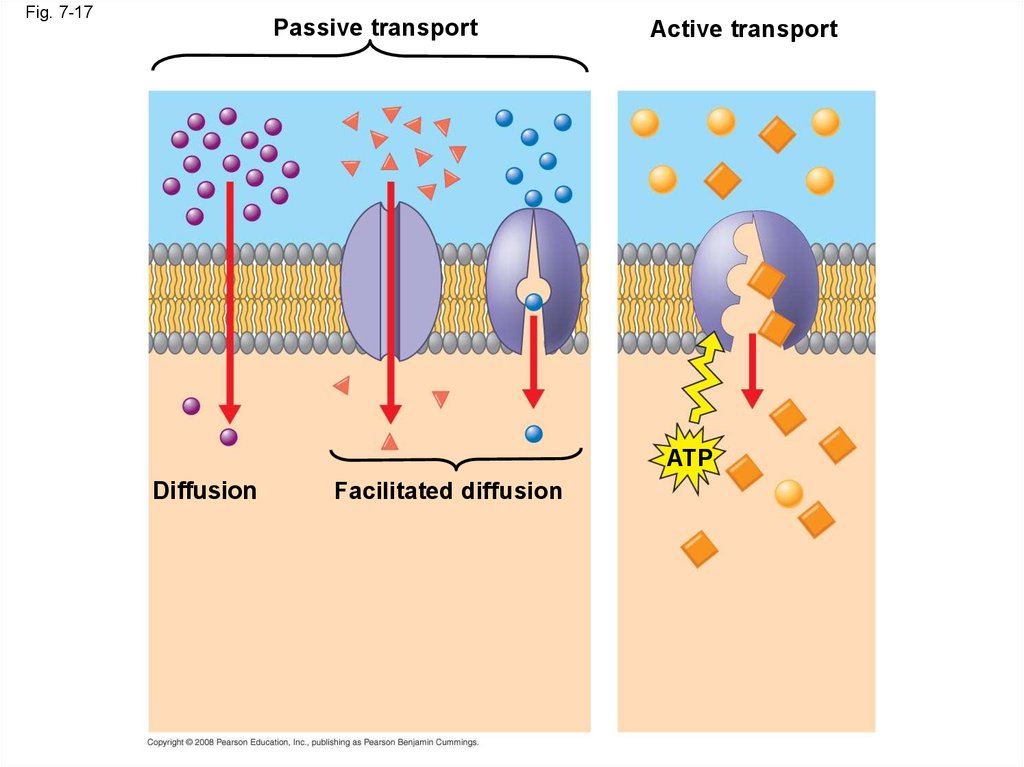
34. Concept 7.3: Passive transport is diffusion of a substance across a membrane with no energy investment
• Diffusion is the tendency for molecules tospread out evenly into the available space
• Although each molecule moves randomly,
diffusion of a population of molecules may
exhibit a net movement in one direction
• At dynamic equilibrium, as many molecules
cross one way as cross in the other direction
Animation: Membrane Selectivity
Copyright © 2008 Pearson Education, Inc., publishing as Pearson Benjamin Cummings
Animation: Diffusion
35.
Fig. 7-11Molecules of dye
Membrane (cross section)
WATER
Net diffusion
Net diffusion
Equilibrium
(a) Diffusion of one solute
Net diffusion
Net diffusion
(b) Diffusion of two solutes
Net diffusion
Net diffusion
Equilibrium
Equilibrium
36.
Fig. 7-11aMolecules of dye
Membrane (cross section)
WATER
Net
diffusion
(a) Diffusion of one solute
Net
diffusion
Equilibrium
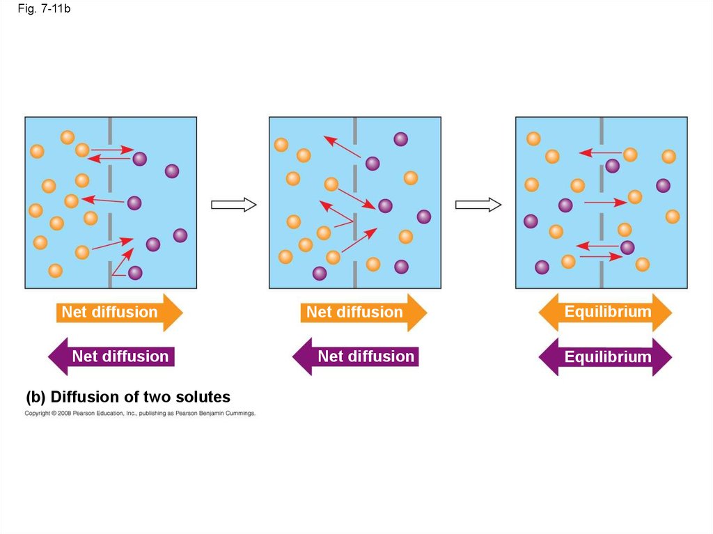
37.
• Substances diffuse down their concentrationgradient, the difference in concentration of a
substance from one area to another
• No work must be done to move substances
down the concentration gradient
• The diffusion of a substance across a biological
membrane is passive transport because it
requires no energy from the cell to make it
happen
Copyright © 2008 Pearson Education, Inc., publishing as Pearson Benjamin Cummings
38.
Fig. 7-11bNet diffusion
Net diffusion
(b) Diffusion of two solutes
Net diffusion
Net diffusion
Equilibrium
Equilibrium
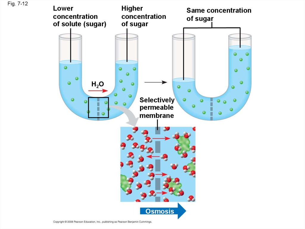
39. Effects of Osmosis on Water Balance
• Osmosis is the diffusion of water across aselectively permeable membrane
• Water diffuses across a membrane from the
region of lower solute concentration to the
region of higher solute concentration
Copyright © 2008 Pearson Education, Inc., publishing as Pearson Benjamin Cummings
40.
Fig. 7-12Lower
concentration
of solute (sugar)
Higher
concentration
of sugar
H2O
Selectively
permeable
membrane
Osmosis
Same concentration
of sugar
41. Water Balance of Cells Without Walls
• Tonicity is the ability of a solution to cause acell to gain or lose water
• Isotonic solution: Solute concentration is the
same as that inside the cell; no net water
movement across the plasma membrane
• Hypertonic solution: Solute concentration is
greater than that inside the cell; cell loses
water
• Hypotonic solution: Solute concentration is
less than that inside the cell; cell gains water
Copyright © 2008 Pearson Education, Inc., publishing as Pearson Benjamin Cummings
42.
Fig. 7-13Hypotonic solution
H2O
Isotonic solution
H2O
H2O
Hypertonic solution
H2O
(a) Animal
cell
Lysed
H2O
Normal
H2O
Shriveled
H2O
H2O
(b) Plant
cell
Turgid (normal)
Flaccid
Plasmolyzed
43.
• Hypertonic or hypotonic environments createosmotic problems for organisms
• Osmoregulation, the control of water balance,
is a necessary adaptation for life in such
environments
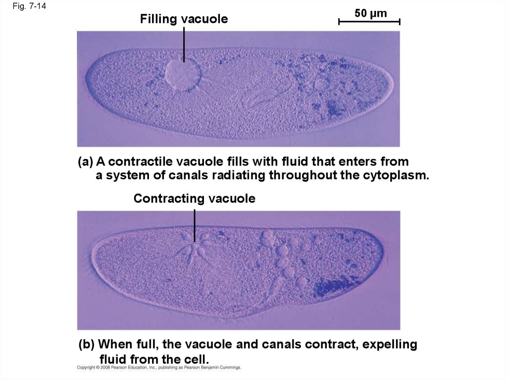
• The protist Paramecium, which is hypertonic to
its pond water environment, has a contractile
vacuole that acts as a pump
Video: Chlamydomonas
Copyright © 2008 Pearson Education, Inc., publishing as Pearson Benjamin Cummings
Video: Paramecium Vacuole
44.
Fig. 7-14Filling vacuole
50 µm
(a) A contractile vacuole fills with fluid that enters from
a system of canals radiating throughout the cytoplasm.
Contracting vacuole
(b) When full, the vacuole and canals contract, expelling
fluid from the cell.
45. Water Balance of Cells with Walls
• Cell walls help maintain water balance• A plant cell in a hypotonic solution swells until
the wall opposes uptake; the cell is now turgid
(firm)
• If a plant cell and its surroundings are isotonic,
there is no net movement of water into the cell;
the cell becomes flaccid (limp), and the plant
may wilt
Copyright © 2008 Pearson Education, Inc., publishing as Pearson Benjamin Cummings
46.
• In a hypertonic environment, plant cells losewater; eventually, the membrane pulls away
from the wall, a usually lethal effect called
plasmolysis
Video: Plasmolysis
Video: Turgid Elodea
Animation: Osmosis
Copyright © 2008 Pearson Education, Inc., publishing as Pearson Benjamin Cummings
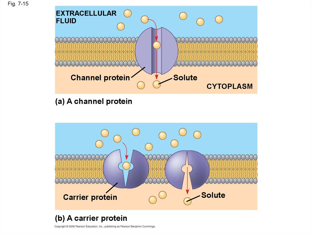
47. Facilitated Diffusion: Passive Transport Aided by Proteins
• In facilitated diffusion, transport proteinsspeed the passive movement of molecules
across the plasma membrane
• Channel proteins provide corridors that allow a
specific molecule or ion to cross the membrane
• Channel proteins include
– Aquaporins, for facilitated diffusion of water
– Ion channels that open or close in response
to a stimulus (gated channels)
Copyright © 2008 Pearson Education, Inc., publishing as Pearson Benjamin Cummings
48.
Fig. 7-15EXTRACELLULAR
FLUID
Channel protein
Solute
CYTOPLASM
(a) A channel protein
Carrier protein
(b) A carrier protein
Solute
49.
• Carrier proteins undergo a subtle change inshape that translocates the solute-binding site
across the membrane
Copyright © 2008 Pearson Education, Inc., publishing as Pearson Benjamin Cummings
50.
• Some diseases are caused by malfunctions inspecific transport systems, for example the
kidney disease cystinuria
Copyright © 2008 Pearson Education, Inc., publishing as Pearson Benjamin Cummings
51. Concept 7.4: Active transport uses energy to move solutes against their gradients
• Facilitated diffusion is still passive because thesolute moves down its concentration gradient
• Some transport proteins, however, can move
solutes against their concentration gradients
Copyright © 2008 Pearson Education, Inc., publishing as Pearson Benjamin Cummings
52. The Need for Energy in Active Transport
• Active transport moves substances againsttheir concentration gradient
• Active transport requires energy, usually in the
form of ATP
• Active transport is performed by specific
proteins embedded in the membranes
Animation: Active Transport
Copyright © 2008 Pearson Education, Inc., publishing as Pearson Benjamin Cummings
53.
• Active transport allows cells to maintainconcentration gradients that differ from their
surroundings
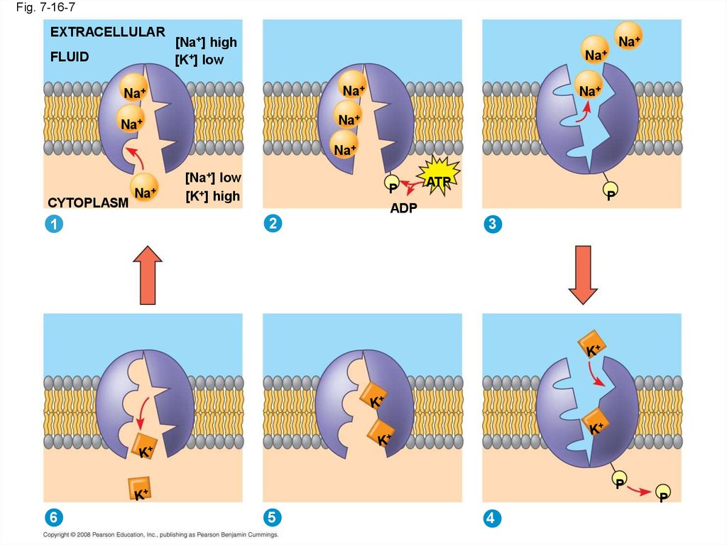
• The sodium-potassium pump is one type of
active transport system
Copyright © 2008 Pearson Education, Inc., publishing as Pearson Benjamin Cummings
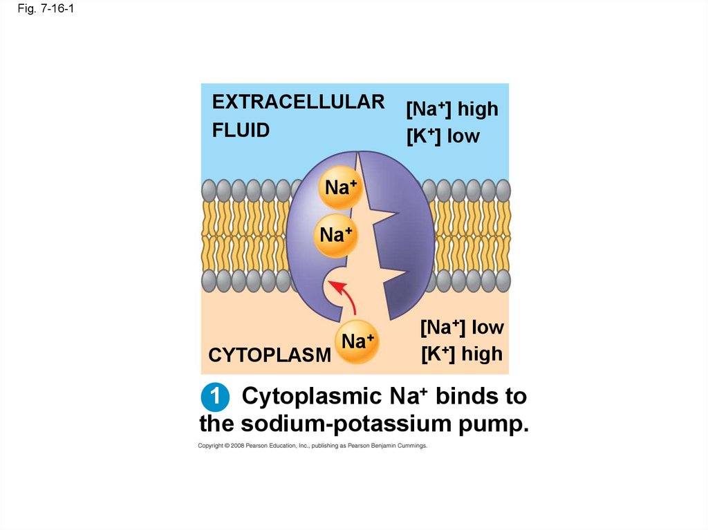
54.
Fig. 7-16-1EXTRACELLULAR
FLUID
[Na+] high
[K+] low
Na+
Na+
CYTOPLASM
Na+
[Na+] low
[K+] high
1 Cytoplasmic Na+ binds to
the sodium-potassium pump.
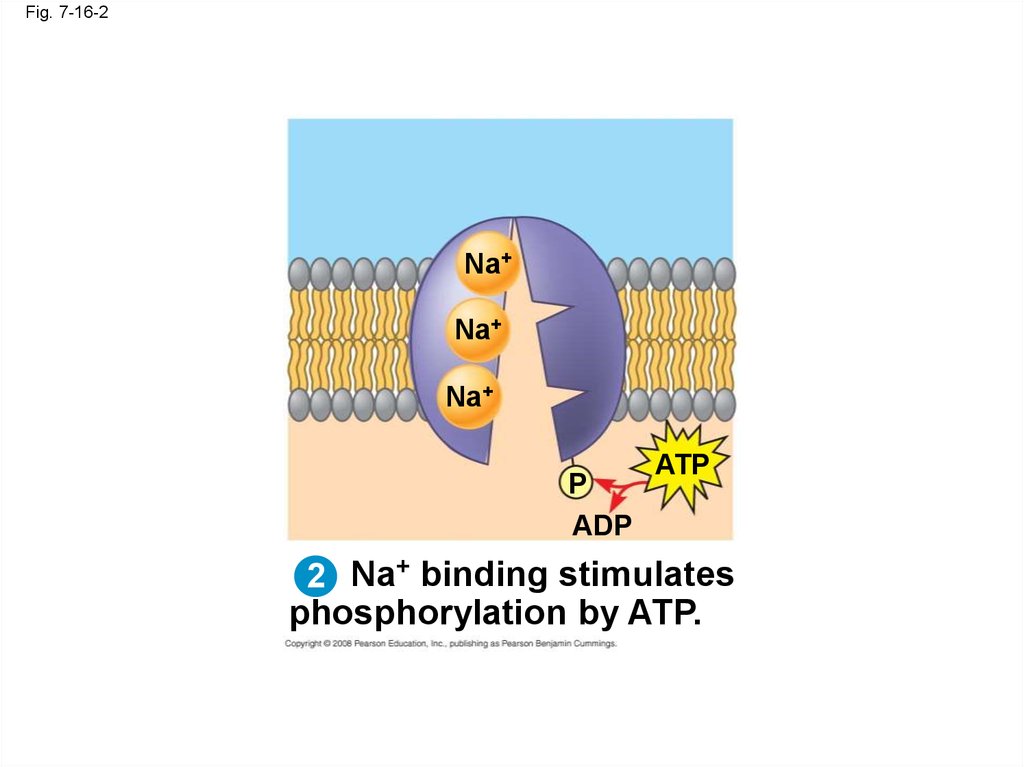
55.
Fig. 7-16-2Na+
Na+
Na+
P
ADP
ATP
2 Na+ binding stimulates
phosphorylation by ATP.
56.
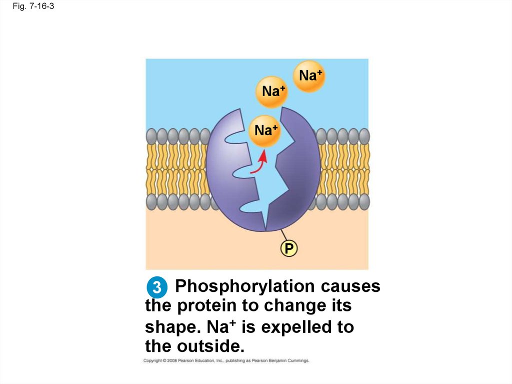
Fig. 7-16-3Na+
Na+
Na+
P
3 Phosphorylation causes
the protein to change its
shape. Na+ is expelled to
the outside.
57.
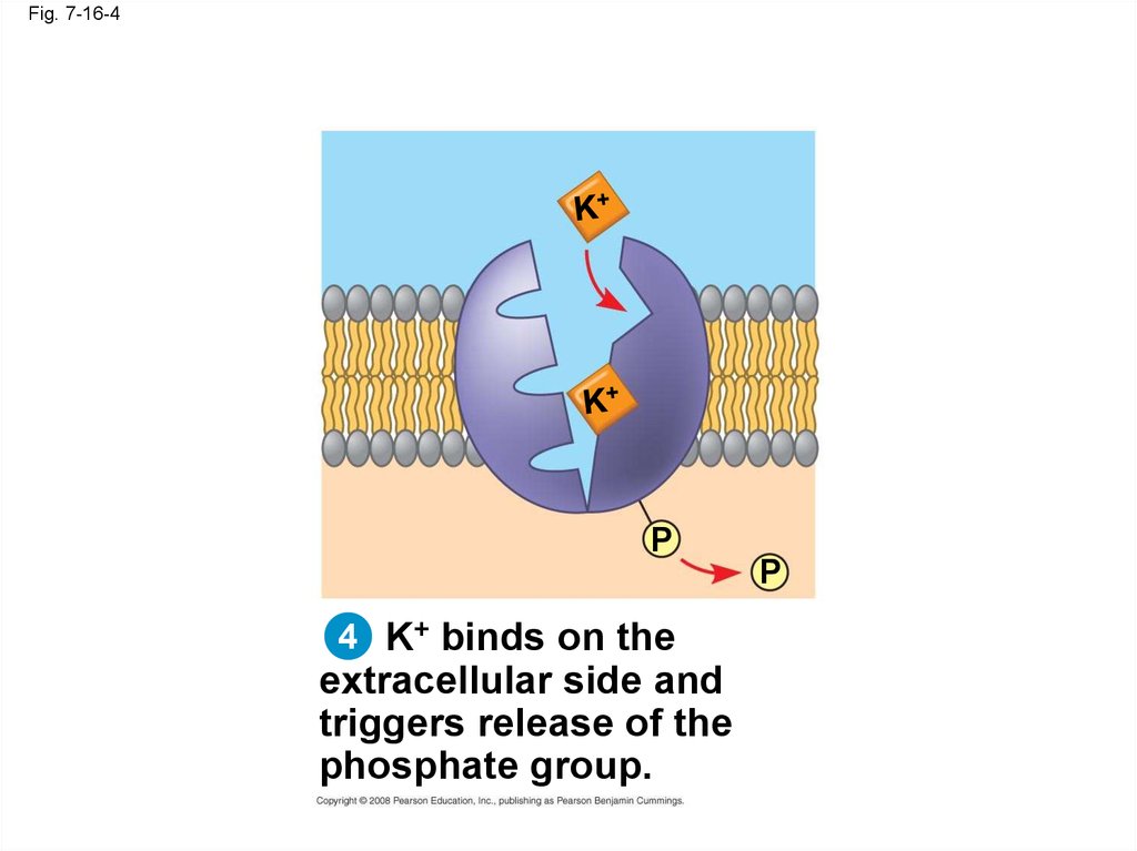
Fig. 7-16-4P
P
4 K+ binds on the
extracellular side and
triggers release of the
phosphate group.
58.
Fig. 7-16-55 Loss of the phosphate
restores the protein’s original
shape.
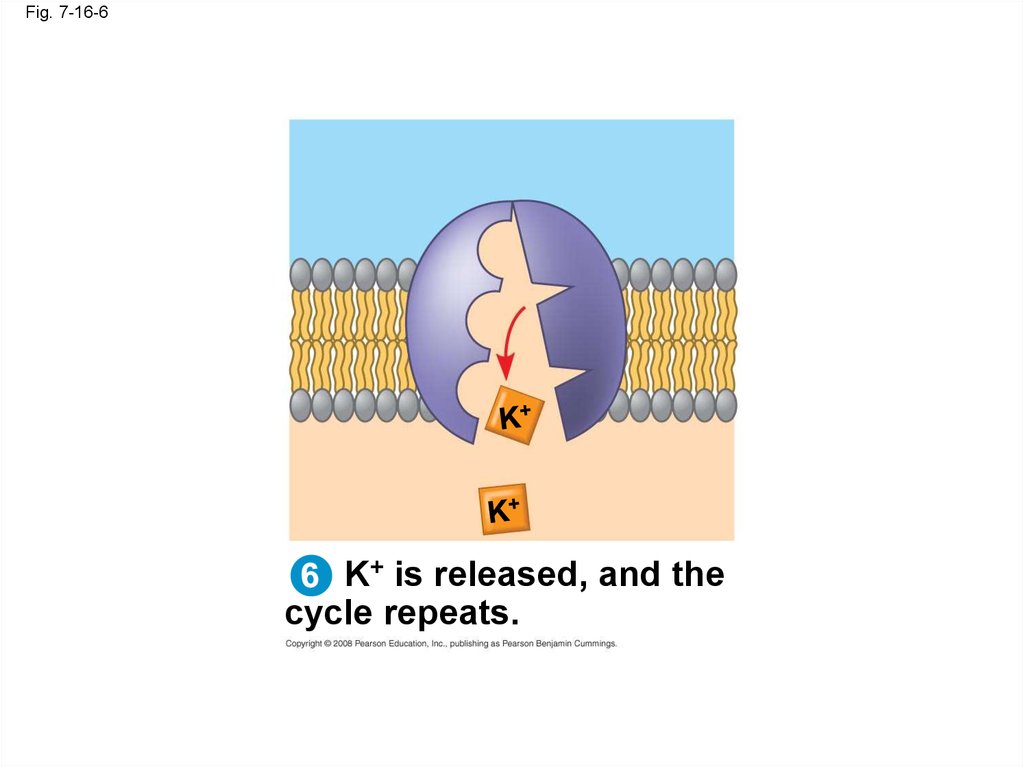
59.
Fig. 7-16-6K+ is released, and the
cycle repeats.
60.
Fig. 7-16-7EXTRACELLULAR
FLUID
Na+
[Na+] high
[K+] low
Na+
Na+
Na+
Na+
Na+
Na+
Na+
CYTOPLASM
1
Na+
[Na+] low
[K+] high
P
ADP
2
ATP
P
3
P
P
6
5
4
61.
Fig. 7-17Passive transport
Active transport
ATP
Diffusion
Facilitated diffusion
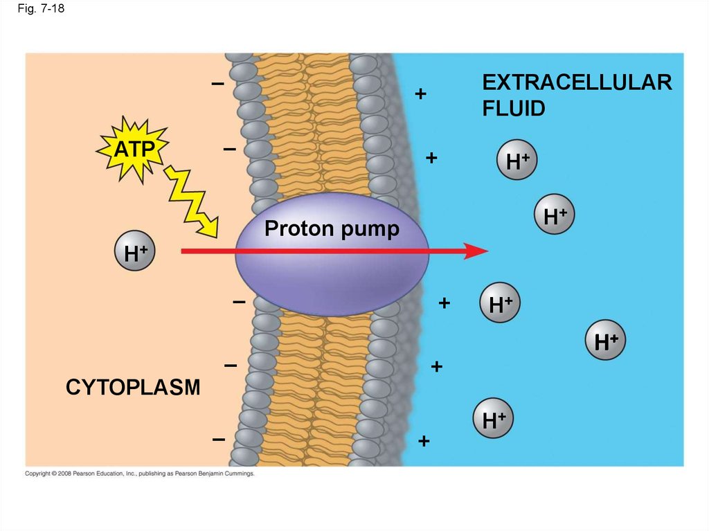
62. How Ion Pumps Maintain Membrane Potential
• Membrane potential is the voltage differenceacross a membrane
• Voltage is created by differences in the
distribution of positive and negative ions
Copyright © 2008 Pearson Education, Inc., publishing as Pearson Benjamin Cummings
63.
• Two combined forces, collectively called theelectrochemical gradient, drive the diffusion
of ions across a membrane:
– A chemical force (the ion’s concentration
gradient)
– An electrical force (the effect of the membrane
potential on the ion’s movement)
Copyright © 2008 Pearson Education, Inc., publishing as Pearson Benjamin Cummings
64.
• An electrogenic pump is a transport proteinthat generates voltage across a membrane
• The sodium-potassium pump is the major
electrogenic pump of animal cells
• The main electrogenic pump of plants, fungi,
and bacteria is a proton pump
Copyright © 2008 Pearson Education, Inc., publishing as Pearson Benjamin Cummings
65.
Fig. 7-18–
ATP
EXTRACELLULAR
FLUID
+
–
+
H+
H+
Proton pump
H+
–
+
H+
H+
–
+
CYTOPLASM
–
H+
+
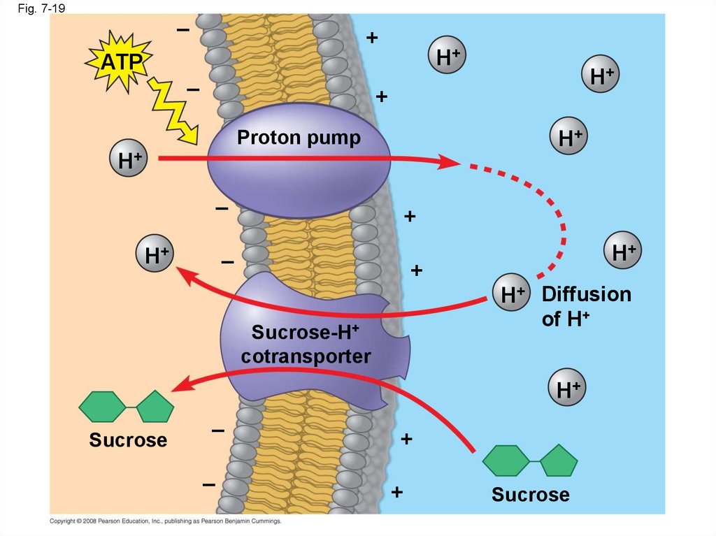
66. Cotransport: Coupled Transport by a Membrane Protein
• Cotransport occurs when active transport of asolute indirectly drives transport of another
solute
• Plants commonly use the gradient of hydrogen
ions generated by proton pumps to drive active
transport of nutrients into the cell
Copyright © 2008 Pearson Education, Inc., publishing as Pearson Benjamin Cummings
67.
Fig. 7-19–
+
H+
ATP
–
H+
+
H+
Proton pump
H+
–
H+
+
–
H+
+
H+ Diffusion
of H+
Sucrose-H+
cotransporter
H+
Sucrose
–
–
+
+
Sucrose
68. Concept 7.5: Bulk transport across the plasma membrane occurs by exocytosis and endocytosis
• Small molecules and water enter or leave thecell through the lipid bilayer or by transport
proteins
• Large molecules, such as polysaccharides and
proteins, cross the membrane in bulk via
vesicles
• Bulk transport requires energy
Copyright © 2008 Pearson Education, Inc., publishing as Pearson Benjamin Cummings
69. Exocytosis
• In exocytosis, transport vesicles migrate to themembrane, fuse with it, and release their
contents
• Many secretory cells use exocytosis to export
their products
Animation: Exocytosis
Copyright © 2008 Pearson Education, Inc., publishing as Pearson Benjamin Cummings
70. Endocytosis
• In endocytosis, the cell takes in macromoleculesby forming vesicles from the plasma membrane
• Endocytosis is a reversal of exocytosis, involving
different proteins
• There are three types of endocytosis:
– Phagocytosis (“cellular eating”)
– Pinocytosis (“cellular drinking”)
– Receptor-mediated endocytosis
Animation: Exocytosis and Endocytosis Introduction
Copyright © 2008 Pearson Education, Inc., publishing as Pearson Benjamin Cummings
71.
• In phagocytosis a cell engulfs a particle in avacuole
• The vacuole fuses with a lysosome to digest
the particle
Animation: Phagocytosis
Copyright © 2008 Pearson Education, Inc., publishing as Pearson Benjamin Cummings
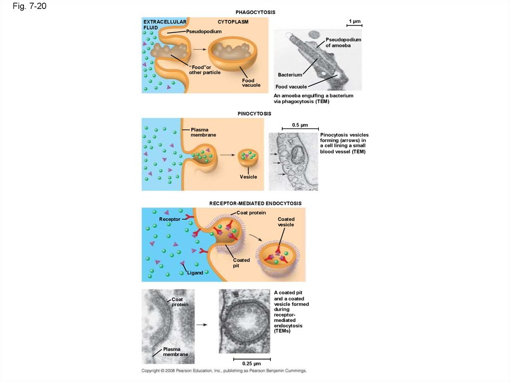
72.
Fig. 7-20PHAGOCYTOSIS
1 µm
CYTOPLASM
EXTRACELLULAR
FLUID
Pseudopodium
Pseudopodium
of amoeba
“Food”or
other particle
Bacterium
Food
vacuole
Food vacuole
An amoeba engulfing a bacterium
via phagocytosis (TEM)
PINOCYTOSIS
0.5 µm
Plasma
membrane
Pinocytosis vesicles
forming (arrows) in
a cell lining a small
blood vessel (TEM)
Vesicle
RECEPTOR-MEDIATED ENDOCYTOSIS
Coat protein
Receptor
Coated
vesicle
Coated
pit
Ligand
A coated pit
and a coated
vesicle formed
during
receptormediated
endocytosis
(TEMs)
Coat
protein
Plasma
membrane
0.25 µm
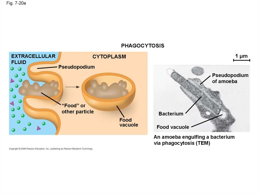
73.
Fig. 7-20aPHAGOCYTOSIS
EXTRACELLULAR
FLUID
1 µm
CYTOPLASM
Pseudopodium
Pseudopodium
of amoeba
“Food” or
other particle
Bacterium
Food
vacuole
Food vacuole
An amoeba engulfing a bacterium
via phagocytosis (TEM)
74.
• In pinocytosis, molecules are taken up whenextracellular fluid is “gulped” into tiny vesicles
Animation: Pinocytosis
Copyright © 2008 Pearson Education, Inc., publishing as Pearson Benjamin Cummings
75.
Fig. 7-20bPINOCYTOSIS
0.5 µm
Plasma
membrane
Pinocytosis vesicles
forming (arrows) in
a cell lining a small
blood vessel (TEM)
Vesicle
76.
• In receptor-mediated endocytosis, binding ofligands to receptors triggers vesicle formation
• A ligand is any molecule that binds specifically
to a receptor site of another molecule
Animation: Receptor-Mediated Endocytosis
Copyright © 2008 Pearson Education, Inc., publishing as Pearson Benjamin Cummings
77.
Fig. 7-20cRECEPTOR-MEDIATED ENDOCYTOSIS
Coat protein
Receptor
Coated
vesicle
Coated
pit
Ligand
A coated pit
and a coated
vesicle formed
during
receptormediated
endocytosis
(TEMs)
Coat
protein
Plasma
membrane
0.25 µm
78.
Fig. 7-UN1Channel
protein
Passive transport:
Facilitated diffusion
Carrier
protein
79.
Fig. 7-UN2Active transport:
ATP
80.
Fig. 7-UN3“Cell”
0.03 M sucrose
0.02 M glucose
Environment:
0.01 M sucrose
0.01 M glucose
0.01 M fructose
81.
Fig. 7-UN482. You should now be able to:
1. Define the following terms: amphipathicmolecules, aquaporins, diffusion
2. Explain how membrane fluidity is influenced
by temperature and membrane composition
3. Distinguish between the following pairs or
sets of terms: peripheral and integral
membrane proteins; channel and carrier
proteins; osmosis, facilitated diffusion, and
active transport; hypertonic, hypotonic, and
isotonic solutions
Copyright © 2008 Pearson Education, Inc., publishing as Pearson Benjamin Cummings
83.
4. Explain how transport proteins facilitatediffusion
5. Explain how an electrogenic pump creates
voltage across a membrane, and name two
electrogenic pumps
6. Explain how large molecules are transported
across a cell membrane
Copyright © 2008 Pearson Education, Inc., publishing as Pearson Benjamin Cummings