


 biology
Similar presentations:


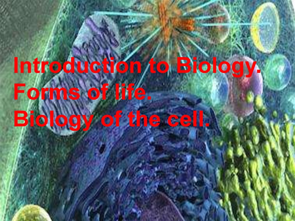
Introduction to Biology. Forms of life. Biology of the cell
1.
Introduction to Biology.Forms of life.
Biology of the cell.
2.
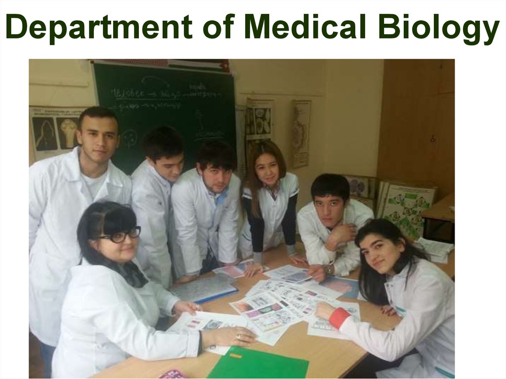
Department of Medical Biology3.
Biology teacher Svetlana4.
5.
6.
Biology teacher Tatyana7.
Biology teacher Anna8.
9.
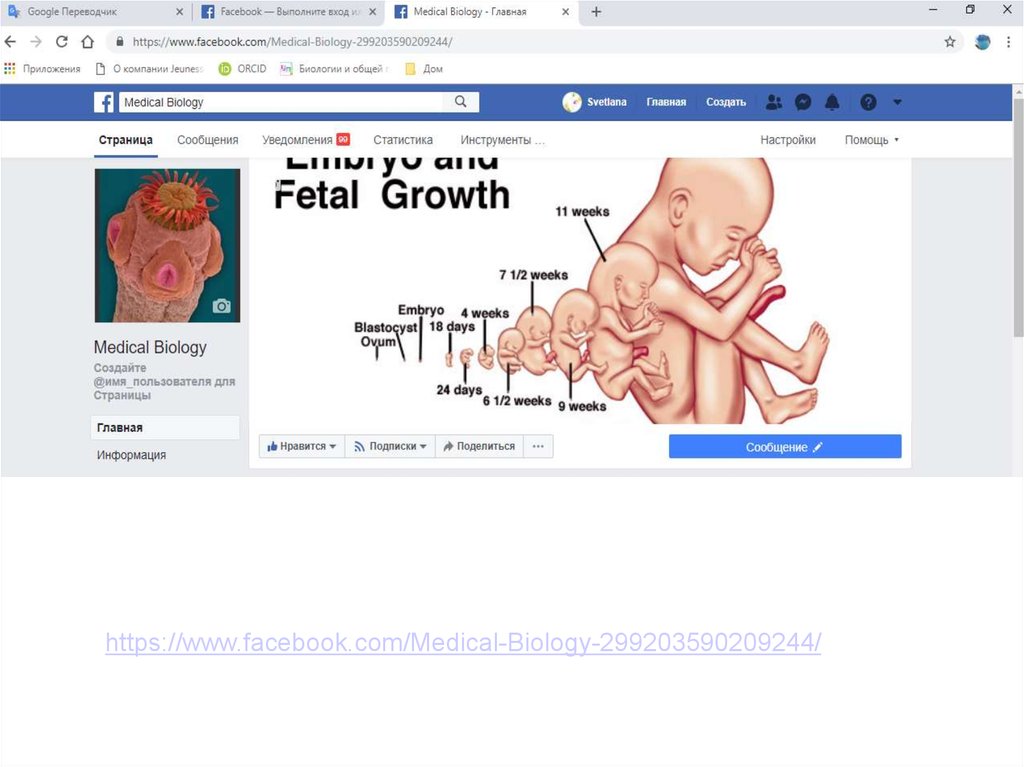
https://www.facebook.com/Medical-Biology-299203590209244/10.
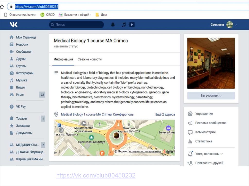
https://vk.com/club8045023211.
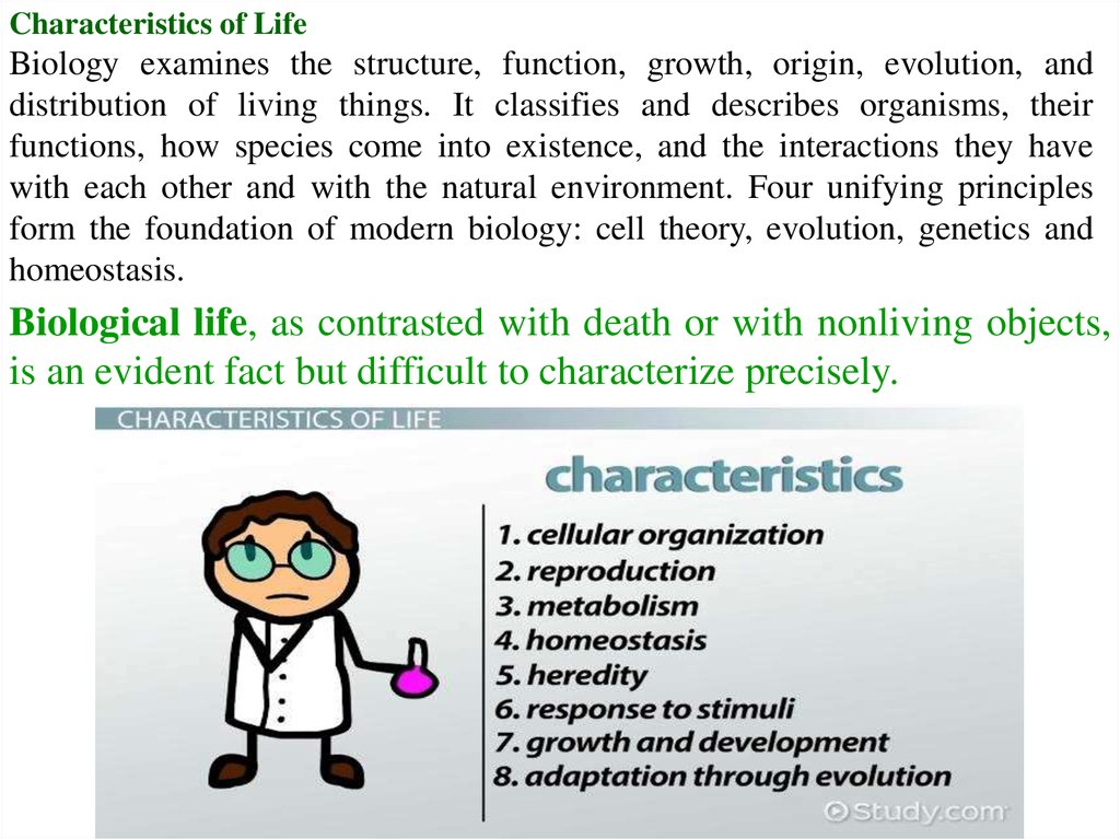
Characteristics of LifeBiology examines the structure, function, growth, origin, evolution, and
distribution of living things. It classifies and describes organisms, their
functions, how species come into existence, and the interactions they have
with each other and with the natural environment. Four unifying principles
form the foundation of modern biology: cell theory, evolution, genetics and
homeostasis.
Biological life, as contrasted with death or with nonliving objects,
is an evident fact but difficult to characterize precisely.
12.
13.
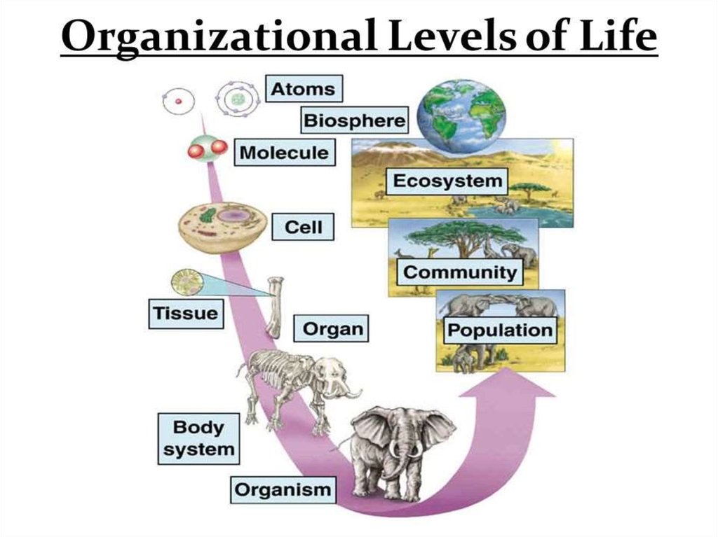
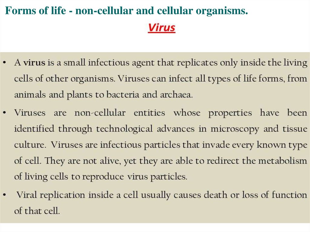
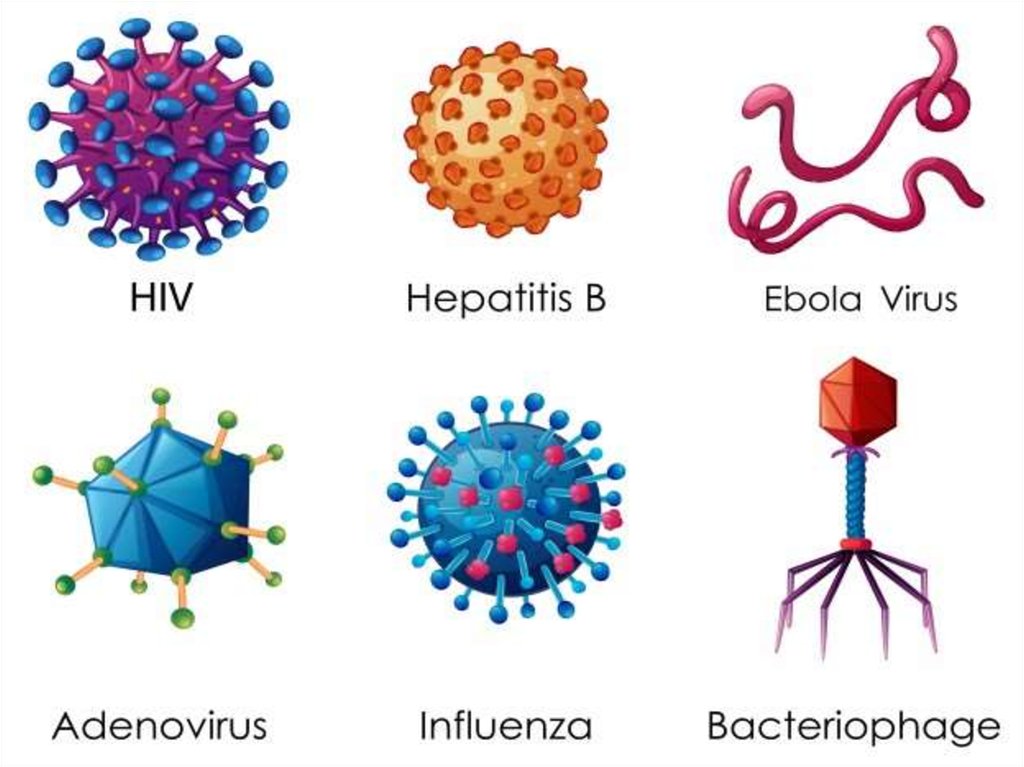
Forms of life - non-cellular and cellular organisms.14.
15.
16.
17.
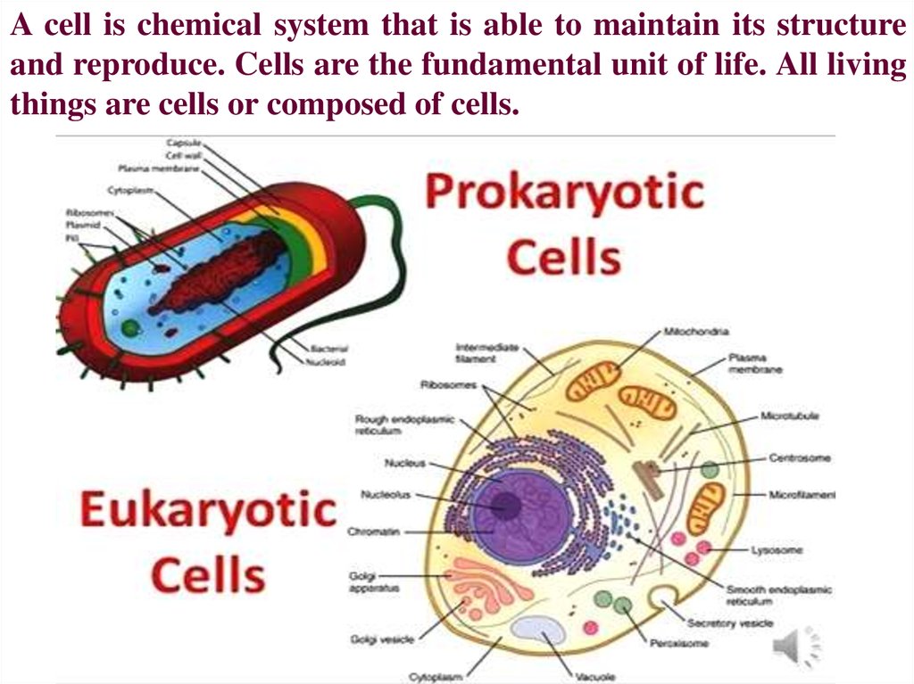
A cell is chemical system that is able to maintain its structureand reproduce. Cells are the fundamental unit of life. All living
things are cells or composed of cells.
18.
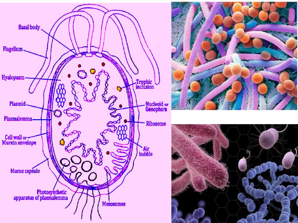
The first life on Earth came in the form of a prokaryotic cell. For two billion yearsprokaryotic cells were the only living things on Earth and spread to almost every corner
of the planet. Today they are still the most abundant and diverse organisms on Earth and
more prokaryotes are found in one handful of soil than all the humans that have ever
existed.
A prokaryotic cell is one of the two types of cells that make up all the trillions of
organisms that live on Earth, the other type being eukaryotic cells. Although prokaryotic
cells appear far less advanced than eukaryotic cells, prokaryotic organisms outperform
eukaryotes in many ways.
19.
20.
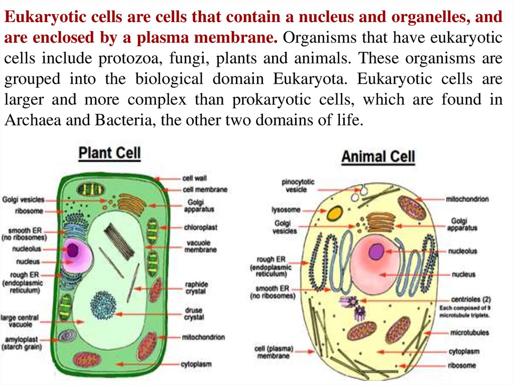
Eukaryotic cells are cells that contain a nucleus and organelles, andare enclosed by a plasma membrane. Organisms that have eukaryotic
cells include protozoa, fungi, plants and animals. These organisms are
grouped into the biological domain Eukaryota. Eukaryotic cells are
larger and more complex than prokaryotic cells, which are found in
Archaea and Bacteria, the other two domains of life.
21.
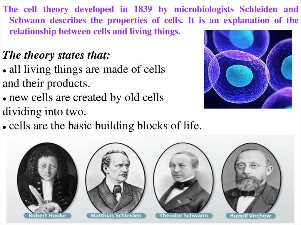
The cell theory developed in 1839 by microbiologists Schleiden andSchwann describes the properties of cells. It is an explanation of the
relationship between cells and living things.
The theory states that:
all living things are made of cells
and their products.
new cells are created by old cells
dividing into two.
cells are the basic building blocks of life.
22.
23.
24.
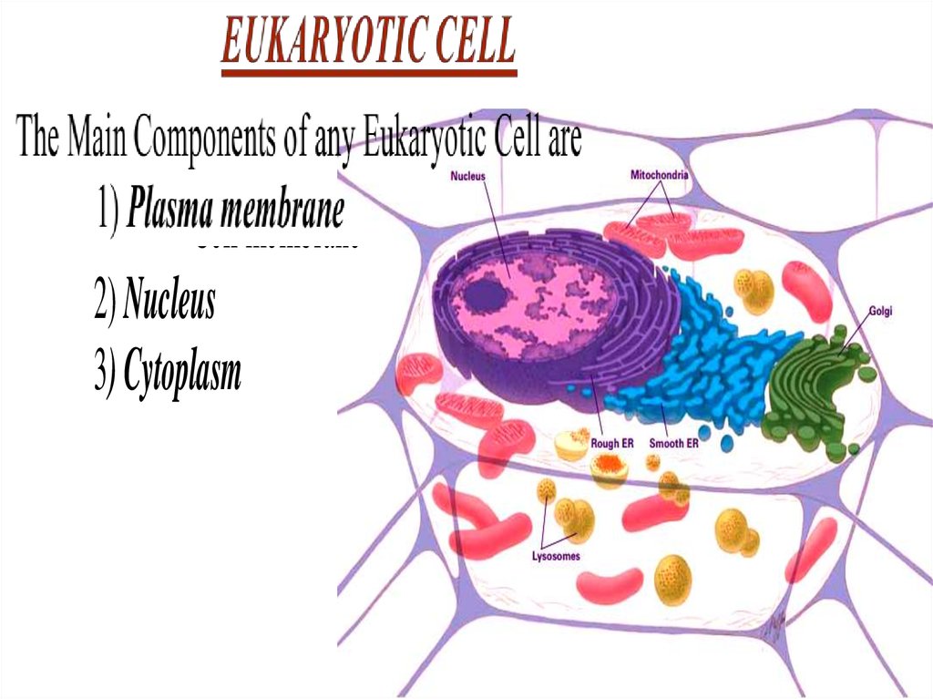
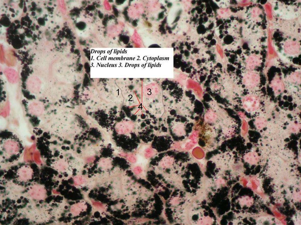
1) Plasma membrane- Cytoplasmic membrane
- Plasmalemma
- Cell membrane
2) Nucleus
3) Cytoplasm
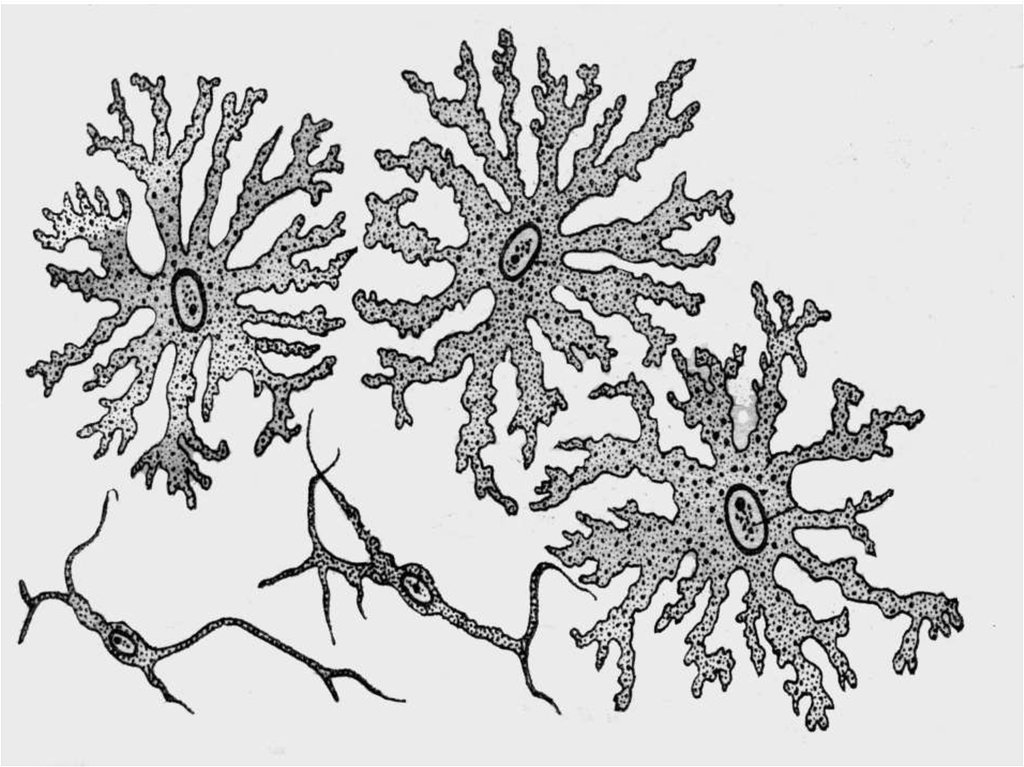
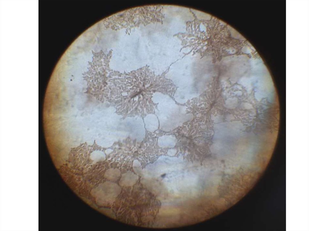
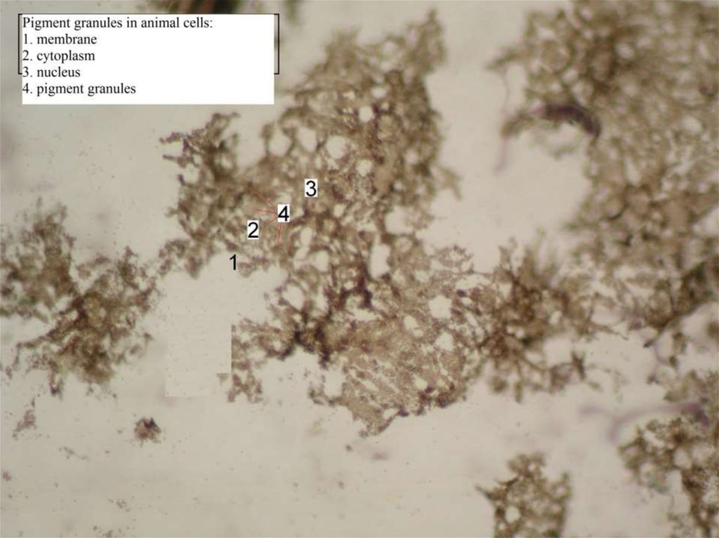
25. Representative Animal Cell
26.
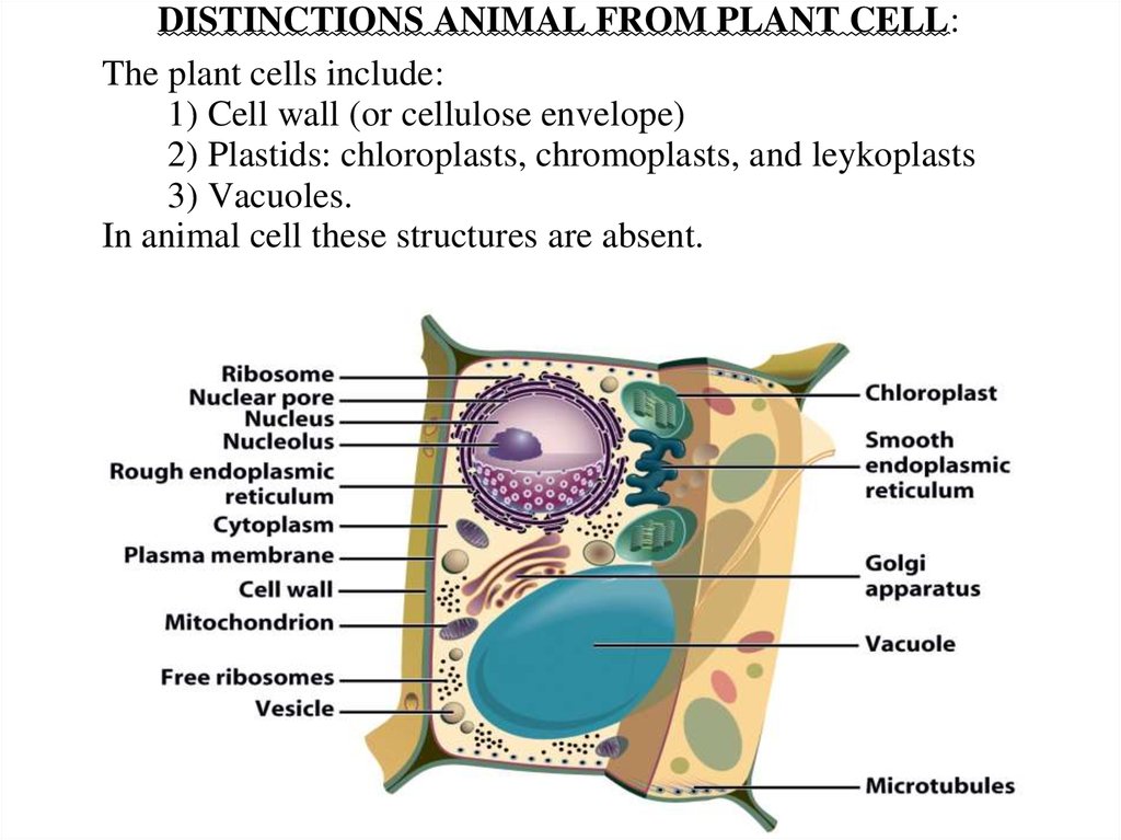
DISTINCTIONS ANIMAL FROM PLANT CELL:The plant cells include:
1) Cell wall (or cellulose envelope)
2) Plastids: chloroplasts, chromoplasts, and leykoplasts
3) Vacuoles.
In animal cell these structures are absent.
27.
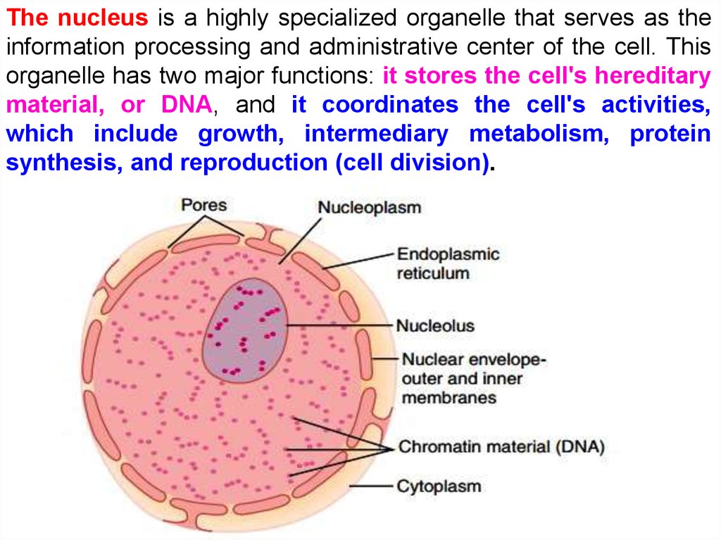
The nucleus is a highly specialized organelle that serves as theinformation processing and administrative center of the cell. This
organelle has two major functions: it stores the cell's hereditary
material, or DNA, and it coordinates the cell's activities,
which include growth, intermediary metabolism, protein
synthesis, and reproduction (cell division).
28.
29.
30.
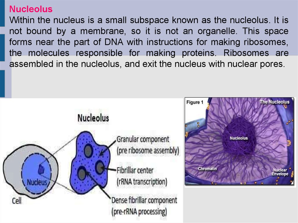
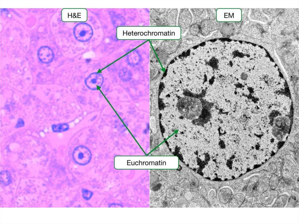
NucleolusWithin the nucleus is a small subspace known as the nucleolus. It is
not bound by a membrane, so it is not an organelle. This space
forms near the part of DNA with instructions for making ribosomes,
the molecules responsible for making proteins. Ribosomes are
assembled in the nucleolus, and exit the nucleus with nuclear pores.
31.
32.
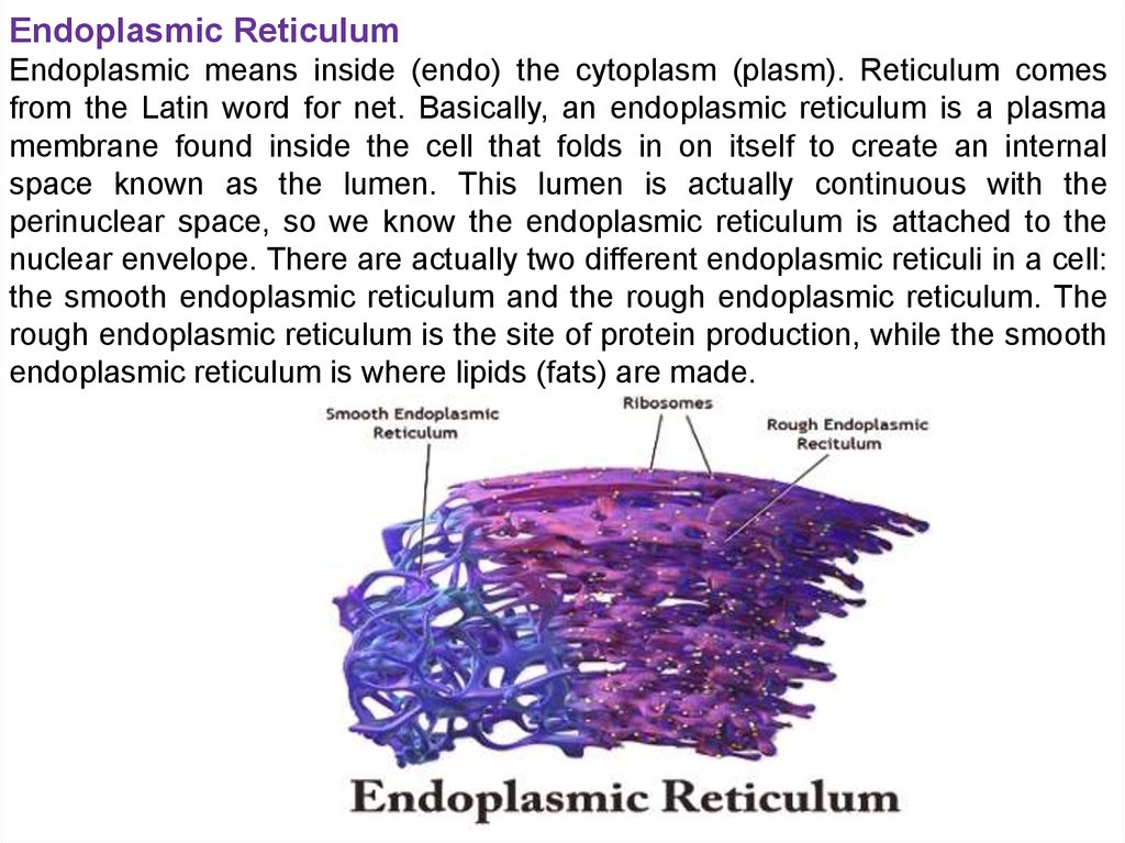
Endoplasmic ReticulumEndoplasmic means inside (endo) the cytoplasm (plasm). Reticulum comes
from the Latin word for net. Basically, an endoplasmic reticulum is a plasma
membrane found inside the cell that folds in on itself to create an internal
space known as the lumen. This lumen is actually continuous with the
perinuclear space, so we know the endoplasmic reticulum is attached to the
nuclear envelope. There are actually two different endoplasmic reticuli in a cell:
the smooth endoplasmic reticulum and the rough endoplasmic reticulum. The
rough endoplasmic reticulum is the site of protein production, while the smooth
endoplasmic reticulum is where lipids (fats) are made.
33.
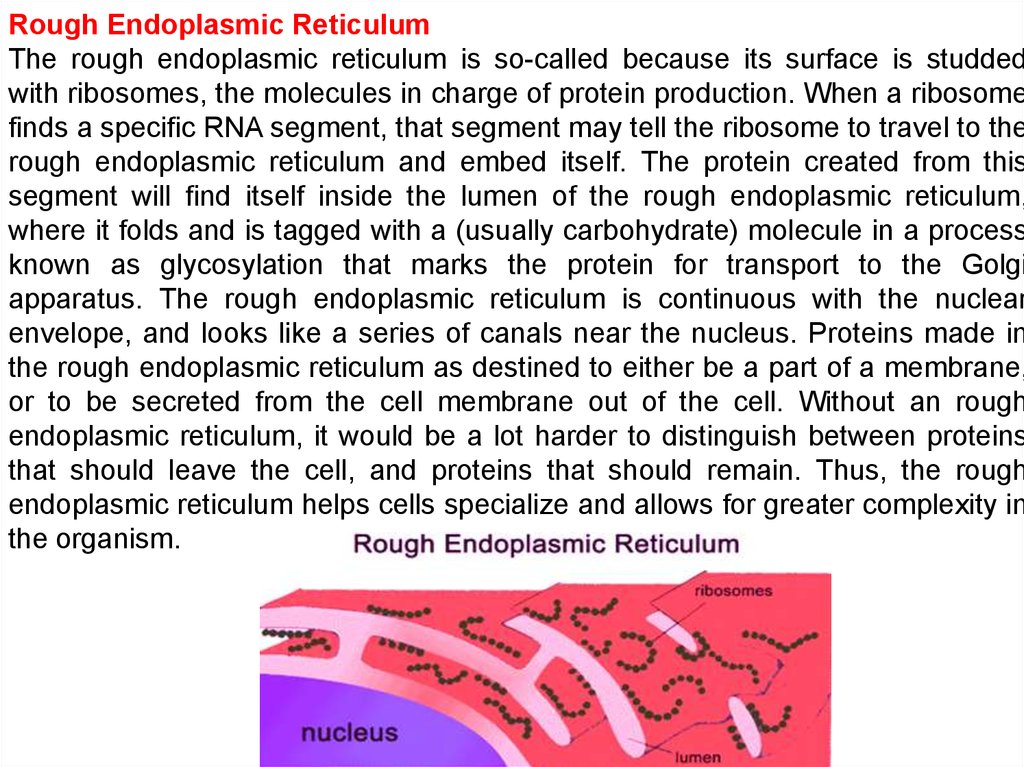
Rough Endoplasmic ReticulumThe rough endoplasmic reticulum is so-called because its surface is studded
with ribosomes, the molecules in charge of protein production. When a ribosome
finds a specific RNA segment, that segment may tell the ribosome to travel to the
rough endoplasmic reticulum and embed itself. The protein created from this
segment will find itself inside the lumen of the rough endoplasmic reticulum,
where it folds and is tagged with a (usually carbohydrate) molecule in a process
known as glycosylation that marks the protein for transport to the Golgi
apparatus. The rough endoplasmic reticulum is continuous with the nuclear
envelope, and looks like a series of canals near the nucleus. Proteins made in
the rough endoplasmic reticulum as destined to either be a part of a membrane,
or to be secreted from the cell membrane out of the cell. Without an rough
endoplasmic reticulum, it would be a lot harder to distinguish between proteins
that should leave the cell, and proteins that should remain. Thus, the rough
endoplasmic reticulum helps cells specialize and allows for greater complexity in
the organism.
34.
Smooth Endoplasmic ReticulumThe smooth endoplasmic reticulum makes lipids and steroids, instead of being
involved in protein synthesis. These are fat-based molecules that are important
in energy storage, membrane structure, and communication (steroids can act
as hormones). The smooth endoplasmic reticulum is also responsible for
detoxifying the cell. It is more tubular than the rough endoplasmic reticulum,
and is not necessarily continuous with the nuclear envelope. Every cell has a
smooth endoplasmic reticulum, but the amount will vary with cell function. For
example, the liver, which is responsible for most of the body’s detoxification,
has a larger amount of smooth endoplasmic reticulum.
35.
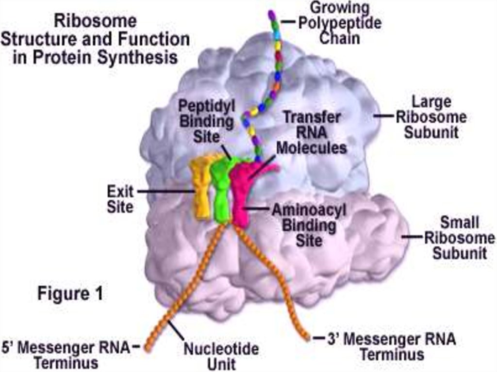
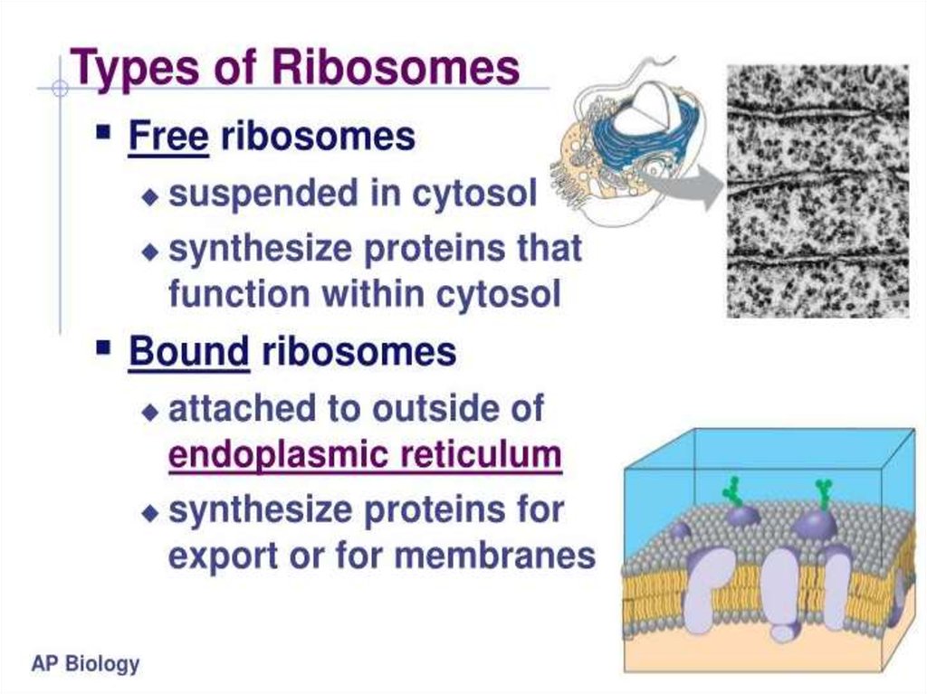
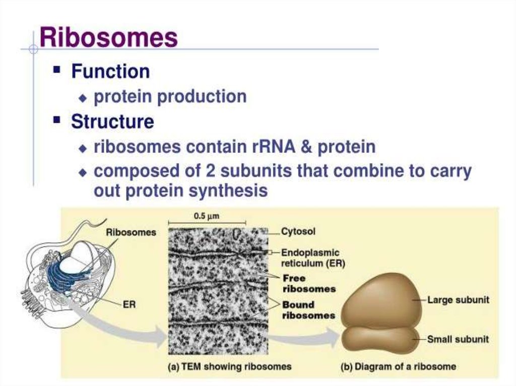
Ribosome:Situated in two areas of the cytoplasm.
They are seen scattered in the cytoplasm and a few are connected to the
endoplasmic reticulum.
Whenever joined to the ER they are called the rough endoplasmic reticulum.
The free and the bound ribosomes are very much alike in structure and are
associated with protein synthesis.
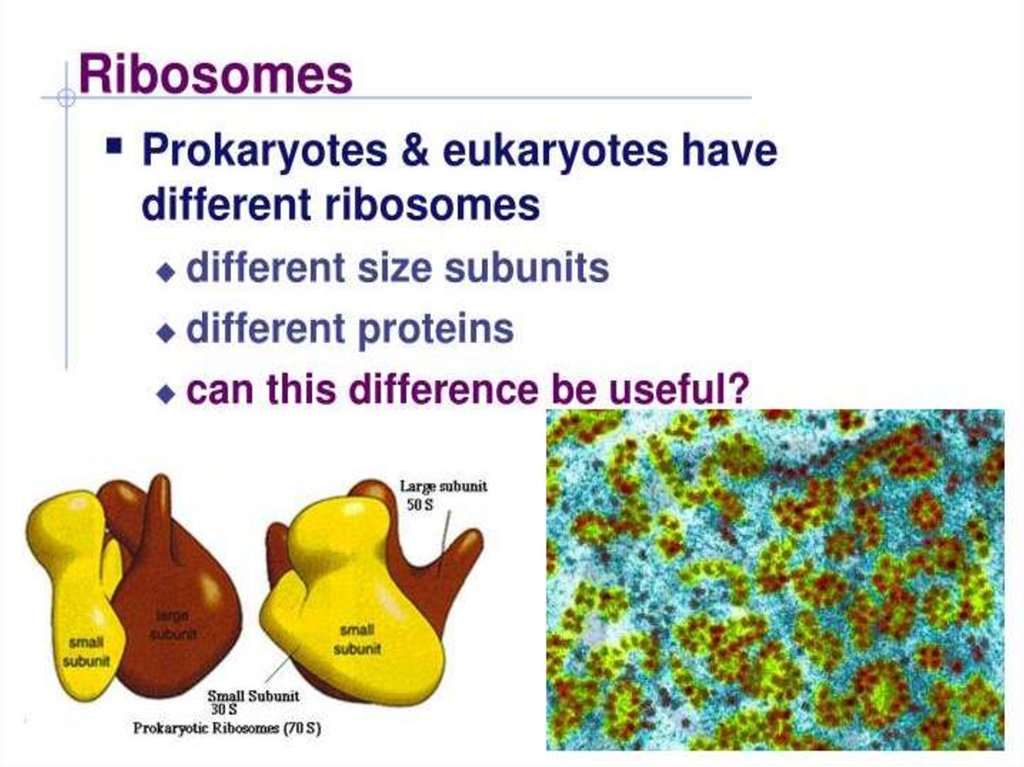
Around 37 to 62% of ribosome is comprised of rRNA and the rest is proteins.
Prokaryotes have 70S ribosomes respectively subunits comprising the little
subunit of 30S and the bigger subunit of 50S. Eukaryotes have 80S
ribosomes respectively comprising of little (40S) and substantial (60S)
subunits.
36.
37.
38.
39.
40.
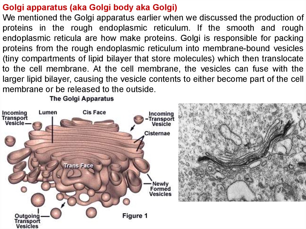
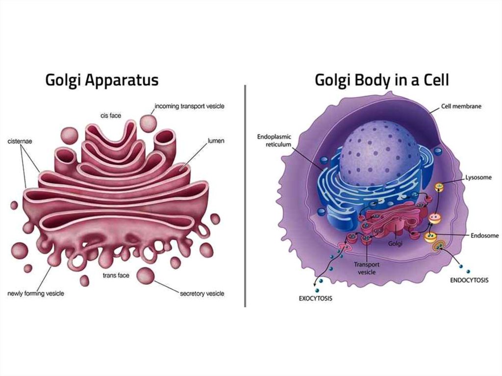
Golgi apparatus (aka Golgi body aka Golgi)We mentioned the Golgi apparatus earlier when we discussed the production of
proteins in the rough endoplasmic reticulum. If the smooth and rough
endoplasmic reticula are how make proteins. Golgi is responsible for packing
proteins from the rough endoplasmic reticulum into membrane-bound vesicles
(tiny compartments of lipid bilayer that store molecules) which then translocate
to the cell membrane. At the cell membrane, the vesicles can fuse with the
larger lipid bilayer, causing the vesicle contents to either become part of the cell
membrane or be released to the outside.
41.
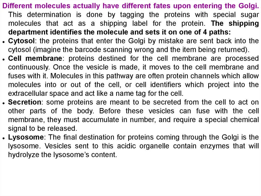
Different molecules actually have different fates upon entering the Golgi.This determination is done by tagging the proteins with special sugar
molecules that act as a shipping label for the protein. The shipping
department identifies the molecule and sets it on one of 4 paths:
Cytosol: the proteins that enter the Golgi by mistake are sent back into the
cytosol (imagine the barcode scanning wrong and the item being returned).
Cell membrane: proteins destined for the cell membrane are processed
continuously. Once the vesicle is made, it moves to the cell membrane and
fuses with it. Molecules in this pathway are often protein channels which allow
molecules into or out of the cell, or cell identifiers which project into the
extracellular space and act like a name tag for the cell.
Secretion: some proteins are meant to be secreted from the cell to act on
other parts of the body. Before these vesicles can fuse with the cell
membrane, they must accumulate in number, and require a special chemical
signal to be released.
Lysosome: The final destination for proteins coming through the Golgi is the
lysosome. Vesicles sent to this acidic organelle contain enzymes that will
hydrolyze the lysosome’s content.
42.
43.
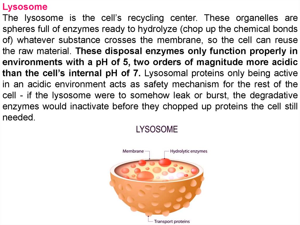
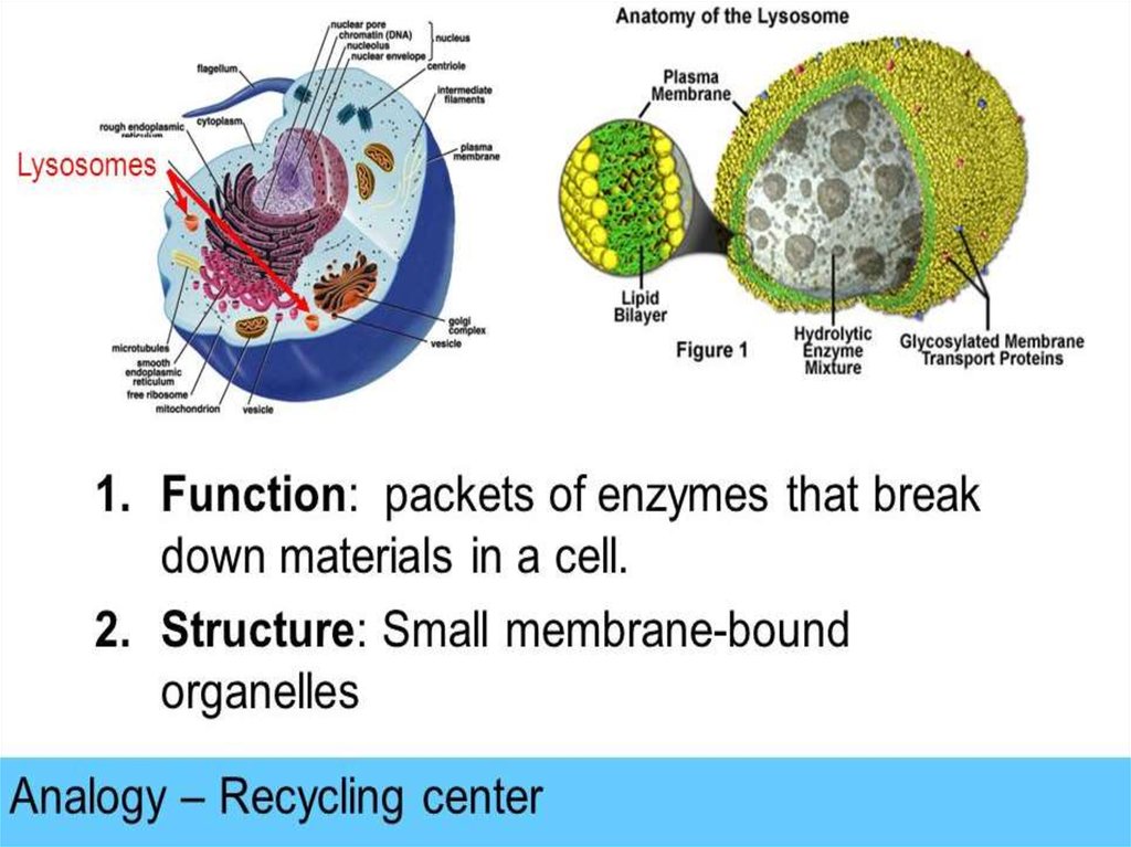
LysosomeThe lysosome is the cell’s recycling center. These organelles are
spheres full of enzymes ready to hydrolyze (chop up the chemical bonds
of) whatever substance crosses the membrane, so the cell can reuse
the raw material. These disposal enzymes only function properly in
environments with a pH of 5, two orders of magnitude more acidic
than the cell’s internal pH of 7. Lysosomal proteins only being active
in an acidic environment acts as safety mechanism for the rest of the
cell - if the lysosome were to somehow leak or burst, the degradative
enzymes would inactivate before they chopped up proteins the cell still
needed.
44.
45.
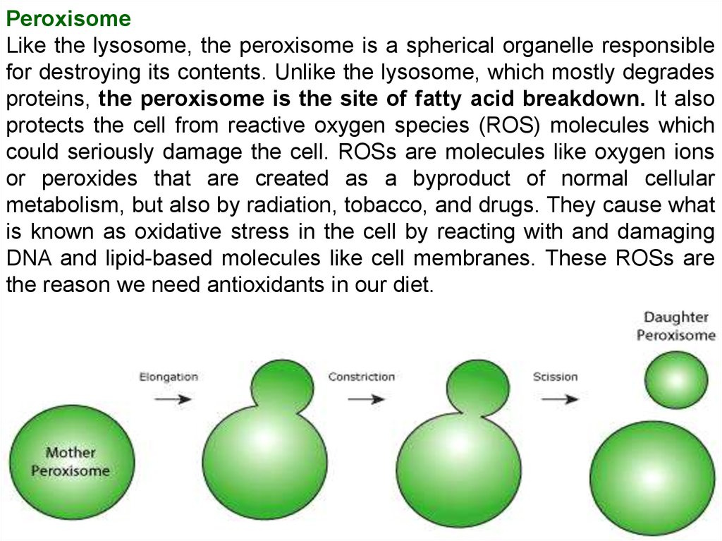
PeroxisomeLike the lysosome, the peroxisome is a spherical organelle responsible
for destroying its contents. Unlike the lysosome, which mostly degrades
proteins, the peroxisome is the site of fatty acid breakdown. It also
protects the cell from reactive oxygen species (ROS) molecules which
could seriously damage the cell. ROSs are molecules like oxygen ions
or peroxides that are created as a byproduct of normal cellular
metabolism, but also by radiation, tobacco, and drugs. They cause what
is known as oxidative stress in the cell by reacting with and damaging
DNA and lipid-based molecules like cell membranes. These ROSs are
the reason we need antioxidants in our diet.
46.
Key ConceptsMitochondria are semi‐autonomous organelles that are descendants of
endosymbiotic bacteria.
Mitochondria play a pivotal role in cellular energy production through the
mitochondria‐housed pathways of citric acid cycle, fatty acid oxidation,
respiration and oxidative phosphorylation (OXPHOS).
Mitochondria have an important anabolic role in cellular metabolism, as they
are fundamental for the synthesis of several amino acids, nucleobases and
enzymatic cofactors such as haem and Fe‐S clusters.
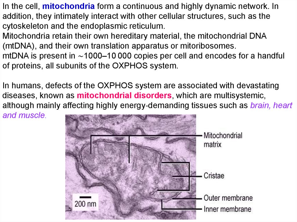
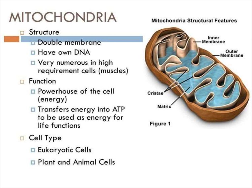
Mitochondria
are membrane‐bound organelles. They have two distinct
membranes: the outer and the inner membrane. The inner membrane is highly
impermeable to ions and forms an extensive series of invaginations called
cristae.
47.
In the cell, mitochondria form a continuous and highly dynamic network. Inaddition, they intimately interact with other cellular structures, such as the
cytoskeleton and the endoplasmic reticulum.
Mitochondria retain their own hereditary material, the mitochondrial DNA
(mtDNA), and their own translation apparatus or mitoribosomes.
mtDNA is present in ∼1000–10 000 copies per cell and encodes for a handful
of proteins, all subunits of the OXPHOS system.
In humans, defects of the OXPHOS system are associated with devastating
diseases, known as mitochondrial disorders, which are multisystemic,
although mainly affecting highly energy‐demanding tissues such as brain, heart
and muscle.
48.
49.
50. Cell Walls
• Found in plants, fungi, & many protists• Surrounds plasma membrane
Cell Wall Differences
Plants – mostly
cellulose
Fungi – contain chitin
51. Cytoskeleton
Filaments & fibersMade of 3 fiber types
– Microfilaments
– Microtubules
– Intermediate filaments
3 functions:
– mechanical support
– anchor organelles
– help move substances
52. Cilia & Flagella
Cilia & FlagellaProvide motility
Cilia
– Short
– Used to move substances
outside human cells
Flagella
– Whip-like extensions
– Found on sperm cells
Basal bodies like
centrioles
53. Centrioles
Pairs of microtubular structuresPlay a role in cell division
54.
55.
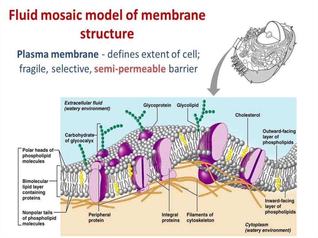
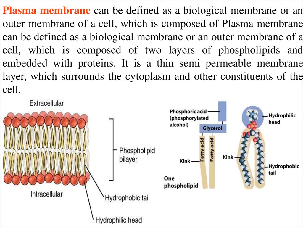
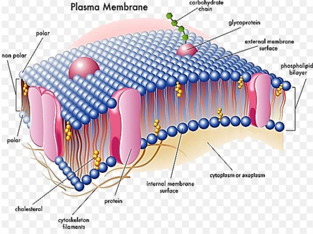
Plasma membrane can be defined as a biological membrane or anouter membrane of a cell, which is composed of Plasma membrane
can be defined as a biological membrane or an outer membrane of a
cell, which is composed of two layers of phospholipids and
embedded with proteins. It is a thin semi permeable membrane
layer, which surrounds the cytoplasm and other constituents of the
cell.
56.
57.
58.
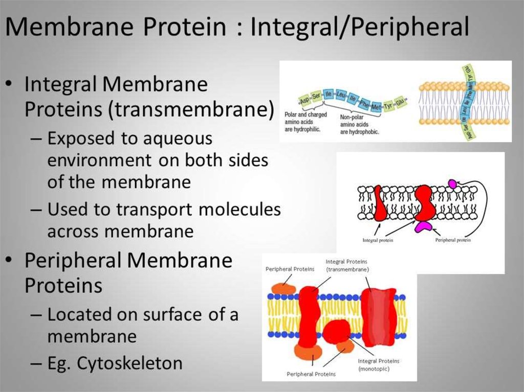
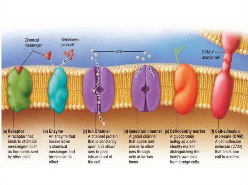
59. Membrane Proteins
1. Channels or transporters– Move molecules in one direction
2. Receptors
– Recognize certain chemicals
60. Membrane Proteins
3. Glycoproteins– Identify cell type
4. Enzymes
– Catalyze production of substances
61.
62.
63.
64.
65.
66.
67.
68.
69.
70.
71.
72.
73.
74.
75.
76.
77.
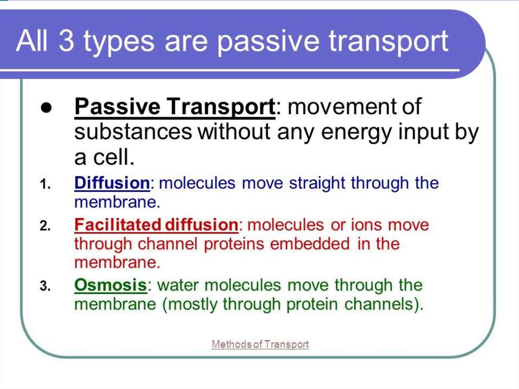
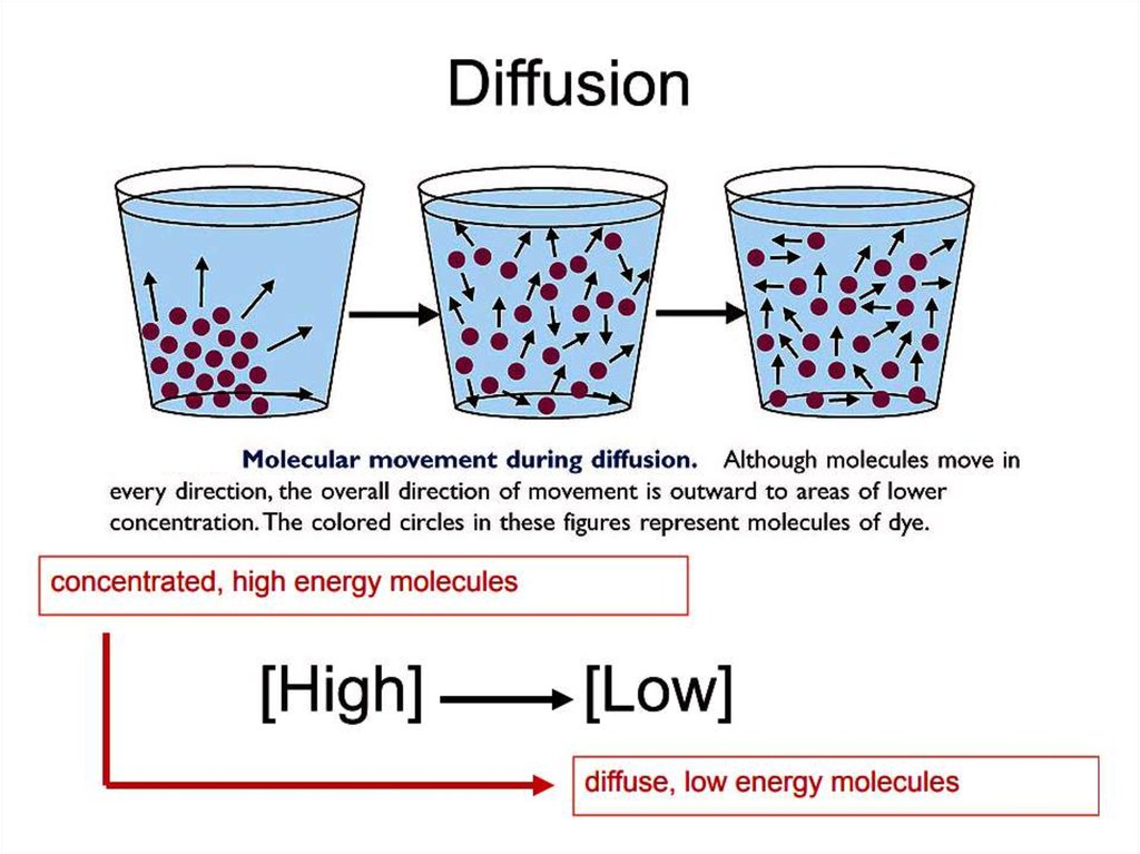
DiffusionDiffusion is a passive process of transport. A single substance tends to move
from an area of high concentration to an area of low concentration until the
concentration is equal across the space. You are familiar with diffusion of
substances through the air. For example, think about someone opening a
bottle of perfume in a room filled with people. The perfume is at its highest
concentration in the bottle and is at its lowest at the edges of the room. The
perfume vapor will diffuse, or spread away, from the bottle, and gradually,
more and more people will smell the perfume as it spreads. Materials move
within the cell’s cytosol by diffusion, and certain materials move through the
plasma membrane by diffusion. Diffusion expends no energy. Rather the
different concentrations of materials in different areas are a form of potential
energy, and diffusion is the dissipation of that potential energy as materials
move down their concentration gradients, from high to low.
78.
79.
Several factors affect the rate of diffusion.Extent of the concentration gradient: The greater the difference in
concentration, the more rapid the diffusion. The closer the
distribution of the material gets to equilibrium, the slower the rate of
diffusion becomes.
Mass of the molecules diffusing: More massive molecules move
more slowly, because it is more difficult for them to move between
the molecules of the substance they are moving through; therefore,
they diffuse more slowly.
Temperature: Higher temperatures increase the energy and
therefore the movement of the molecules, increasing the rate of
diffusion.
Solvent density: As the density of the solvent increases, the rate of
diffusion decreases. The molecules slow down because they have
a more difficult time getting through the denser medium.
80.
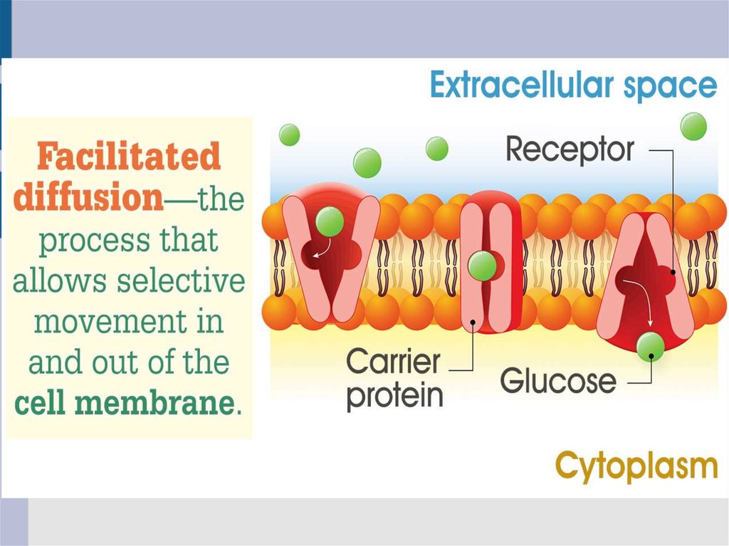
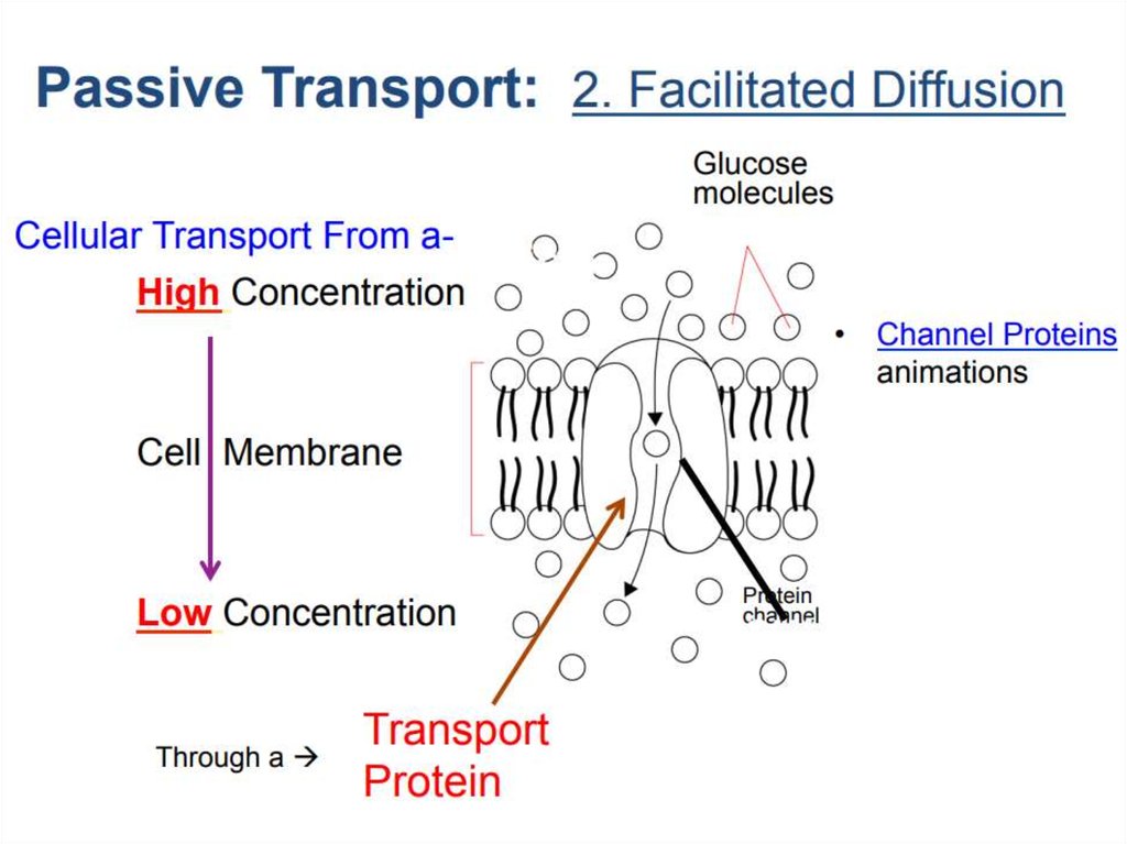
Facilitated transportIn facilitated transport, also called facilitated diffusion, material
moves across the plasma membrane with the assistance of
transmembrane proteins down a concentration gradient (from high to
low concentration) without the expenditure of cellular energy.
However, the substances that undergo facilitated transport would
otherwise not diffuse easily or quickly across the plasma membrane.
The solution to moving polar substances and other substances across
the plasma membrane rests in the proteins that span its surface. The
material being transported is first attached to protein or glycoprotein
receptors on the exterior surface of the plasma membrane. This allows
the material that is needed by the cell to be removed from the
extracellular fluid. The substances are then passed to specific integral
proteins that facilitate their passage, because they form channels or
pores that allow certain substances to pass through the membrane.
The integral proteins involved in facilitated transport are collectively
referred to as transport proteins, and they function as either channels
for the material or carriers.
81.
82.
83.
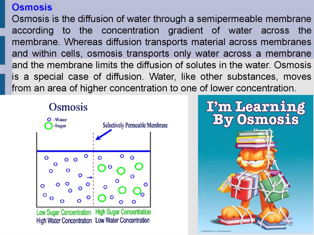
OsmosisOsmosis is the diffusion of water through a semipermeable membrane
according to the concentration gradient of water across the
membrane. Whereas diffusion transports material across membranes
and within cells, osmosis transports only water across a membrane
and the membrane limits the diffusion of solutes in the water. Osmosis
is a special case of diffusion. Water, like other substances, moves
from an area of higher concentration to one of lower concentration.
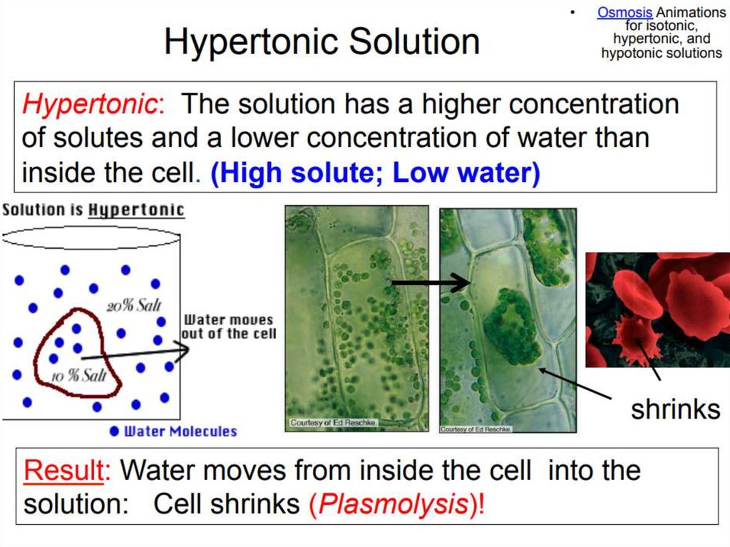
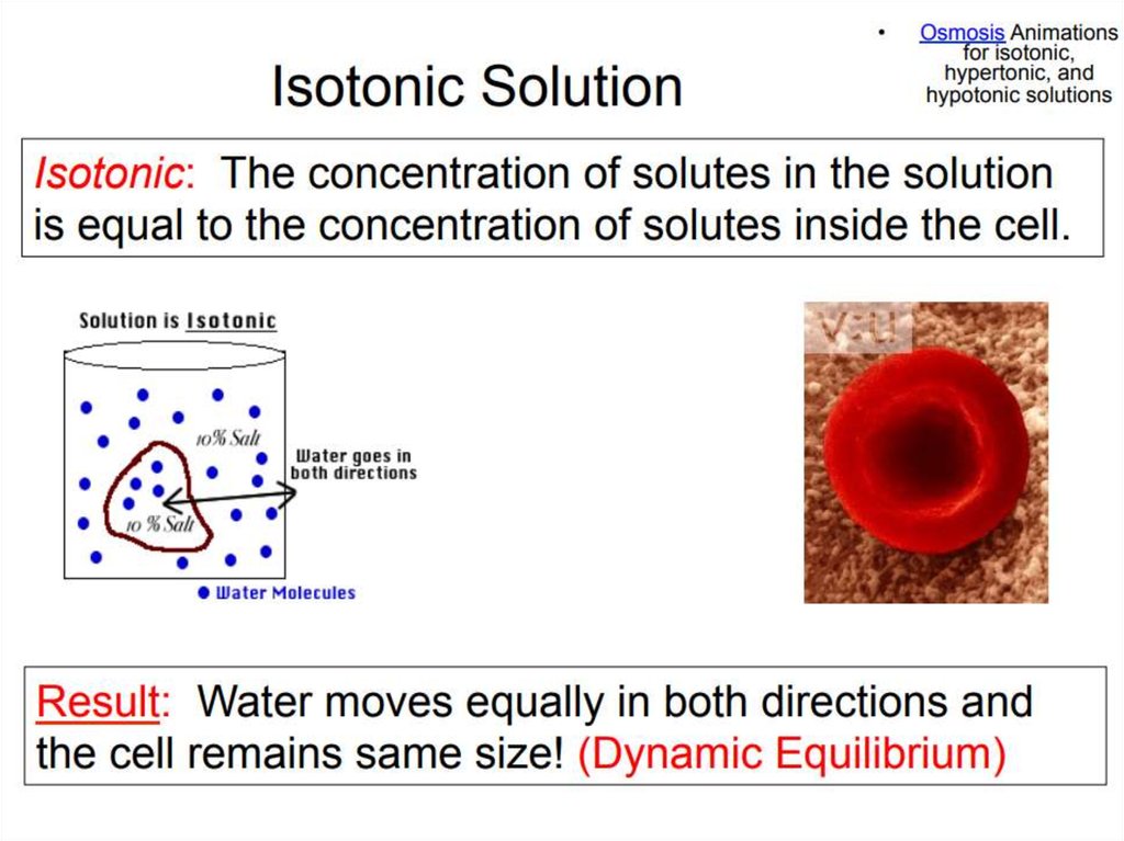
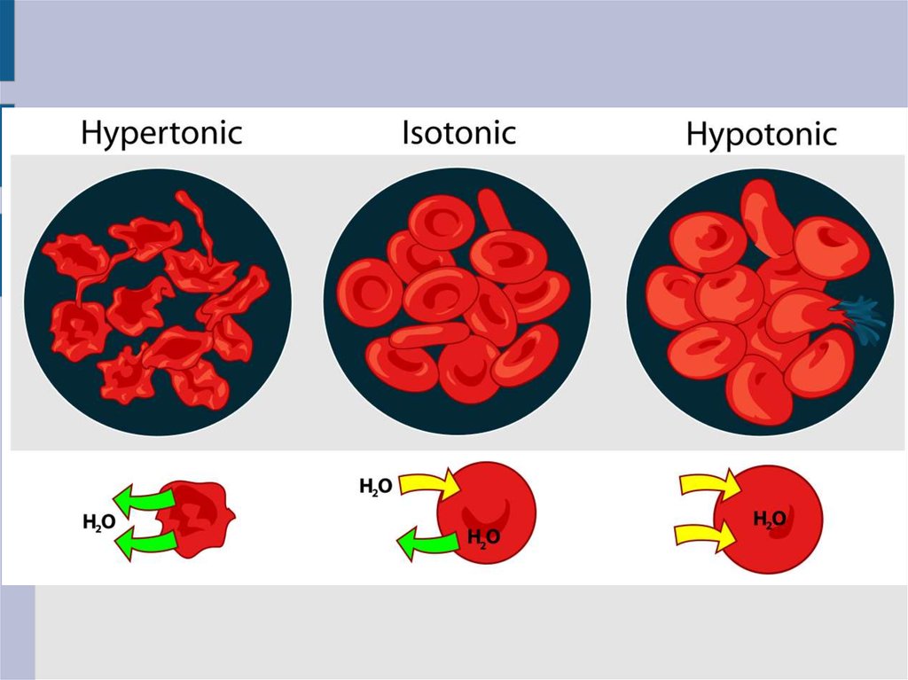
84. Solution Differences & Cells
Solution Differences & Cellssolvent + solute = solution
Hypotonic
– Solutes in cell more than outside
– Outside solvent will flow into cell
Isotonic
– Solutes equal inside & out of cell
Hypertonic
– Solutes greater outside cell
– Fluid will flow out of cell
85.
86.
87.
88.
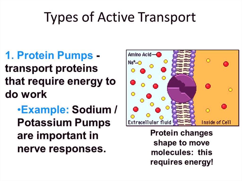
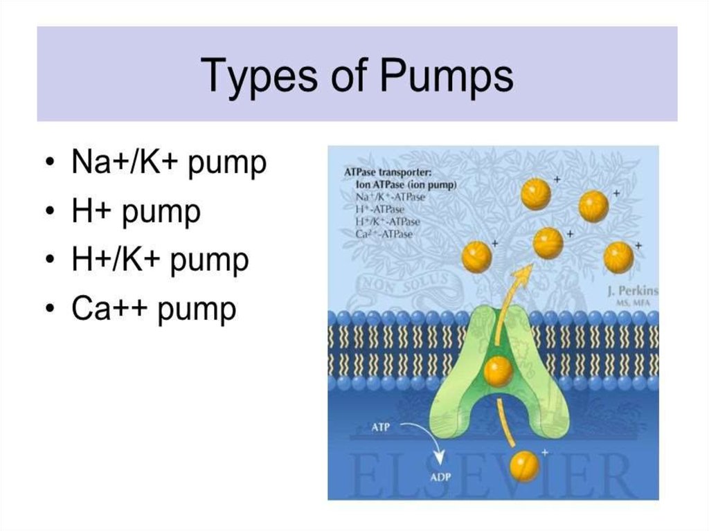
89. Active Transport
Molecular movementRequires energy (against gradient)
Example is sodium-potassium pump
90.
91.
92.
93.
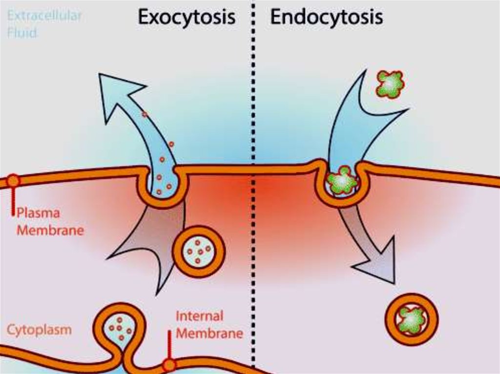
94. Endocytosis
Movement of large material– Particles
– Organisms
– Large molecules
Movement is into cells
Types of endocytosis
– bulk-phase (nonspecific)
– receptor-mediated (specific)
95. Process of Endocytosis
Plasma membrane surrounds materialEdges of membrane meet
Membranes fuse to form vesicle
96. Forms of Endocytosis
Phagocytosis – cell eatingPinocytosis – cell drinking