


 Potential")


")
 # 2")
 # 3")


 biology
Similar presentations:


Sensory Receptors
1. Sensory Receptors
Mikulyak A.I.2. Sensory Receptors: Introduction
Each type of sensory receptor responds to a particular modality of stimulus bycausing the production of action potentials in a sensory neuron.
These impulses are conducted to parts of the brain that provide the proper
interpretation of the sensory information when that specific neural pathway is
activated.
Our perceptions of the world—its textures, colors, and sounds; its warmth, smells,
and tastes—are created by the brain from electrochemical nerve impulses delivered
to it from sensory receptors.
These receptors transduce (change) different forms of energy in the “real world” into
the energy of nerve impulses that are conducted into the central nervous system by
sensory neurons. Different modalities (forms) of sensation—sound, light, pressure,
and so forth—result from differences in neural pathways and synaptic connections.
3. Categories of Sensory Receptors
Sensory receptors can be categorized on the basis of structure or variousfunctional criteria.
Structurally, the sensory receptors may be the dendritic endings of sensory
neurons. These dendritic endings may be free, such as those that respond to
pain and temperature, or encapsulated within nonneural structures, such as
those that respond to pressure. The photoreceptors in the retina of the eyes (rods
and cones) are highly specialized neurons that synapse with other neurons in the
retina. In the case of taste buds and of hair cells in the inner ears, modified
epithelial cells respond to an environmental stimulus and activate sensory
neurons.
4. Functional Categories
Sensory receptors can be grouped according to the type of stimulus energy they transduce.These categories include
•chemoreceptors, which sense chemical stimuli in the environment or the blood (e.g., the
taste buds, olfactory epithelium, and the aortic and carotid bodies);
•photoreceptors—the rods and cones in the retina of the eye;
•thermoreceptors, which respond to heat and cold; and
•mechanoreceptors, which are stimulated by mechanical deformation of the receptor cell
membrane (e.g., touch and pressure receptors in the skin and hair cells within the inner ear).
•nociceptors are pain receptors that depolarize in response to stimuli that accompany tissue
damage. These stimuli include noxiously high heat or pressure, acid, and a variety of
chemicals such as bradykinin, prostaglandins, nitric oxide, adenosine, and ATP.
5. Functional Categories # 2
Receptors also can be grouped according to the type of sensory information theydeliver to the brain.
Proprioceptors include the muscle spindles, Golgi tendon organs, and joint
receptors. These provide a sense of body position and allow fine control of skeletal
movements.
Cutaneous (skin) receptors include
•touch and pressure receptors,
•heat and cold receptors, and
•pain receptors.
The receptors that mediate sight, hearing, equilibrium, taste, and smell are grouped
together as the special senses.
6. Functional Categories # 3
In addition, receptors can be grouped intoexteroceptors, which respond to stimuli from outside of the body (such as those
involved in touch, vision, and hearing), and
interoceptors, which respond to internal stimuli. Interoceptors are found in many
organs, and include mechanoreceptors and chemoreceptors. An example of
mechanoreceptors are those in blood vessels that respond to stretch induced by
changes in blood pressure, and chemoreceptors include those that monitor blood
pH or oxygen concentration in the regulation of breathing.
7. Tonic and Phasic Receptors
Some receptors respond with a burst of activity when a stimulus is first applied,but then quickly decrease their firing rate— adapt to the stimulus—if the stimulus
is maintained. Receptors with this response pattern are called phasic receptors.
An example of a phasic receptor is a pacinian corpuscle (a pressure receptor).
Some other phasic receptors respond with a quick, short burst of impulses when
a stimulus is first applied, and then with another quick short burst of impulses
when the stimulus is removed. These phasic receptors thus provide information
regarding the “on” and “off” of a stimulus. Those receptors that maintain their
higher firing rate the entire time that a stimulus is applied are known as tonic
receptors.
8. Adaptation of Receptors
9. Relation between Stimulus Intensity and the Receptor Potential
The amplitude increases rapidly at firstbut then progressively less rapidly at high
stimulus strength.
In turn, the frequency of repetitive action
potentials transmitted from sensory
receptors increases approximately in
proportion to the increase in receptor
potential.
Putting this principle together with the
data in Figure, one can see that very
intense stimulation of the receptor
causes progressively less and less
additional increase in numbers of action
potentials. This exceedingly important
principle is applicable to almost all
sensory receptors. It allows the receptor
to be sensitive to very weak sensory
experience and yet not reach a maximum
ring rate until the sensory experience is
extreme. This feature allows the receptor
to have an extreme range of response,
from very weak to very intense.
10. Law of Specific Nerve Energies
Stimulation of a sensory nerve fiber produces only one sensation—touch, or cold, or pain,and so on.
According to the law of specific nerve energies, the sensation characteristic of each
sensory neuron is that produced by its normal stimulus, or adequate stimulus. Also,
although a variety of different stimuli may activate a receptor, the adequate stimulus requires
the least amount of energy to do so. The adequate stimulus for the photoreceptors of the
eye, for example, is light, where a single photon can have a measurable effect. If these
receptors are stimulated by some other means—such as by the high pressure produced by a
punch to the eye—a flash of light (the adequate stimulus) may be perceived.
The effect of paradoxical cold provides another example of the law of specific nerve
energies. When the tip of a cold metal rod is touched to the skin, the perception of cold
gradually disappears as the rod warms to body temperature. Then, when the tip of a rod
heated to 45C is applied to the same spot, the sensation of cold is perceived once again.
This paradoxical cold is produced because the heat slightly damages receptor endings, and
by this means produces an “injury current” that stimulates the receptor.
11. Generator (Receptor) Potential
The electrical behavior of sensory nerve endings is similar to that of the dendrites ofother neurons. In response to an environmental stimulus, the sensory endings produce
local graded changes in the membrane potential. In most cases, these potential changes
are depolarizations that are analogous to the excitatory postsynaptic potentials (EPSPs).
In the sensory endings, however, these potential changes in response to stimulation are
called receptor, or generator, potentials because they serve to generate action
potentials in response to the sensory stimulation. Because sensory neurons are
pseudounipolar, the action potentials produced in response to the generator potential are
conducted continuously from the periphery into the CNS.
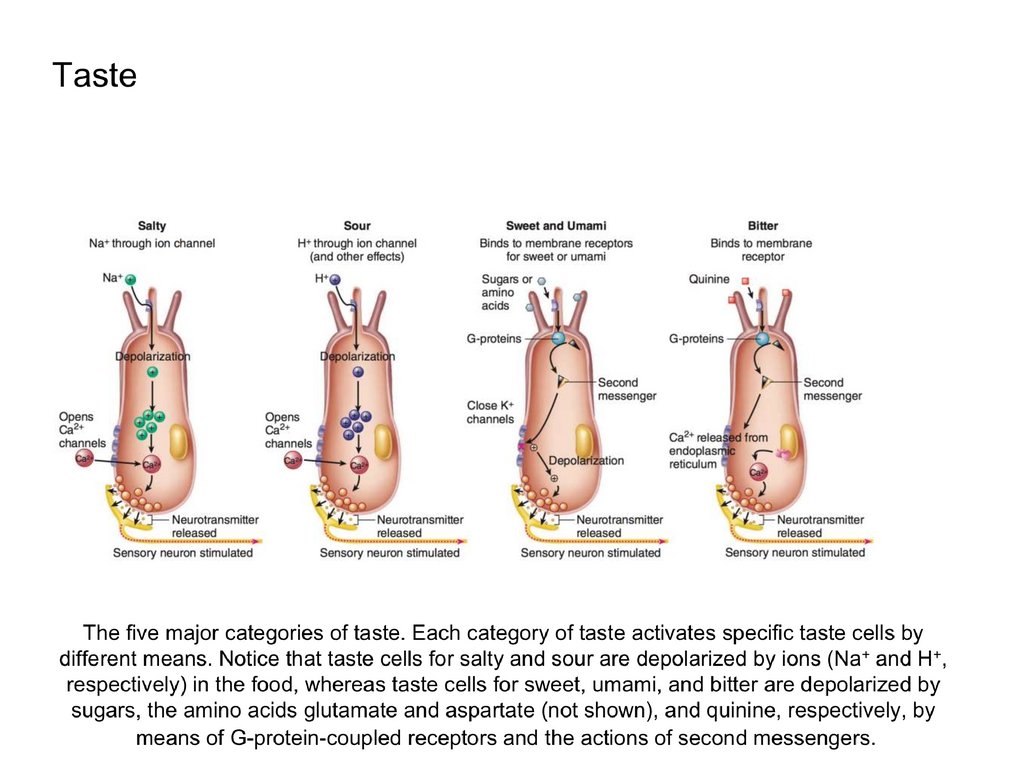
12. Taste
Gustation, the sense of taste, is evoked by receptors that consist of barrel-shapedtaste buds. Located primarily on the dorsal surface of the tongue, each taste bud
consists of 50 to 100 specialized epithelial cells with long microvilli that extend
through a pore in the taste bud to the external environment, where they are bathed in
saliva.
These sensory epithelial cells are not neurons, they behave like neurons; they
become depolarized when stimulated appropriately, produce action potentials, and
release neurotransmitters that stimulate sensory neurons associated with the taste
buds.
Because of this, some scientists classify the taste cells as neuroepithelial cells.
Taste buds are located mainly within epithelial papillae.
These include
•fungiform papillae on the anterior surface of the tongue;
•circumvallate papillae on the posterior surface of the tongue;
•foliate papillae on the sides of the tongue.
13.
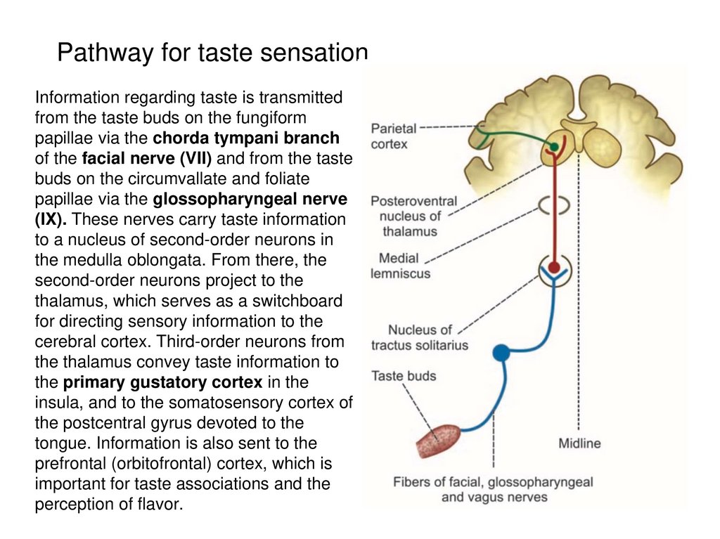
Pathway for taste sensationInformation regarding taste is transmitted
from the taste buds on the fungiform
papillae via the chorda tympani branch
of the facial nerve (VII) and from the taste
buds on the circumvallate and foliate
papillae via the glossopharyngeal nerve
(IX). These nerves carry taste information
to a nucleus of second-order neurons in
the medulla oblongata. From there, the
second-order neurons project to the
thalamus, which serves as a switchboard
for directing sensory information to the
cerebral cortex. Third-order neurons from
the thalamus convey taste information to
the primary gustatory cortex in the
insula, and to the somatosensory cortex of
the postcentral gyrus devoted to the
tongue. Information is also sent to the
prefrontal (orbitofrontal) cortex, which is
important for taste associations and the
perception of flavor.
14.
15. Smell
The receptors responsible for olfaction are located in the olfactory epithelium.The olfactory apparatus consists of receptor cells (which are bipolar neurons), supporting
(sustentacular) cells, and basal stem cells.
The stem cells generate new receptor cells every one to two months to replace the neurons
damaged by exposure to the environment.
The supporting cells are epithelial cells rich in enzymes that oxidize hydrophobic volatile
odorants, thereby making these molecules less lipid-soluble and thus less able to penetrate
membranes and enter the brain.
Each bipolar sensory neuron has one dendrite that projects into the nasal cavity, where it
terminates in a knob containing cilia. It is the plasma membrane covering the cilia that
contains the receptor proteins that bind to odorant molecules. The axon of each olfactory
neuron thereby conveys information relating only to the specific odorant molecule that
stimulated that neuron.
The olfactory receptors are G-protein-coupled receptors. This means that before the odorant
molecule binds to its receptor, the receptor is associated with the three G-protein subunits (a,
b, and g). When an odorant molecule binds to its receptor, these subunits dissociate, move in
the plasma membrane to adenylate cyclase, and activate this enzyme. Adenylate (or adenyl)
cyclase catalyzes the conversion of ATP into cyclic AMP (cAMP) and PPi (pyrophosphate).
The cAMP serves as a second messenger, opening ion channels that allow inward diffusion
of Na+ and Ca2+. This produces a graded depolarization, the receptor potential, which then
stimulates the production of action potentials.
Up to 50 G-proteins may be associated with a single receptor protein.
16.
The processing of olfactory information begins in the olfactory bulb, where the bipolarsensory neurons synapse with neurons located in spherically shaped arrangements
called glomeruli. Evidence suggests that each glomerulus receives input from one
type of olfactory receptor. Identification of an odor is improved by inhibition provided
by GABA released from periglomerular cells that surround the glomerulus and make
dendrodendritic synapses with the second-order neurons within the glomerulus
(termed mitral and tufted cells).
The mitral and tufted neurons of the olfactory glomeruli in the olfactory bulb send
axons through the lateral olfactory tracts to numerous brain regions of the frontal and
medial temporal lobes that comprise the primary olfactory cortex. There are
interconnections between these regions and the amygdala, hippocampus, and other
structures of the limbic system. For example, the piriform cortex, a pear-shaped
region at the medial junction of the frontal and temporal lobes, receives projections
from the olfactory bulb and makes reciprocal connections with the prefrontal cortex
and amygdala, among other structures.
The prefrontal cortex receives information regarding taste as well as smell; perhaps
this is why olfactory stimulation during eating can be perceived as taste rather than
smell.
17. Smell
The neural pathway for olfaction.The olfactory epithelium contains receptor neurons that synapse with neurons in the olfactory bulb of
the cerebral cortex.
The synapses occur in rounded structures called glomeruli.
Secondary neurons, known as tufted cells and mitral cells, transmit impulses from the olfactory bulb to
the olfactory cortex in the medial temporal lobes.
Each glomerulus receives input from only one type of olfactory receptor, regardless of where
those receptors are located in the olfactory epithelium.
18. VESTIBULAR APPARATUS AND EQUILIBRIUM
The sense of equilibrium, which provides orientation with respect to gravity, is due to thefunction of an organ called the vestibular apparatus. The vestibular apparatus and a
snail-shaped structure called the cochlea, which is involved in hearing, form the inner ear
within the temporal bones of the skull.
The vestibular apparatus consists of two parts:
• the otolith organs, which include the utricle and saccule,
• the semicircular canals
The sensory structures of the vestibular apparatus and cochlea are located within the
membranous labyrinth, a tubular structure that is filled with a fluid called endolymph.
Endolymph is unlike any other extracellular fluid: it has a higher K+ concentration (higher
even than in the intra-cellular compartment) and much lower concentrations of Na+ and
Ca2+ than do other extracellular fluids.
The membranous labyrinth is located within a bony cavity in the skull, the bony labyrinth.
Within this cavity, between the membranous labyrinth and the bone, is a fluid called
perilymph. Unlike endolymph, perilymph is fairly typical of extracellular fluids such as
cerebrospinal fluid.
19. The cochlea and vestibular apparatus of the inner ear
The vestibular apparatus consists of the utricle and saccule (together called theotolith organs) and the three semicircular canals. The base of each semicircular
canal is expanded into an ampulla that contains sensory hair cells
20. Sensory Hair Cells of the Vestibular Apparatus
The utricle and saccule provide information about linear acceleration—changes in velocitywhen traveling horizontally or vertically. We therefore have a sense of acceleration and
deceleration when riding in a car or when skipping rope. A sense of rotational, or angular,
acceleration is provided by the semicircular canals, which are oriented in three planes like the
faces of a cube. This helps us maintain balance when turning the head, spinning, or tumbling.
The receptors for equilibrium are modified epithelial cells. They are known as hair cells
because each cell contains 20 to 50 hairlike extensions. These are actually modified microvilli
called stereocilia arranged in rows of increasing height. Touching the stereocilia of the tallest
row is an even taller true cilium called a kinocilium.
When the stereocilia are bent in the direction of the kinocilium, the plasma membrane is
depressed and ion channels for K+ are opened, allowing K+ to passively enter and depolarize
the hair cell. This causes the hair cell to release a synaptic transmitter that stimulates the
dendrites of sensory neurons that are part of the vestibulocochlear nerve (VIII).
When the stereocilia are bent in the opposite direction, the membrane of the hair cell
becomes hyperpolarized and, as a result, releases less synaptic transmitter. In this way, the
frequency of action potentials in the sensory neurons that innervate the hair cells carries
information about the direction of movements that cause the hair cell processes to bend.
21. Utricle and Saccule
The otolith organs, the utricle and saccule, each have a patch of specializedepithelium called a macula that consists of hair cells and supporting cells. The hair
cells project into the endolymphfilled membranous labyrinth, with their hairs
embedded in a gelatinous otolithic membrane.
The otolithic membrane contains microscopic crystals of calcium carbonate
(otoliths) from which it derives its name (oto = ear; lith = stone). These stones
increase the mass of the membrane, which results in a higher inertia (resistance to
change in movement).
Because of the orientation of their hair cell processes into the otolithic membrane,
the utricle is more sensitive to horizontal acceleration and the saccule is more
sensitive to vertical acceleration.
22. Semicircular Canals
The three semicircular canals project in three different planes at nearly right anglesto each other.
Each canal contains an inner extension of the membranous labyrinth called a
semicircular duct, and at the base of each duct is an enlarged swelling called the
ampulla.
The crista ampullaris, an elevated area of the ampulla, is where the sensory hair
cells are located. The processes of these cells are embedded in a gelatinous
membrane, the cupula, which has a higher density than that of the surrounding
endolymph. Like a sail in the wind, the cupula can be pushed in one direction or the
other by movements of the endolymph.
23. Neural Pathways
Stimulation of hair cells in the vestibular apparatus activates sensory neurons ofthe vestibulocochlear nerve (VIII). These fibers transmit impulses to the
cerebellum and to the vestibular nuclei of the medulla oblongata.
The vestibular nuclei, in turn, send fibers to the oculomotor center of the brain
stem and to the spinal cord.
Neurons in the oculomotor center control eye movements, and neurons in the
spinal cord stimulate movements of the head, neck, and limbs.
Movements of the eyes and body produced by these pathways serve to maintain
balance and “track” the visual field during rotation.
24. THE EARS AND HEARING
Sound waves are alternating zones of high and low pressure traveling in a medium, usuallyair or water.
These waves are characterized by their frequency and intensity.
The frequency is measured in hertz (Hz), which is the modern designation for cycles per
second (cps). The pitch of a sound is directly related to its frequency—the greater the
frequency of a sound, the higher its pitch.
The intensity, or loudness, of a sound is directly related to the amplitude of the sound waves
and is measured in units called decibels (dB). A sound that is barely audible—at the
threshold of hearing—has an intensity of zero decibels. Every 10 decibels indicates a tenfold
increase in sound intensity; a sound is 10 times louder than threshold at 10 dB, 100 times
louder at 20 dB, a million times louder at 60 dB, and 10 billion times louder at 100 dB.
The ear of a trained, young individual can hear sound over a frequency range of 20 to 20,000
Hz, yet still can distinguish between two pitches that have only a 0.3% difference in
frequency.
The human ear can detect differences in sound intensities of only 0.1 to 0.5 dB, while the
range of audible intensities covers 12 orders of magnitude (1012), from the barely audible to
the limits of painful loudness. Human hearing is optimal at sound intensities of 0 to 80 dB.
25. Outer Ear
Sound waves are funneled by the pinna, or auricle, into the external auditory meatus.These two structures form the outer ear.
The external auditory meatus channels the sound waves to the eardrum, or
tympanic membrane. Sound waves in the external auditory meatus produce
extremely small vibrations of the tympanic membrane; movements of the eardrum
during speech (with an average sound intensity of 60 dB) are estimated to be about
the diameter of a molecule of hydrogen.
26. Middle Ear
The middle ear is the cavity between the tympanic membrane on the outer sideand the cochlea on the inner side.
Within this cavity are three middle-ear ossicles—the malleus (hammer), incus
(anvil), and stapes (stirrup). The malleus is attached to the tympanic membrane,
so that vibrations of this membrane are transmitted via the malleus and incus to
the stapes. The stapes, in turn, is attached to a membrane in the cochlea called
the oval window, which thus vibrates in response to vibrations of the tympanic
membrane.
The fact that vibrations of the tympanic membrane are transferred through three
bones instead of just one affords protection. If the sound is too intense, the
ossicles may buckle. This protection is increased by the action of the stapedius
muscle, which attaches to the neck of the stapes. When sound becomes too
loud, the stapedius muscle contracts and dampens the movements of the stapes
against the oval window. This action helps to prevent nerve damage within the
cochlea. If sounds reach high amplitudes very quickly, however the stapedius
muscle may not respond soon enough to prevent nerve damage.
27. A medial view of the middle ear
The locations of auditory muscles, attached to the middle-ear ossicles, are indicated.28. Cochlea
Encased within the dense temporal bone of the skull is an organ called thecochlea, about 34 mm long (the size of a pea) and shaped like the shell of
a snail. Together with the vestibular apparatus, it composes the inner ear.
Vibrations of the stapes and oval window displace perilymph fluid within a
part of the bony labyrinth known as the scala vestibuli, which is the upper
of three chambers within the cochlea. The lower of the three chambers is
also a part of the bony labyrinth and is known as the scala tympani. The
middle chamber of the cochlea is a part of the membranous labyrinth called
the cochlear duct, or scala media. Like the cochlea as a whole, the
cochlear duct coils to form three turns, similar to the basal, middle, and
apical portions of a snail shell. Because the cochlear duct is a part of the
membranous labyrinth, it contains endolymph rather than perilymph.
The perilymph of the scala vestibuli and scala tympani is continuous at the
apex of the cochlea because the cochlear duct ends blindly, leaving a small
space called the helicotrema between the end of the cochlear duct and the
wall of the cochlea. Vibrations of the oval window produced by movements
of the stapes cause pressure waves within the scala vestibuli, which pass to
the scala tympani.
29. Cochlea # 2
Movements of perilymph within the scala tympani, in turn, travel to the base ofthe cochlea where they cause displacement of a membrane called the round
window into the middle-ear cavity. This occurs because fluid, such as perilymph,
cannot be compressed; an inward movement of the oval window is thus
compensated for by an outward movement of the round window.
Sound waves transmitted through perilymph from the scala vestibuli to the scala
tympani thus produce displacement of the vestibular membrane and the basilar
membrane. Although the movement of the vestibular membrane does not directly
contribte to hearing, displacement of the basilar membrane is central to pitch
discrimination. Each sound frequency produces maximum vibrations at a different
region of the basilar membrane. Sounds of higher frequency (pitch) cause
maximum vibrations of the basilar membrane closer to the stapes.
30. A cross section of the cochlea
In this view, its three turns and its three compartments—the scala vestibuli, cochlearduct (scala media), and scala tympani—can be seen.
31. Spiral Organ (Organ of Corti)
The sensory hair cells are located on the basilar membrane with their “hairs” projectinginto the endolymph of the cochlear duct. The hairs are actually stereocilia, which are
large, specialized microvilli arranged in bundles. The stereocilia increase in size
stepwise within each bundle, as they do in the vestibular apparatus; however, unlike the
case in the vestibular apparatus, the cochlear hair cells lack kinocilia.
There are two categories of hair cells, inner and outer.
Inner hair cells, about 3,500 per cochlea, form one row that extends the length of the
basilar membrane. The hair bundles on the inner hair cells are mechanosensory: they
transform sound waves in cochlear fluid into nerve impulses. Their stereocilia are
interconnected near their tips with filaments that are coupled to mechanotransduction
channels in the plasma membrane. These channels open when the stereocilia within
each bundle are bent in the direction of their tallest members, allowing the movement of
K+ across the plasma membrane as will be described shortly. Each of the inner hair cells
is innervated by 6–20 sensory neurons of cranial nerve VIII from the spiral ganglion,
which transmit sound information to the brain. The number of synapses with afferent
neurons depends on the location of the inner hair cells along the basilar membrane, with
those in the middle having the greatest number of synapses and the highest sensitivity to
sound.
32. Spiral Organ (Organ of Corti) # 2
There are also about 11,000 outer hair cells arranged in multiple rows: three rows in the basilarturn, four in the middle turn, and five in the apical turn of the cochlea. The outer hair cells are
innervated primarily by motor neurons that originate in the olivary nuclei of the medulla oblongata.
These depolarize or hyperpolarize the outer hair cells, causing them to shorten when they are
depolarized or lengthen when they are hyperpolarized. Such movements are believed to aid the
sensory function of the inner hair cells.
The stereocilia of the hair cells are embedded in a gelatinous tectorial membrane (tectum = roof,
covering), which overhangs the hair cells within the cochlear duct. The association of the basilar
membrane, inner hair cells with sensory fibers, and tectorial membrane forms a functional unit
called the spiral organ, or organ of Corti. When the cochlear duct is displaced by pressure
waves of perilymph, a shearing force is created between the basilar membrane and the tectorial
membrane. This causes the stereocilia to bend, and this mechanical process opens K+ channels
in the plasma membrane covering the tops of the stereocilia.
These K+ channels face endolymph, which uniquely has a high concentration of K+ similar to that
of the intracellular compartment. Also, the endolymph of the cochlea (but not the vestibular
apparatus) has an amazingly high positive potential: 1100 mV. Combined with the negative
resting membrane potential of the hair cells, this produces an extremely steep electrochemical
gradient favoring the entry of K+.
So, when the K+ channels in the bent stereocilia open, K+ moves passively down its
electrochemical gradient into the hair cells. This depolarizes the hair cells and stimulates them to
release glutamate, which stimulates the associated sensory neurons. The K+ that entered the hair
cells at their apical surface can then move passively out through channels in their basal surface,
which face perilymph in the scala tympani. Perilymph, as previously mentioned, has a low K+
concentration typical of extracellular fluids.
33. Spiral Organ (Organ of Corti) # 3
The greater the displacement of the basilar membrane and the bending of thestereocilia, the greater the amount of transmitter released by the inner hair cell,
and therefore the greater the generator potential produced in the sensory neuron.
By this means, a greater bending of the stereocilia will increase the frequency of
action potentials produced by the fibers of the cochlear nerve that are stimulated
by the hair cells. Experiments suggest that the stereocilia need bend only 0.3
nanometers to be detected at the threshold of hearing! A greater bending will
result in a higher frequency of action potentials, which will be perceived as a
louder sound.
As mentioned earlier, traveling waves in the basilar membrane reach a peak in
different regions, depending on the pitch (frequency) of the sound. High-pitched
sounds produce a peak displacement closer to the base, while sounds of lower
pitch cause peak displacement further toward the apex. Those neurons that
originate in hair cells located where the displacement is greatest will be
stimulated more than neurons that originate in other regions. This mechanism
provides a neural code for pitch discrimination.
34. Neural Pathways for Hearing
Sensory neurons in the spiral ganglion of each ear send their axons in thevestibulocochlear nerve (VIII) to one of two cochlear nuclei in the junction of the
medulla and pons of the brain stem. Neurons in the cochlear nuclei send axons
either directly to the inferior colliculi of the midbrain or to the superior olive, a
collection of brain stem nuclei. Axons from the superior olive pass through the
lateral lemniscus to the inferior colliculus. Whatever the route, all auditory paths
synapse in the inferior colliculus. Neurons in the inferior colliculus then send
axons to the medial geniculate body of the thalamus, which in turn projects to the
auditory cortex of the temporal lobe.
The cochlea is a frequency analyzer, in that different frequencies (pitches) of
sound stimulate different sensory neurons that innervate the basilar membrane.
This is because hair cells located in different places along the basilar membrane
are most effectively stimulated by different frequencies of sound. This is known
as the place theory of pitch, and has been previously described. Sensory
neurons stimulated by low-frequency sounds, and those stimulated by highfrequency sounds, project their axons to different regions of the cochlear nucleus.
The cochlear nucleus displays a tonotopic organization, in that different regions
represent different “tones” (pitches). This separation of neurons by pitch is
preserved in the tonotopic organization of the auditory cortex, which allows to
perceive the different pitches of sounds.
35. THE EYES AND VISION
The eyes transduce energy in the electromagnetic spectrum into nerve impulses. Only alimited part of this spectrum can excite the photoreceptors—electromagnetic energy with
wavelengths between 400 and 700 nanometers (1 nm = 10-9 m, or one-billionth of a
meter) constitutes visible light.
Light of longer wavelengths in the infrared regions of the spectrum is felt as heat but does
not have sufficient energy to excite the photoreceptors.
WALL OF THE EYEBALL
Wall of the eyeball is composed of three layers