


 biology
Similar presentations:


Muscle tissue
1. Muscle tissue
Lecture N72.
• Muscle tissue satisfy requirement of thebody in movement.
3. Classification – The 3 types of muscle tissue:
1. skeletal• groups:
• Striated
2. cardiac
3. smooth
• Smooth
4. Why do muscles contract?
• Muscle cells have contractile proteins actin and myosin,and some another .
The interaction of actin and myosin mediates
the contraction of muscle cells.
5. Why do muscles contract?
Actin and myosin form myofilaments:
Myosin - thick, dark and Anisotropic (A)
Actin – thin, light and Isotropic (I)
Actin and myosin form special organelles –
myofibrils, responsible for muscle
contraction.
6. SMOOTH MUSCLE
7.
• Locations: walls of visceral holloworgans
(stomach).
Functions: involuntary movement -(peristaltics)
(The innervation -- by autonomic nervous
system)
8. SMOOTH MUSCLE
• Unit – spindle shapedcell -- myocyte
• Individual cells are
organized in sheath
• In hollow organs
forms layers
Contraction is usually
slow.
SMOOTH
MUSCLE
9. Origin of smooth muscle
• Smooth muscle cells arise frommesenchymal cells.
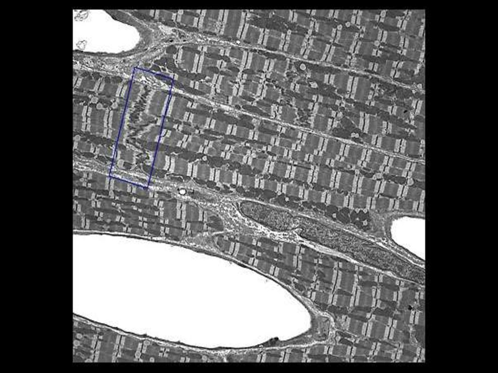
10. Striated muscles
11. See: regular organization of the myofibrils gives rise to the cross-striation, which characterises skeletal and cardiac muscle.
12. CARDIAC MUSCLE
• Locations: heart• Function:
involuntary,
rhythmic
contraction
• Unit –
cardiomyocyte
(cell)
13. Cardiac muscle cells:
3 types:• Contractile,
• Conducting
• Secretory
14. CARDIAC MUSCLE
cardiac muscle cells arecylindrical,
connect end-by-end,
and form “functional
fiber”, which
often branch at acute
angles.
15. CARDIAC MUSCLE
• They are connectedby special junction intercalated discs –
consisting of
gap junctions
and
desmosomes.
16.
17. SKELETAL MUSCLE
18. Location
• Muscles associatedwith the skeleton
(are connected to
bones by tendons).
• Platisma and mimic
muscles
• Voluntary
sphincters of inner
organs
19. SKELETAL MUSCLE
• --- is innervated by the somaticnervous system – voluntary!!
• ---- consists of very long tubular cells
(also called muscle fibres).
20. SKELETAL MUSCLE
Nuclei:• Skeletal muscle
fibres contain many
nuclei
(up to several
hundred )
placed beneath the
plasma membrane
21. Nuclei:
MyofibrilsMechanism of contraction:
Sliding filaments model
22. Myofibrils Mechanism of contraction: Sliding filaments model
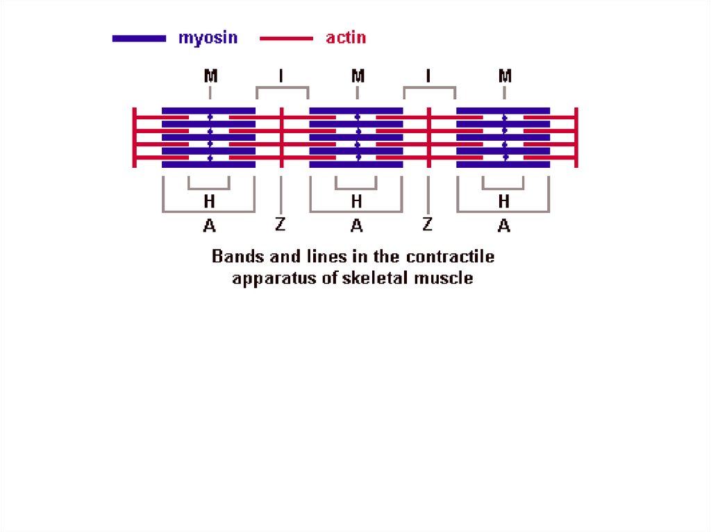
• Myofibrils has some bands and lines depending on thedistribution and interconnection of myofilaments -- :
• I-band - actin filaments only,
• A-band - myosin filaments which may overlap with
actin filaments
• T or Z-line -- band of connections between actin
filaments; zone of apposition of actin filaments
belonging to two neighboring sarcomeres;
• M-line - band of connections between myosin
filaments.
• H-band - zone of myosin filaments only (no overlap
with actin filaments) within the A-band
23.
24.
25.
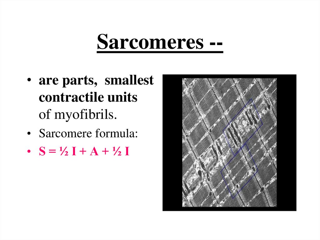
Sarcomeres -• are parts, smallestcontractile units
of myofibrils.
• Sarcomere formula:
• S=½I+A+½I
26. Sarcomeres --
Sarcomere formula after contraction• S=A
• (- ½ I, - ½ I, - H)
27. Sarcomere formula after contraction
Mechanism of contraction28. Mechanism of contraction
Origin of skeletal muscle• The myoblasts of all skeletal muscle fibres
originate from the paraxial mesoderm myotome.
29. Origin of skeletal muscle
• 1. Myoblasts undergo frequent divisionsand coalesce with the formation of a
multinucleated, syncytial muscle fibre or
myotube. The nuclei of the myotube are
still located centrally in the muscle fibre.
• 2. In the course of the synthesis of the
myofilaments and myofibrils, the nuclei are
gradually displaced to the periphery of the
cell.
30.
Regeneration. Satellite cells• Satellite cells are small cells which are
closely apposed to muscle fibers within the
basal lamina which surrounds the muscle
fiber.
• Satellite cells are believed to represent
persistent myoblasts. They may regenerate
muscle fibers in case of damage.