


 medicine
Similar presentations:


")


Chronic middle suppurative otitis. Nonsuppurative pathology of the chronic ear: sensoneural deafness
1. Chronic middle suppurative otitis. Nonsuppurative pathology of the chronic ear: sensoneural deafness.
Zaporozhye - 20112.
The actuality of the theme.Chronic purulent middle otitis is the most frequent disease
of the ear and you can meet it in 20-25 per cents of cases
among the all pathology of ENT organs. But unsymptomatically
taking chronic otitis, especially epitympanitis, can suddenly
causes the hard intracranial complications (meningitis, sepsis,
brains abscesses, etc).
An expressed hardness of a hearing, the unpurulent ear’s
diseases are in 98 percents of observations. The loss of
hearing is accompanied by agonizing noise in the ears and
reflected on the human ability to work, his moral condition. A
child, who lost in hearing early, usually can’t study to speak.
When he grows, he becomes deaf mute. All these factors
determine a social importance of the problem of unpurulent
ear’s diseases.
2
3.
Chronic suppurative otitis media is a common disease.Chronic suppurative otitis media is characterized by persistent
perforation of the tympanic membrane, periodic or permanent
otopyorrhoea, and hearing loss of various degrees.
Aetiology and pathogenesis. The disease is usually
secondary to acute suppurative otitis which can persist during
several months for various reasons. Among frequent causes of
conversion of acute otitis media into the chronic form is a
severe acute pathological process in the middle ear, which
depends on virulence and the character of infection,
decreased resistance of the body associated with chronic
specific or non-specific infection. Pathology of the upper
airways is also important for the onset of the disease.
According to the clinical course and gravity, chronic
suppurative otitis media is classified as mesotympanitis and
epitympanitis.
3
4.
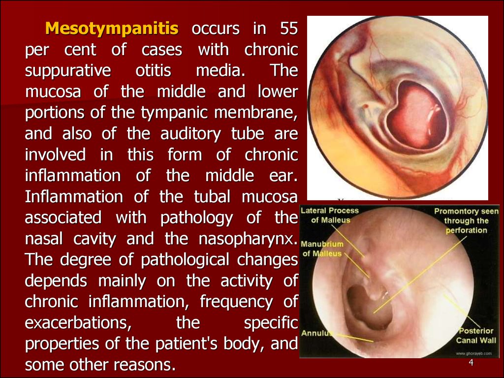
Mesotympanitis occurs in 55per cent of cases with chronic
suppurative otitis media. The
mucosa of the middle and lower
portions of the tympanic membrane,
and also of the auditory tube are
involved in this form of chronic
inflammation of the middle ear.
Inflammation of the tubal mucosa
associated with pathology of the
nasal cavity and the nasopharynx.
The degree of pathological changes
depends mainly on the activity of
chronic inflammation, frequency of
exacerbations,
the
specific
properties of the patient's body, and
some other reasons.
4
5.
Otoscopy in mesotympanitis reveals intact flaccid part of thetympanic membrane and the presence of a perforation in the
tense part. The perforation can be round, oblong, beanshaped; it can vary in size from punctate to an opening
occupying almost the whole area of the tense part, a narrow
band remaining by the circumference.
Subjective symptoms are indistinct. Patients complain of
periodical or constant otopyorrhoea and impaired hearing
function. In rare cases the patients complain of tinnitus and
vertigo. Pain in the ear arises only during exacerbation or due
to development of secondary diseases of the ear, such as
diffuse otitis extema or circumscribed otitis extema. Discharge
from the ear is mucopurulent. The discharge is usually
odourless. It can be meagre or profuse (in exacerbation). The
hearing function is impaired as in affection of the conduction
system.
5
6.
The discharge from the ear can persist for years withoutcausing any serious complications. Otopyorrhoea can stop
spontaneously and recur only during exacerbation caused by
common cold, water in the ear, respiratory diseases, diseases
of the nose, nasopharynx, paranasal sinuses, etc.
Despite the benign course of mesotympanitis, severe
intracranial complications can sometimes occur. They can be
caused by caries of the promontorial wall, polyps, and
granulation.
Diagnosis is based on the anamnestic, clinical, and otoscopic
findings (persistent central perforation). Mesotympanitis should
be differentiated from epitympanitis. The distinguishing signs
of mesotympanitis are persistent central perforation of the
tense part of the eardrum, mucous, mucopurulent, or (less
frequently) purulent odourless discharge. The odour indicates
involvement of the bone (malignization of the disease).
6
7.
Prognosis is usually favourable, provided a systematic andrational general and local treatment is given. But it is difficult
to improve the hearing function, and in this respect the
physician should be careful in his prognosis. Hearing improves
in most cases after cessation of otopyorrhoea.
Treatment includes prevention of pus retention in the
middle and external ear and action on the microflora and the
inflamed mucosa with disinfectants and astringent
preparations. Local treatment includes daily irrigation of the
ear with the following warm solutions: 3 per cent hydrogen
peroxide,
and antibiotics, after preliminary testing the
microflora for sensitivity to them. In the presence of
perforation
in
the
tympanic
membrane,
endaural
administration of medicinal preparations is effective: 1.5-2 ml
of medicinal solution is instilled in the external acoustic meatus
and the tragus is then pressed rhythmically by the finger for
7
10-15 seconds to pump the liquid into the middle ear.
8.
If the patient feels the taste of the medicine in the mouth, itindicates that the solution has passed the middle ear and
entered the auditory tube.
Local treatment includes also direct instillation of the
following solutions: antibiotic solutions, antibiotics should be
injected intramuscularly only during exarcerbation.
Minor surgical operations are sometimes necessary:
treatment of small granulations or polyps with trichloroacetic
acid, a 40 per cent silver nitrate solution;; removal of large
granulations using a conchotome, or a curet; and removal of
polyps using an aural snare. Physiotherapy is also necessary. It
includes UV-therapy and UHF on the ear in the absence of
polyps or granulation. General envigorating measures are
recommended: rational nutrition, hardening of the body,
climatotherapy, and the like.
8
9.
Epitympanitis(atticitis).
The
inflammation is mainly localized in the
epitympanum, the attic of the tympanum.
A perforation is usually present in the
lateral wall of the epitympanum. Atticitis
is characterized by affection of the
mucosa and the bony tissue of the middle
ear walls and the mastoid process. Caries
or cholesteatoma can destroy the wall of
the middle ear thus causing a severe
intracranial or general complication. The
main otoscopic sign of the pathology is
persistent marginal perforation in the
upper (flaccid) portion of the tympanic
membrane. If the process is destructive,
pus has a putrid odour specific for
epitympanitis
9
10.
A sample of cholesteatoma or pus can be extracted fromthe attic on the tip of the probe. Probing detects the presence
of granulation (and determines its location) and can also reveal
the presence of labyrinthine fistula.
Cholesteatomatous
epitympanitis.
Cholesteatoma
causes
vast
destruction
in
the
temporal
bone.
Cholesteatomatous masses can sometimes be seen during
otoscopy through a perforation in the tympanic membrane.
Cholesteatoma increases in size gradually and constantly due
to desquamation of the epidermis, fills in the attic and the
antrum and then destroys the bone. As a result the
cholesteatoma can reach the meninges, destroy the bony
capsule of the labyrinth, the wall of the canal for the facial
nerve, almost the entire mastoid process, and thus expose the
cerebellar meninges and the wall of the sigmoid sinus.
Purulation of cholesteatomatous mass can extend to the
10
intracranial tissues to cause intracranial pathology.
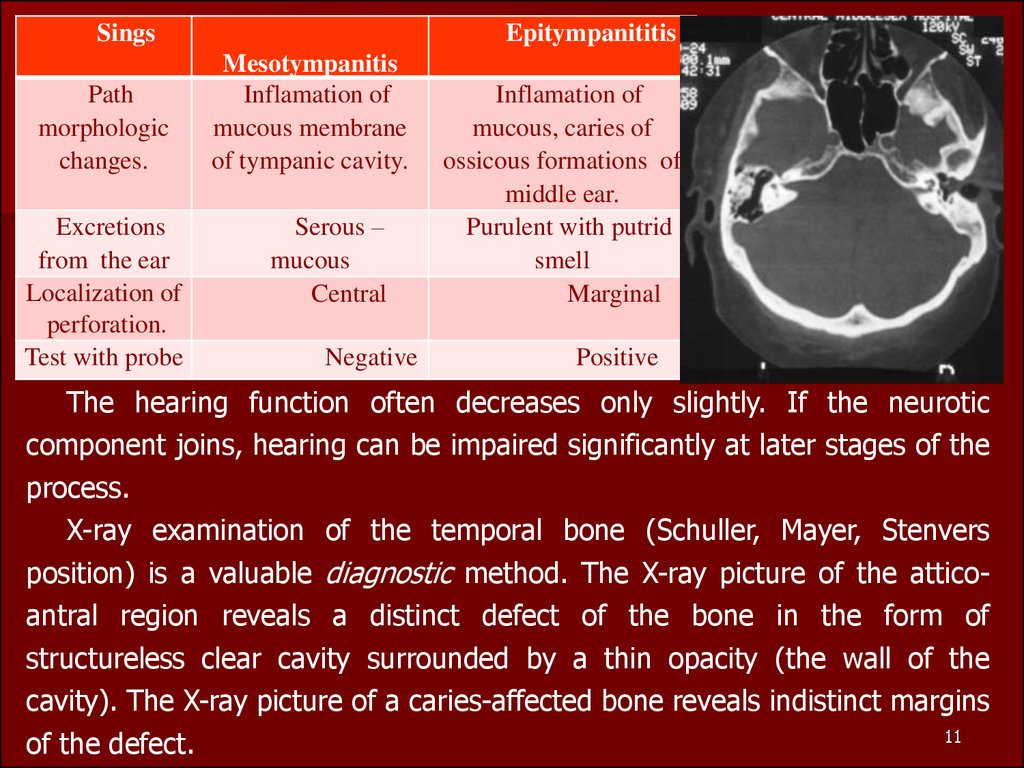
11.
SingsPath
morphologic
changes.
Excretions
from the ear
Localization of
perforation.
Test with probe
Epitympanititis
Mesotympanitis
Inflamation of
mucous membrane
of tympanic cavity.
Serous –
mucous
Central
Negative
Inflamation of
mucous, caries of
ossicous formations of
middle ear.
Purulent with putrid
smell
Marginal
Positive
The hearing function often decreases only slightly. If the neurotic
component joins, hearing can be impaired significantly at later stages of the
process.
X-ray examination of the temporal bone (Schuller, Mayer, Stenvers
position) is a valuable diagnostic method. The X-ray picture of the atticoantral region reveals a distinct defect of the bone in the form of
structureless clear cavity surrounded by a thin opacity (the wall of the
cavity). The X-ray picture of a caries-affected bone reveals indistinct margins
11
of the defect.
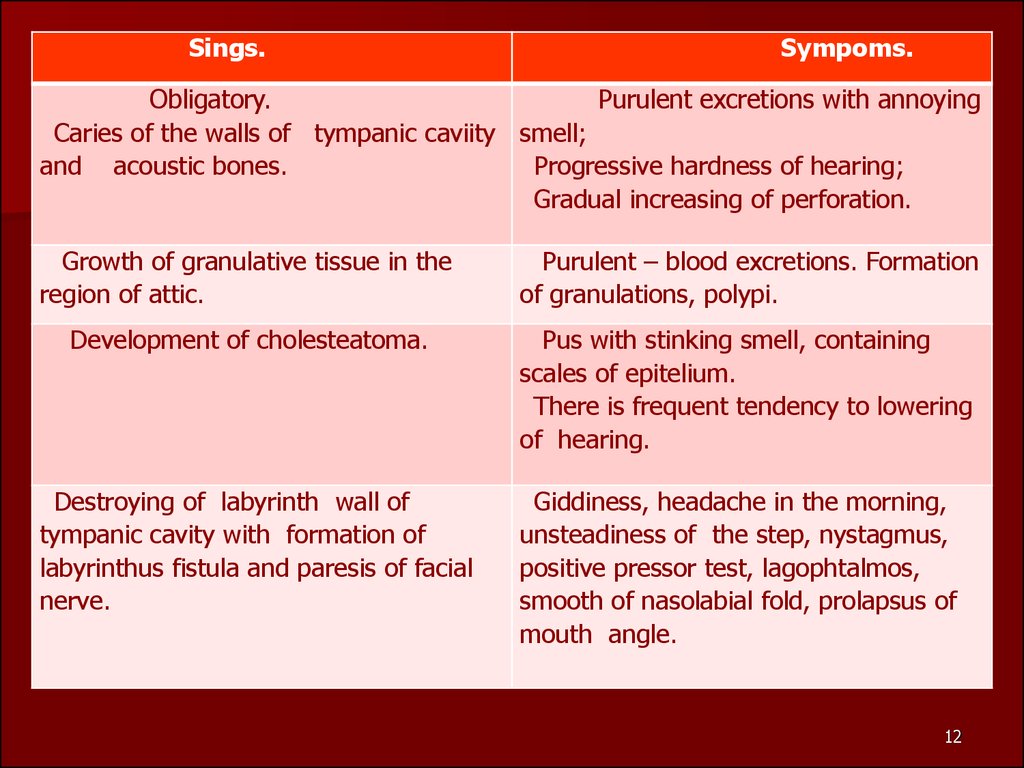
12.
Sings.Sympoms.
Obligatory.
Purulent excretions with annoying
Caries of the walls of tympanic caviity smell;
and acoustic bones.
Progressive hardness of hearing;
Gradual increasing of perforation.
Growth of granulative tissue in the
region of attic.
Purulent – blood excretions. Formation
of granulations, polypi.
Development of cholesteatoma.
Pus with stinking smell, containing
scales of epitelium.
There is frequent tendency to lowering
of hearing.
Destroying of labyrinth wall of
tympanic cavity with formation of
labyrinthus fistula and paresis of facial
nerve.
Giddiness, headache in the morning,
unsteadiness of the step, nystagmus,
positive pressor test, lagophtalmos,
smooth of nasolabial fold, prolapsus of
mouth angle.
12
13.
Treatment of chronic suppurative epitympanitis is moredifficult than of chronic suppurative mesotympanitis.
Conservative treatment is effective in cases with anterior
epitympanitis. Local treatment includes daily irrigation of the
attic by attic needle with the following warm solutions: ….
Conservative treatment is usually ineffective in cases with the
medial and posterior location of the marginal perforation in
the superior parts of the tympanic membrane. A surgical
intervention is necessary in such cases.
The radical operation. The radical operation essentially
consists in the tympanic cavity, the epitympanic recess, the
antrum with the remaining mastoid cells and the external
auditory meatus being thrown into one wide cavity. A
thorough removal of carious bone and the cholesteatoma will
ensure free pus drainage through the auditory canal and
prevent possible intracranial complications.
13
14.
The operation begins with openingthe antrum, as in mastoidectomy; next
follows the removal of the upper
section of the posterior bony wall of
the external auditory meatus and the
external wall of the attic. Here, in the
depth of the operative cavity, great
care must be taken to avoid injury to
the facial nerve, as the descending
knee of the facial nerve canal is located
in the depth of the posterior bony wall
of the auditory meatus. The concluding
stage of the operation is removal of all
necrotic auditory ossicles apart from
the stapes. Polyps, granulations and
carious bone are carefully removed
with a curette.
14
15.
The operation is rounded off with aplastic repair in order that the walls of the
operative cavity may later be overgrown
with epidermis. For this purpose one or two
flaps are cut out of the skin of the posterior
wall and roof of the external auditory
meatus and are transplanted on to the
lower or upper parts of the wound.
The operation area is packed with a
tampon soaked in iodoform or antibiotic
solution. Dry dressing is first applied on the
sixth to eighth day following the operation,
provided there is no fever or pain in the
wound. The postoperative treatment is
rather complicated and normally continues
for at least six to eight weeks.
15
16.
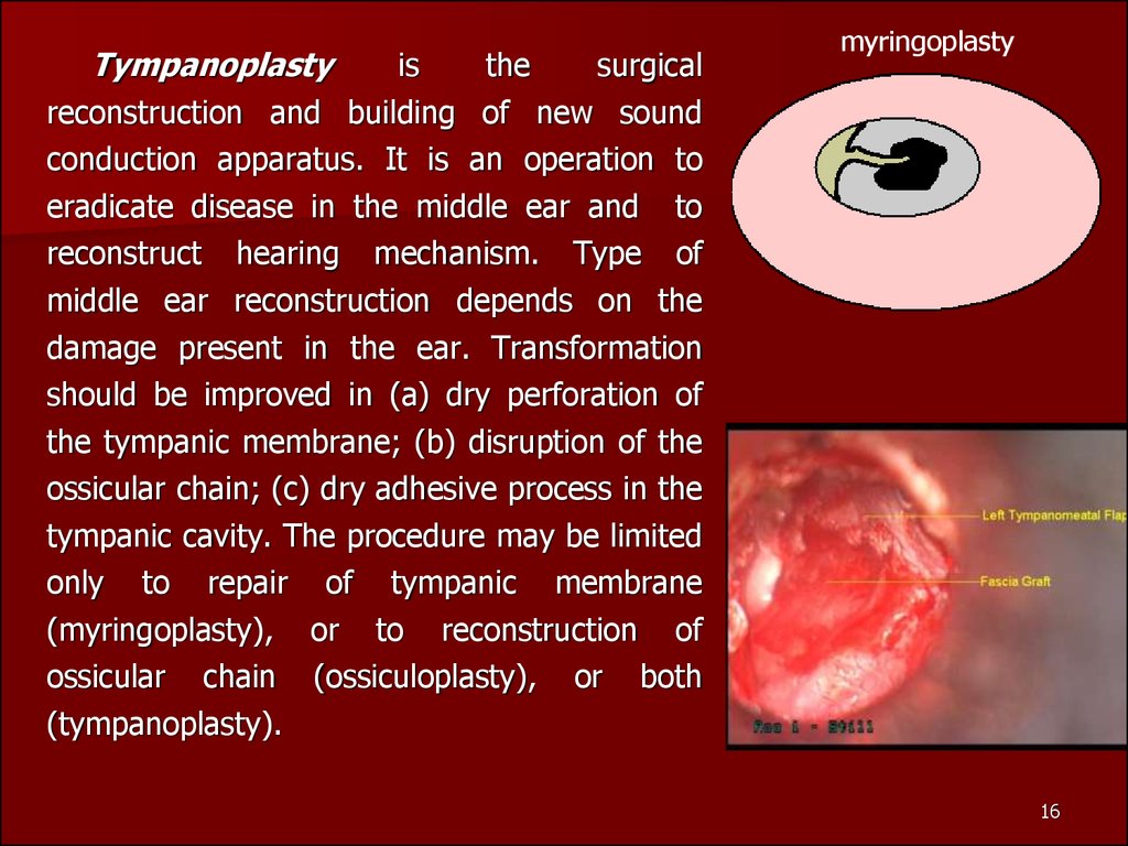
Tympanoplastyis
the
surgical
reconstruction and building of new sound
conduction apparatus. It is an operation to
eradicate disease in the middle ear and to
reconstruct hearing mechanism. Type of
middle ear reconstruction depends on the
damage present in the ear. Transformation
should be improved in (a) dry perforation of
the tympanic membrane; (b) disruption of the
ossicular chain; (c) dry adhesive process in the
tympanic cavity. The procedure may be limited
only to repair of tympanic membrane
(myringoplasty), or to reconstruction of
ossicular chain (ossiculoplasty), or both
(tympanoplasty).
myringoplasty
16
17.
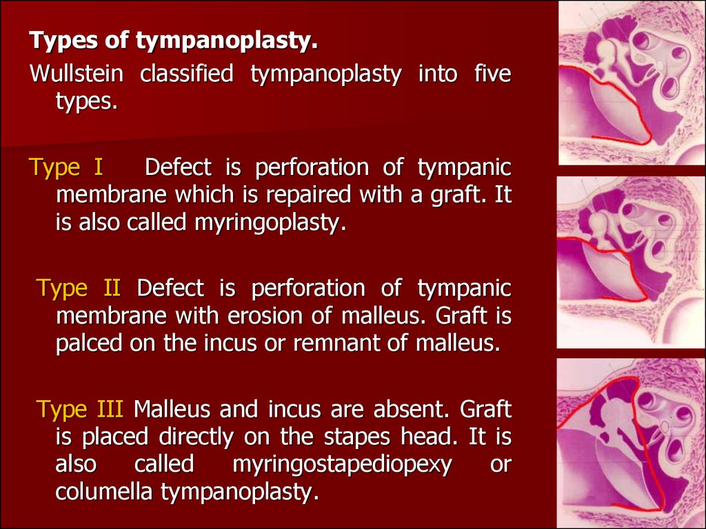
Types of tympanoplasty.Wullstein classified tympanoplasty into five
types.
Type I
Defect is perforation of tympanic
membrane which is repaired with a graft. It
is also called myringoplasty.
Type II Defect is perforation of tympanic
membrane with erosion of malleus. Graft is
palced on the incus or remnant of malleus.
Type III Malleus and incus are absent. Graft
is placed directly on the stapes head. It is
also
called
myringostapediopexy
or
columella tympanoplasty.
17
18.
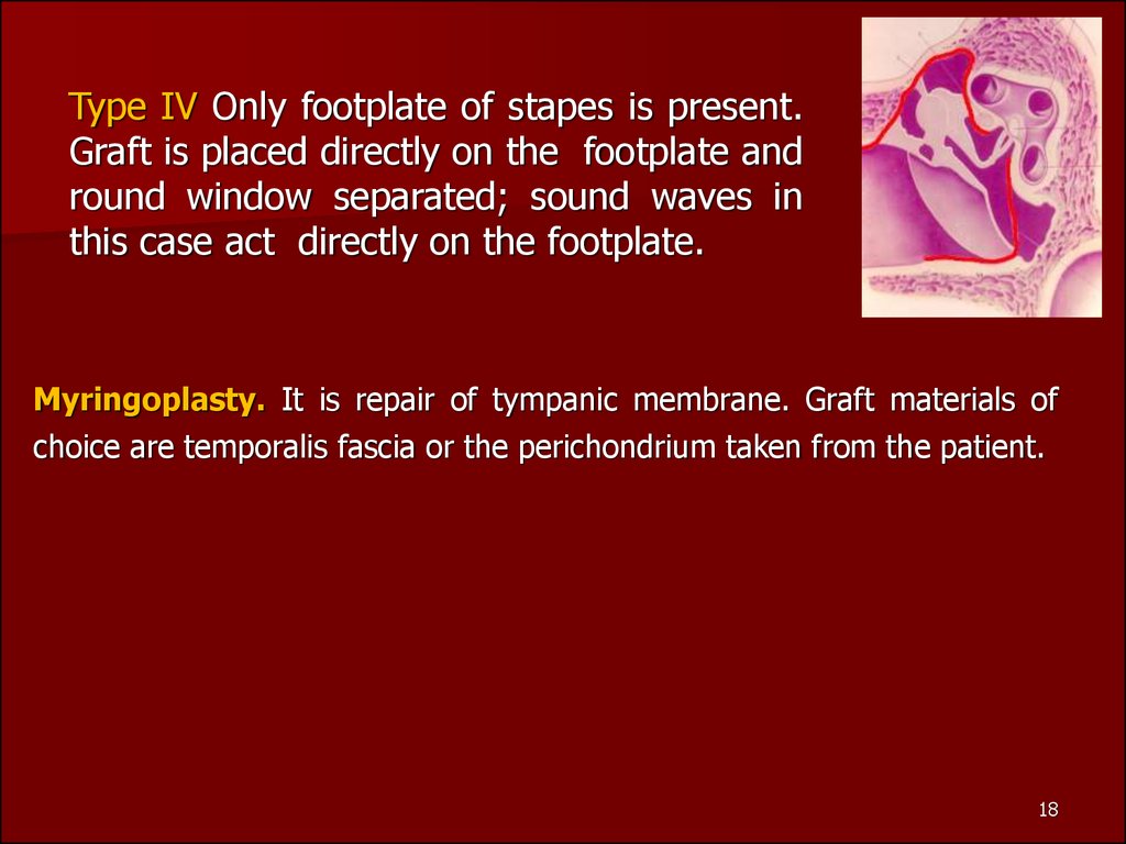
Type IV Only footplate of stapes is present.Graft is placed directly on the footplate and
round window separated; sound waves in
this case act directly on the footplate.
Myringoplasty. It is repair of tympanic membrane. Graft materials of
choice are temporalis fascia or the perichondrium taken from the patient.
18
19.
VESTIBULOCOCHLEAR(COCHLEAR)
NEURITIS.
Neuritis of the vestibulocochlear (auditory) nerve is a collective
term implying affection of any part of the auditory apparatus,
beginning with the neuroepithelial cells of the spiral organ to
the transverse temporal (Heschl's) gyri. It may be present at
birth (congenital) or start later in life (delayed onset or
acquired). Common causes of acquired SNHL include :
1. Infections of labyrinth. Most common causes of the
disease are infectious diseases such as influenza, measles.
2. Trauma to labyrinth or VIII-th nerve, e.g. fractures of
temporal bone.
3. Noise induced hearing loss (acoustic, vibrational.
4. Ototoxic drugs or industrial poisoning.Degenerative
changes in the cells of the organ of hearing prevail in toxic
neuritis caused by medicamentous poisoning (streptomycin,
monomycin, kanamycin).
19
20.
4. Presbycusis.6. Acoustic neuroma.
7. Sudden hearing loss (vessel etiology).
8. Familial progressive SNHL.
9. Systemic disorders, e.g. diabetes, cardiovascular pathology,
hypothyroidism, kidney disease, autoimmune disorders, multiple sclerosis,
as.
A. INFLAMMATIONS OF LABYRINTH
1. Viral labyrinthitis. Viruses usually reach the inner ear by
blood stream affecting stria vascularis and then the endolymph
and organ of corti. Several other viruses, e.g. rubella, herpes
zoster, herpes simplex, influenza and Epstein-Barr are clinically
known to cause deafness but direct proof of their invasion of
labyrinth is lacking.
20
21.
2. Bacterial. Bacterial infections reach labyrinth through themiddle ear (tympanogenic) or through CSF (meningogenic).
Sensorineural deafness following meningitis is a well known
clinical entity.
B.
FAMILIAL
HEARING LOSS
PROGRESSIVE
SENSORINEURAL
It is a genetic disorder in which there is progressive
degeneration of the cochlea startingin late childhood or early
adult life. Deafness is bilateral with flat or basin-shaped
audiogram but an excellent speech discrimination.
C. OTOTOXICITY
1. Aminoglycoside antibiotics. Streptomycin, gentamicin and
tobramycin are primarily vestibulotoxic.They selectively destroy
type I hair cells of the crista ampullaris but, administered in
large doses, can damage the cochlea also.
21
22.
They cause selective destruction of outer hair cells, startingat the basal coil and progressing onto the apex of cochlea.
Symptoms of ototoxicity - hearing loss, tinnitus and/or
giddiness, may manifest during the treatment or after
completion of treatment (delayed toxicity).
2. Diuretics. Furosemide and ethacrinic acid are called loop
diuretics as they block transport of sodium and chloride ions in
the ascending loop of Henle. They are known to cause oedema
and cysticchangesinthe stria vascularis of the cochlear duct.
The effect, in most cases, is reversible but permanent damage
may occur.
3. Salicylates. Symptoms of salicylate ototoxicity are tinnitus
and bilateral sensorineural hearing loss particularly affecting
higher frequencies.
22
23.
Hearing loss due to salicylates is reversible after the drug isdiscontinued.
4. Quinine. Ototoxic symptoms due to quinine are tinnitus
and sensorineural hearing loss, both of which arc reversible.
The symptoms generally appear wilh prolonged medication but
may occur with smaller doses in those who are susceptible.
Congenital deafness and hypoplasia of of cochlea have been
reported in children whose mothers received this drug duringthe first trimester of pregnancy.
5.Topical ear drops. Topical use of drugs in the middle ear
can also cause damage to the cochlea by absorption through
oval and round windows. Deafness has occurred with the use
of chlorhexidine which was used in the preparation of ear
canal before surgery or use of eardrops containing
aminoglycoside anitbiotics, e.g. neomycin and gentamycin.
23
24.
D. NOISE TRAUMA. Hearing loss associated with exposureto noise has been well-known in boiler makers, iron- and
copper-smiths and artillary men. Lately noise trauma has
assumed greater significance because of its being anoccupational hazard, the compensations asked for, and the
responsibilities thrust upon the employer and the employee to
conserve hearing. Hearing loss caused by excessive noise can
be divided into two groups:
Acoustic trauma. Permanent damage to hearing can be
caused by a single brief exposure to very intense sound, e.g.
an explosion, gunfire or a powerful cracker. Noise level in rifle
or a gun fire may reach 140-170 dB SPL. Sudden loud sound
may damage outer hair cells, disrupt the organ of Corti and
rupture the Reissner's membrane. A severe blast may
concomitantly rupture tympanic membrane and disrupt
ossicular chain.
24
25.
F. PRESBYCUSIS. Sensorineural hearing loss associatedwith physiological aging process in the ear is called
presbycusis. It usually manifests at the age of 65 years but
may do so early if there is hereditary predispostion, chronic
noise exposure or generalised vascular disease. Patients of
presbycusis have great difficulty in hearing in the presence of
background noise though they may hear well in quiet
surroundings. They may complain of speech being heard but
not understood. Curtailment of smoking and stimulants like tea
and coffee may help to decrease tinnitus.
Symptoms. Vestibulocochlear neuritis is characterized by
two main symptoms: permanent noise of varied pitch in the
ears due to inflammatory and degenerative process and
vascular disorders, and impaired hearing which is characterized
by inadequate perception of high-pitch sounds and shortened
bone conduction.
25
26.
Less frequently the patients complain of permanent ortransient buzzing (ringing) noise in the ears (tinnitus). If
neuritis further progresses, impaired hearing can turn into
complete deafness. Complete deafness is a total loss of
auditory sensitivity. A rapidly progressing hearing loss is often
attended by symptoms of irritation of the vestibular apparatus;
these are, first of all, vomiting, vertigo, and absence of the
sense of balance. A spontaneous nystagmus can develop.
Diagnosis. A thoroughly collected anamnesis and also
clinical findings are important for diagnosis of vestibulocochlear
neuritis. Tuning-fork and audiometric tests are of leading
importance in topical diagnosis. Hearing disorders associated
with neuritis should be differentiated from perceptive disorders
due to brain tumor, haemorrhage into the internal ear, and
some other affections.
26
27.
Characteristics of sensorinural hearing loss are :1. A positive Rinne test, i.e. air conduction better than bone
conduction.
2. Weber lateralised to better ear.
4. More often involves high frequencies.
5. No gap between air and bone conduction curve on
audiometry
6. Loss may exceed 60 dB. 7. Speech discrimination is poor.
Treatment of infectious neuritis should be aimed at elimination
and neutralization of causes of the disease. We should
prescribe the most rational treatment, which is able to
remove the consequences of actions on to the internal ear.
All the remedies are effective only in the first few weeks
from the beginning of the disease before degenerative
changes in the cochlea. That’s why patients with acute
hardness of hearing need in urgent hospitalization.
27
28.
A doctor prescribes to these patients a confinement to bed,a limit of salt and a liquid food, sedative remedies and active
etiotropic treatment.
1) Complex vitamins In — В1, В6, В12, vitamins A and Є;
2) Cocarboxylase (50—100 Mg I./v. or i/m daily N 10-2
3)Agents which improve microcirculation (Angio protektors
and Disagreegants): Trentalum, Cavintonum.
4) Agents which improve conductivity of a nervous tissue
(Anticholesterase preparations): galantaminum (0,5 % 1,0
subcutaneously N 10).
5) Antihistamine preparations (Suprastinum, Tavegilum)
6) Anticoagulants. In the first days of treatment use a
heparin on 5 000 from i/m 2 times for days, then a dose
depends on indicators coagulogrem.
7)Corticosteroids. 60 mg of Prednisolonum a day throughout
28
2-3 weeks with daily dose depression.
29.
Treatment of toxic neuritis first of all includes prevention offurther ingress of toxins into the body and their immediate
withdrawal from the body. Diuretics and sudorifics should be
given. In cases with acute streptomycin intoxication unithiol
should immediately be administered in combination with
vitamins B group. Unithiol should be injected intramuscularly
or subcutaneously, 1 ml of a 5 per cent solution per 10 kg
body weight of the patient.
During the first day unithiol is administered 3-4 times;
during the second day, 2-3 times; and during the next seven
days, 1-2 times a day.
Prognosis. Fortunately about half the patients of idiopathic
sensorineural hearing loss recover spontaneously within 15
days. Chances of recovery are poor after 1 month. Younger
patients below 40 and those with moderate losses have better
prognosis.
29
30.
Meniere's diseaseThis is a non-suppurative disease of the inner ear characterized by the
classical triad: (1) attacks of systemic labyrinthine vertigo attended with
nausea and vomiting; (2) unilateral hearing loss; (3) noise in the involved
ear. The disease was first described by Prosper Meniere, a French
physician, in 1861.
30
31.
Fluctuation of hearing is a leading diagnostic sign of the auditorydisorder: the hearing can improve considerably between attacks against the
background of a gradually progressing deafness. During the initial stage of
the disease, the hearing function can be restored completely thus indicating
the absence of organic changes in the vestibulocochlear nerve during this
period. Meniere's disease occurs mostly in the young. Its onset is
characterized by the noise in the ear which is followed (in a few hours or
years) by attacks of systemic vertigo and vegetative disorders. An
important point is that the auditory, rather than vestibular, disorders are
typical for the onset of the disease. When establishing a diagnosis, it is
necessary to take into account the periodicity of attacks, their short
duration, good subjective condition of the patient during remission, etc.
The disease should first of all be differentiated from the vascular and
vestibular syndrome, arachnoiditis, and tumour of the cerebellopontine
angle.
31
32.
Treatment. The polyaetiological origin of the disease accounts for themultitude of methods of treating it. Surgical methods of treatment have
been widely used in the recent decade (the operation for decompression of
endolymphatic sac).
Treatment at acute vestibular dysfunction
The patient lays down in a bed in convenient position. Bright light and sharp
sounds is not supposed.
2. To feet of the patient the heater lays down, and on a cervicooccipital site
Sinapismuses are imposed.
3. Medicamental therapy is referred on reduction of intralabyrinthine
pressure and normalisation of a parity of processes of nervous excitation
and inhibition: i/v. enter 20 ml of 40 % of a solution of a glucose, 5 ml of
0,5 % of a solution of Novocainum; i/v enter 2 ml of 2,5 % of a solution of
Pipolphenum or 1 ml of a solution of aminazine of 1 %; Subcutaneously
enter 1 ml of 0,1 % of a solution of atropine (or 2 ml of a solution
Platiphilinum) and 1 ml of 10 % of a solution of caffeine.
4. Highly effective method of elimination of an attack of illness Meniere is
meathotimpanium novocainic blockade.
32
33. Treatment in the period between attack
1. I/v. driply pour 4 % a solution of a hydrocarbonate of sodium of150-200 ml, on a course of 15 injections.
2. Prescribe Disagreegants and Angioprotectors or preparations, which
beter microcirculation
3. Throughout last years in an arsenal of preparations for treatment of
illness Мeniere the appreciable place was occupied with preparation
Betaserc. Thanks to microcirculation improvement it is immediate in
blood vessels of a cochlea and modulation of excitation of neurones
of medial vestibular kernels. The preparation is prescribed on 16 mg
(2 tablets) by 3 times per day throughout 1-2 months
34.
Surgical treatmentIt is used only when medical treatment fails.
Conservative procedures. They are used in cases when vertigo is
disabling hut hearing is still useful and needs to be preserved. They are:
decompression of endolymphatic sac., ultrasonic destruction of vestibular
labyrinth. Cochlear function is preserved. 2. Destructive procedures. They
totally destroy cochlear and vestibular function and are thus used only
when cochlear function is not serviceable.
Patients with Meniere's disease should abstain from work with moving
mechanisms or in conditions of vibration and noise exceeding 70 dB. Work
at high altitudes is also prohibited.
34
35.
OTOSCLEROSIS Otosclerosis is a frequent cause of deafness (it occursin more than 0.5 per cent of cases). The morphological substrate of
otosclerosis is a circumscribed osteodystrophic process manifested by small
single foci of newgrowths in the bony walls of the right and left labyrinths.
These foci are relatively symmetric in the bony capsules of the internal ear.
They grow to replace gradually the wall of the labyrinthine capsule by a
spongioid or dense bone with a different structure. In most cases the
otosclerotic focus is localized anteriorly to the oval window; as it grows, the
focus extends to the stapedovestibular junction, the anterior limb of the
stapes, which impairs mobility of the stapes thus affecting the hearing
function and causing noise in the ear. Hearing is first impaired in one ear;
then, following months or years, the other ear is involved. This form of
otosclerosis is called clinical. If otosclerotic foci are localized outside the
windows of the labyrinth, the form is called histological; it can only be
detected during histological examination of pathological material.
35
36.
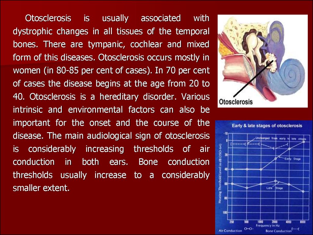
Otosclerosis is usually associated withdystrophic changes in all tissues of the temporal
bones. There are tympanic, cochlear and mixed
form of this diseases. Otosclerosis occurs mostly in
women (in 80-85 per cent of cases). In 70 per cent
of cases the disease begins at the age from 20 to
40. Otosclerosis is a hereditary disorder. Various
intrinsic and environmental factors can also be
important for the onset and the course of the
disease. The main audiological sign of otosclerosis
is considerably increasing thresholds of air
conduction in both ears. Bone conduction
thresholds usually increase to a considerably
smaller extent.
36
37.
Treatment of otosclerosis is surgical. It isactually symptomatic because it does not eliminate
the pathogenic factors of the disease and only
removes to a lesser or greater extent the symptomdeafness and tinnitus. The operation is aimed at
reconstruction of the sound transmission system,
from the ossicles to the perilymph. The mobility of
the base of the stapes in the oval window is
impaired due to the growth of the
otosclerotic focus into the annular
ligament and the base of the stapes
(usually at its anterior pole). The
following
operations
aimed
at
improving the hearing function are
now widely used: Stapedoplasty with
partial or complete stapedectomy, and
Stapedoplasty by a piston method.
37
38.
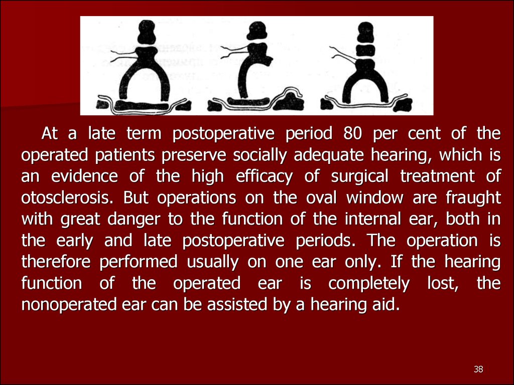
At a late term postoperative period 80 per cent of theoperated patients preserve socially adequate hearing, which is
an evidence of the high efficacy of surgical treatment of
otosclerosis. But operations on the oval window are fraught
with great danger to the function of the internal ear, both in
the early and late postoperative periods. The operation is
therefore performed usually on one ear only. If the hearing
function of the operated ear is completely lost, the
nonoperated ear can be assisted by a hearing aid.
38