


 medicine
Similar presentations:


Otitis Externa
1.
ENTEar
Neck
2.
3.
Otitis ExternaInflammation of the external
auditory canal
Causes: Pseudomonas aeruginosa,
Staphylococcus aureus, Candida and
Aspergillus species
Most common in children 7-14
years old
Symptoms: otalgia, purulent
discharge, pruritis
Risk factors: swimming, humidity,
trauma and others
Exam: pain with movement of ear,
auditory canal is erythematous and
edematous
3
4.
Otitis ExternaDiagnosis: clinical, otoscopy,
culture (for refractory cases),
imaging (mastoiditis)
Treatment:
• Clean the ear canal
• Antibiotic drops (ciprofloxacin)
• Antiseptics, acetic acid
• Control pain: glucocorticoids
4
5.
Malignant (necrotizing) Otitis ExternaInfection of the temporal bone
Dx: CT scan of the temporal bone,
cultures, biopsy of the ear canal
Most common in elderly,
diabetics, immunocompromised
patients
Treatment: intravenous antibiotics
(ciprofloxacin)
Symptoms: severe ear pain, foulsmelling, purulent, CN paralysis
9,10,11
5
6.
ДОБАВИТЬ НИЖНИЙ КОЛОНТИТУЛ6
7.
Acute otitis mediaMost common in children 3 month-3years
Children frequently present with:
• Sudden onset of fever
Symptoms: blocked ear feeling, pain and fever.
Discharge may follow if the TM perforates, with
relief of pain and fever
Causes: adenovirus and enterovirus and the
bacteria H. influenzae, S. pneumoniae, Moraxella
Catarrhalis and β-haemolytic streptococci.
Diagnosis: the redness of the TM. Bulging
eardrum, yellow or white in color with dilated
vessels, decreased movement on pneumatic
otoscopy
• Ear pain
• Fussiness
8.
89.
Acute otitis mediaTreatment
• Analgesics to relieve pain
• Adequate rest in a warm room
• Nasal decongestants for nasal
congestion
• Antibiotics until resolution of all signs
of infection (amoxycillin, doxycycline,
cefaclor)
• Treat associated conditions (e.g.
adenoid hypertrophy)
• Follow-up: review and test hearing
audiometrically
Mild reddening or dullness of the
eardrum and absence of systemic
features (fever and vomiting) antibiotics are not warranted
9
10.
Acute otitis mediaComplications:
• Conductive hearing loss
• Sensorineural hearing loss
• Tympanic membrane perforation
• Retraction pocket
• Mastoiditis
• Petrositis
• Labyrinthitis
10
Perilymphatic fistula
Cholesteatoma
Tympanosclerosis
Cholesterol granuloma
Facial paralysis
Ossicular chain fixation
Ossicular chain discontinuity
11.
Mandy, a 4 year old girl, is due to accompany her parents on a flight to England intwo months time. Her mother is worried about the effect of air travel on Mandy's
ears. Which of the following will NOT increase the likelihood of ear pain during the
flight?
a) A recent cold
b) Nasal congestion
c) Hay fever
d) Recent otitis media
e) Perforation of the ear drum
11
12.
A 8 year old boy with recurrent attacks of otitis media is suspected of developing a glue ear. If hissound conduction is tested, which of the following is most consistent with a unilateral middle ear
effusion?
A) Negative Rinne’s test on the ipsilateral side
B) Positive Rinne’s test on the ipsilateral side
C) Positive Webers and Rinnes test on the ipsilateral side
D) Positive Rinne’s test on the contralateral side
E) Negative Webers test only on the contralateral side
12
13.
ДОБАВИТЬ НИЖНИЙ КОЛОНТИТУЛ13
14.
Chronic otitis mediaSymptoms:
• deafness and discharge without pain
Causes:
• Pseudomonas aeruginosa
• Staphylococcus aureus
• Proteus sp.
• E. coli
• Bacteroides fragilis
Diagnosis: Cl, culture of drainage, imaging
(erosion or abscess)
Safe
• If aural discharge persists for >6 weeks after a
course of antibiotics
Treatment: topical steroid and antibiotic
combination drops, following ear toilet.
Unsafe
• Perforation of the attic region
Treatment: antipseudomonal penicillin or
cephalosporins (children), ear drops &
quinolones (adults)
15.
Chronic otitis mediaSafe Perforation
Unsafe Perforation
• Affects mucosa of the lower front
part of the ME cleft (tubotympanic
portion)
• Threatens the hazard of spread of the
infection intracranially
• Associated with erosion of surrounding
bone
• Central perforation – always a rim of
drum or annulus around the edge
• Involves the vibrating part of the TM
– pars tensa, below the malleolar
folds at the level of the lateral
process of the malleus
ДОБАВИТЬ НИЖНИЙ КОЛОНТИТУЛ
15
16.
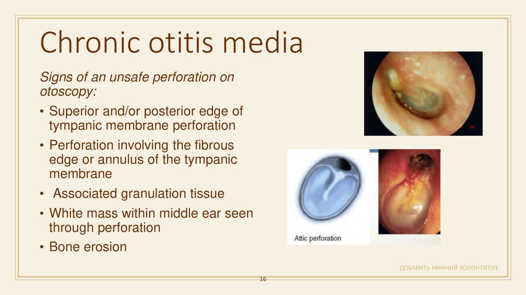
Chronic otitis mediaSigns of an unsafe perforation on
otoscopy:
• Superior and/or posterior edge of
tympanic membrane perforation
• Perforation involving the fibrous
edge or annulus of the tympanic
membrane
• Associated granulation tissue
• White mass within middle ear seen
through perforation
• Bone erosion
ДОБАВИТЬ НИЖНИЙ КОЛОНТИТУЛ
16
17.
A 14-year old teenager is diagnosed with a tympanic membrane perforation secondary tochronic otitis media. Which of the following conditions will not progress to significant
complications if left untreated?
a. Perforation associated with a cholesteatoma
b. Marginal perforation with discharge
c. Continuously discharging central perforation
d. Perforation that is surrounded by granulation tissue
e. Large dry central perforation
ДОБАВИТЬ НИЖНИЙ КОЛОНТИТУЛ
17
18.
CholesteatomaExpanding lesions of the temporal bone
composed of a stratified squamous outer
epithelial lining and a desquamated keratin
center
Symptoms:
• Presenting history (in order of most
common)
• Conductive Hearing Loss
• Foul-smelling ear discharge
Clinically defined as an abnormal extension of
skin into the middle ear and mastoid air cell
spaces
• Persistent otitis media
• Otalgia
• Vertigo
Red flags for cholesteatoma include meningitistype features, cranial nerve deficits,
sensorineural hearing loss and persistent deep
ear pain.
• Facial weakness
ДОБАВИТЬ НИЖНИЙ КОЛОНТИТУЛ
18
19.
CholesteatomaComplications:
• Hearing loss secondary to necrosis of the long process of the incus
• Erosion into the lateral semicircular canal
• Dizziness
• Subperiosteal abscess
• Facial nerve palsy
• Meningitis
• Brain abscess
ДОБАВИТЬ НИЖНИЙ КОЛОНТИТУЛ
19
20.
CholesteatomaDiagnosis: otoscopy, audiogram, CT scan
Treatment:
• Surgery - Mastoidectomy and removal of
cholesteatoma
• Tympanoplasty – an operation to repair a hole
in the eardrum (transcranial or post-auricular
approach)
• Continuous monitoring to look out for
recurrence
20
21.
Audiology21
22.
2223.
Pure tone audiogramConductive hearing loss
Sensorineural hearing loss
23
24.
This pure tone audiogram is recorded from a 12 year old Maori girl complainingof deafness in her right ear. The MOST likely explanation for this problem is:
a) Debris in the external auditory meatus
b) Cholesteatoma
c) Middle ear effusion
d) Toxin-induced nerve damage
e) Necrosis of the ossicular chain
ДОБАВИТЬ НИЖНИЙ КОЛОНТИТУЛ
24
25.
TinnitusExact cause is unknown but is though to be due
to inappropriate activity in the hair cells of the
cochlea
Diagnosis
• Audiological examination by audiologist
• Tympanometry and speech discrimination
• MRI or CT scan (if serious cause suspected or
head injury)
ДОБАВИТЬ НИЖНИЙ КОЛОНТИТУЛ
25
26.
TinnitusObjective Tinnitus
Subjective Tinnitus
Otologic
Presbycusis
• Noise-induced hearing
loss
• Otitis media with effusion
• Menière’s disease
• Otosclerosis
• Cerumen
• Foreign body against TM
Drugs
Vascular
ASA
NSAIDs
Aminoglycosides
Arteriovenous malformation
Antihypertensives
Glomus tympanicum
• Hyper/hypothyroidism
Heavy metals
Glomus jugulare
• Mechanical
Benign intracranial
• Carotid stenosis
• High jugular bulb
hypertension
Arterial bruits:
High-riding carotid artery
Vascular loop
Persistent stapedial artery
26
• Hypertension
• Patulous eustachian tube
• Palatal myoclonus
• Stapedius muscle spasm
27.
TinnitusHolistic approach (options)
Medical (trials of options)
Mainly based on acoustic de-sensitisation:
• Clonazepam 0.5 mg nocte
• Relaxation techniques
• Minerals (e.g. zinc and magnesium)
• Tinnitus retraining therapy (clinical psychologist)
• Betahistine (Serc) 8–16 mg daily (max. 32 mg)
• Cognitive behaviour therapy
• Carbamazepine
• Background ‘noise’ (e.g. music played during night
• Antidepressants
for masking)
• Acute severe tinnitus
• Tinnitus maskers
• Lignocaine 1% IV slowly (up to 5 mL)
• Hearing aids (based on audiologist assessment)
• Consider hypnotherapy
ДОБАВИТЬ НИЖНИЙ КОЛОНТИТУЛ
27
28.
You review a 70-year-old woman who is on multiple medications. For the past fewmonths she has noticed bilateral tinnitus and hearing loss. Which one of the following
medications may be responsible?
A) Lofepramine
B) Ezetimibe
C) Furosemide
D) Tramadol
E) Digoxin
ДОБАВИТЬ НИЖНИЙ КОЛОНТИТУЛ
28
29.
ДОБАВИТЬ НИЖНИЙ КОЛОНТИТУЛ29
30.
Benign paroxysmal positional vertigoAcute vertigo that is induced by changing head position
Diagnosis:
Pathognomonic sign: nystagmus toward the affected
ear on doing a Dix-Hallpike test
Caused by sediment, such as otoconia (calcium
carbonate crystals) that have become free floating
within the inner ear
Treatment:
• Affects all ages, especially the elderly
• The female to male ratio is 2:1
• Avoidance measures: encourage the patient to
move in ways that avoid the attack
• Recurs periodically for several days
• Drugs are not recommended
• Each attack is brief, usually lasts 10–60 seconds, and
subsides rapidly
• Epley repositioning maneuver
• Severe vertigo on getting out of bed
• Brandt-Daroff exercises
• Can occur on head extension and turning head in bed
• For pations: Brandt-Daroff Exercises
• Attacks are not accompanied by vomiting, tinnitus or
deafness (nausea may occur)
ДОБАВИТЬ НИЖНИЙ КОЛОНТИТУЛ
30
31.
A 52-year-old woman presents with dizziness and vertigo when she moves her headtowards right and in extension of head.
She is asymptomatic when she is lying still on the bed and is not moving her head. She
is having these symptoms since last 3 hours. She had similar symptoms 5 years ago
when she recovered in two days. There is no neurological deficit on examination apart
from positional nystagmus.
Which of the following is best management?
a. Hallpike manoeuvre
b. Epley manoeuvre
c. Frusemide
d. Intravenous fluids
e. Steroids
ДОБАВИТЬ НИЖНИЙ КОЛОНТИТУЛ
31
32.
Vestibular neuritis• Usually subsides over a course of several
days or weeks
• Second most common disorder affecting the
labyrinth
• Viral etiology with consequent inflammation of
the vestibular nerve
• Differential diagnoses: cerebellar
hemorrhage and infarction
• Frequently associated with recent flu
symptoms
• Labyrinthitis refers to the simultaneous loss
of hearing and balance in the affected ear
Signs/symptoms:
• Sudden onset of severe rotatory vertigo
• Nausea and vomiting
• Spontaneous nystagmus and diminished VOR
32
33.
Vestibular neuritisTreatment:
• Bed rest, vestibular sedatives and anti-emetics in the first 24-72
hours
• Dimenhydrinate
• Prochlorperazine
• Diazepam
• Short tapering course of oral steroids
• Vestibular adaptation exercises/rehabilitation in the recovery phase
ДОБАВИТЬ НИЖНИЙ КОЛОНТИТУЛ
33
34.
3435.
Meniere’s diseaseSymptoms:
• It is commonest in the 30–50 years age group
• Typical history consists of recurrent
• Triggers: high salt intake, chocolate, alcohol,
smoking, stress, menstrual cycle
attacks of vertigo, tinnitus, and
ipsilateral hearing loss
• Attacks last 30 minutes to several hours.
• There is a variable interval between attacks
(twice a month to twice a year).
• Nausea and vomiting
• SNHL is fluctuating and progressive
• Nystagmus is observed only during an attack
(often to side opposite affected inner ear).
ДОБАВИТЬ НИЖНИЙ КОЛОНТИТУЛ
35
36.
Meniere disease• DxT vertigo + vomiting + tinnitus + sensorineural deafenss → Ménière syndrome
Treatment:
• Low salt diet +/- diuretic for maintenance treatment (hydrochlorothiazide, acetazolamide)
• Vestibular sedative, antiemetic for acute episodes
• Prochloperazine
• Diazepam
• Vasodilators - Betahistine
• Meniette device
• Intra-tympanic therapy – steroids, aminoglycosides
• Surgery for refractive cases
ДОБАВИТЬ НИЖНИЙ КОЛОНТИТУЛ
36
37.
An elderly patient has acute onset unilateral deafness, tinnitis & vertigo. What is thediagnosis?
a) Meniere’s disease
b) Acoustic neuroma
c) Vestibula neuronitis
d) Positional vertigo
ДОБАВИТЬ НИЖНИЙ КОЛОНТИТУЛ
37
38.
A 39-year-old woman arrives at the hospital after her third episode of dizziness.Her first episode was 6 months ago and her most recent episode occurred yesterday. She describes
feeling as if the room was spinning around her. During each of these episodes she has experienced
significant nausea, often accompanied by emesis. Upon further questioning she tells you that she has
been hearing a low rumbling noise in her right ear.
What test is required to confirm your diagnosis?
(A) CT head
(B) MRI head
(C) Audiogram
(D) Tilt table test
(E) No need for further testing
ДОБАВИТЬ НИЖНИЙ КОЛОНТИТУЛ
38
39.
Acoustic neuroma (vestibular schwannomas)• Benign tumour of Schwann cells surrounding
auditory nerve
Symptoms:
• Unilateral progressive SNHL 85%
• Sudden hearing loss 15%
• Usually unilateral
• Tinnitus 56%
• Bilateral tumour associated with Type 2
Neurofibromatosis
• Vertigo 19%
• Midface hypesthesia,
• Cranial nerve V and VII Facial paresis
• Chromosome 22 abnormality
• Diplopia, dysphagia, hoarseness, aspiration,
• Autosomal dominant transmission
cerebellar ataxia
• Hydrocephalus: headache and vomiting
• DxT (unilateral) tinnitus + hearing loss +
unsteady gait → acoustic neuroma
ДОБАВИТЬ НИЖНИЙ КОЛОНТИТУЛ
39
40.
Acoustic neuromaDiagnosis: audiometry (SNHL), MRI, CT
Treatment:
• Conservative: monitoring
• Surgical resection
Translabyrinthine, middle fossa, or suboccipital retrosigmoid
approaches
Stereotactic radiosurgery (gamma knife)
ДОБАВИТЬ НИЖНИЙ КОЛОНТИТУЛ
40
41.
Which of the following is least likely to cause facialnerve palsy?
a) Skull fracture
b) Mastoiditis
c) Chronic parotitis
d) Parotid tumour
e) Acoustic neuroma
ДОБАВИТЬ НИЖНИЙ КОЛОНТИТУЛ
41
42.
ДОБАВИТЬ НИЖНИЙ КОЛОНТИТУЛ42
43.
Otosclerosis• Disease of the bone surrounding the inner
ear and is the most common cause of
conductive hearing loss in the adult with a
normal tympanic membrane
Features:
• Progressive disease
• Develops in the 20s and 30s
• Family history (autosomal dominant)
• Unilateral or bilateral
• The normal middle ear bone is replaced
by vascular, spongy bone that becomes
sclerotic
• Female preponderance
• Stapes footplate is affected
• May progress rapidly during pregnancy
• Conductive hearing loss
ДОБАВИТЬ НИЖНИЙ КОЛОНТИТУЛ
43
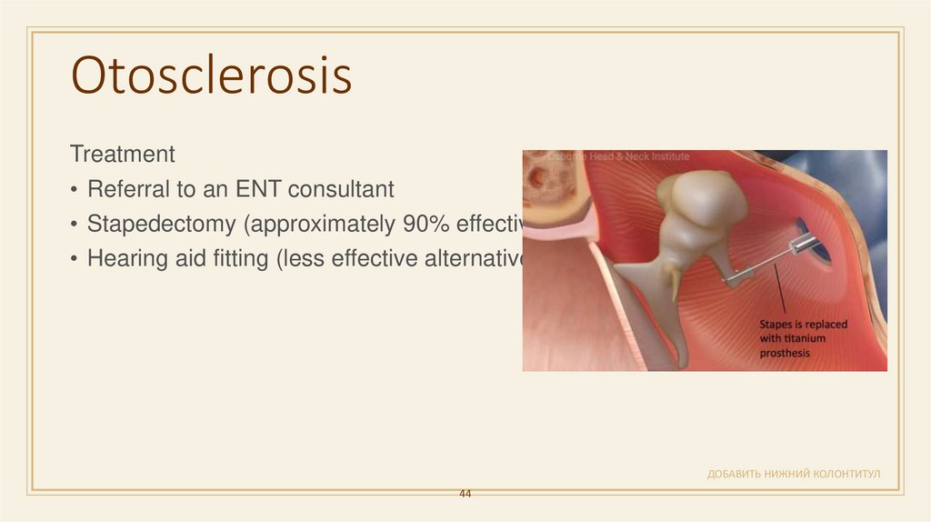
44.
OtosclerosisTreatment
• Referral to an ENT consultant
• Stapedectomy (approximately 90% effective)
• Hearing aid fitting (less effective alternative)
ДОБАВИТЬ НИЖНИЙ КОЛОНТИТУЛ
44
45.
Which of the following is most likely to be associated with otosclerosis?a) Normal tympanic membrane
b) Red & inflamed tympanic membrane
c) Tense & transparent tympanic membrane with fluid level behind
d) Blue gray sclera
e) Obstruction of the Eustachian tube
ДОБАВИТЬ НИЖНИЙ КОЛОНТИТУЛ
45
46.
ДОБАВИТЬ НИЖНИЙ КОЛОНТИТУЛ46
47.
Head and neck massesДОБАВИТЬ НИЖНИЙ КОЛОНТИТУЛ
47
48.
Cystic lesions of the neckДОБАВИТЬ НИЖНИЙ КОЛОНТИТУЛ
48
49.
Cystic hygroma• Commonly involves the posterior cervical
space
May be macrocystic or microcystic
MRI is the gold standard for radiologic
evaluation
ДОБАВИТЬ НИЖНИЙ КОЛОНТИТУЛ
49
50.
Branchial deft cyst• located inferior to the external auditory
meatus or anterior to the sternomastoid
muscle. The opening may
• Most common cystic lesion of the anterior
triangle of
• the neck in children
• discharge mucous. A skin tag or cartilage
remnant may be present. Refer when
diagnosed for excision.
Unilocular, cystic mass displacing the
submandibular
• gland anteriorly and the sternocleidomastoid
• muscle posteriorly
ДОБАВИТЬ НИЖНИЙ КОЛОНТИТУЛ
50
51.
Thyroglossal duct cyst• the most common childhood midline neck
swelling
• Midline lesion anywhere from foramen caecum
and
• the thyroid gland
• It moves with swallowing and tongue
protrusion. It is prone to infection, including
• abscess formation. The cyst and its tract are
best excised before it
• becomes infected.
Moves with protrusion of the tongue
May contain ectopic thyroid tissue
May contain all of the functioning thyroid
Ultrasound, thyroid scans
Surgical excision -- Sistrunk procedure
ДОБАВИТЬ НИЖНИЙ КОЛОНТИТУЛ
51
52.
Carotid body tumor• Diagnosis: ultrasonography with
color Doppler, CT,MRI
• The carotid body is a small, reddishbrown, oval structure, located in the
posteromedial aspect of the carotid
artery bifurcation.
• Carotid body tumors (CBTs) present
most commonly as an asymptomatic
palpable neck mass in the anterior
triangle of the neck
• They are slow-growing tumors that
can remain asymptomatic for many
years.
• Symptoms cranial nerve palsy 10%,
pain, hoarseness, dysphagia, Horner
syndrome, or shoulder drop.
• Treatment: surgery or radiotherapy
ДОБАВИТЬ НИЖНИЙ КОЛОНТИТУЛ
52
53.
SCM tumor• hard painless lump (2–3 cm long) within
sternomastoid muscle
• tight and shortened sternomastoid muscle
• usually not observed at birth
• appears at 20–30 days of age
• associated torticollis—head turned away from
but tilted towards the
• Tumour restricted head rotation to side of
tumour
ДОБАВИТЬ НИЖНИЙ КОЛОНТИТУЛ
53