

 medicine
 biology
Similar presentations:


")


Pulmonary diseases
1.
Pulmonary diseasesTsukanov Egor
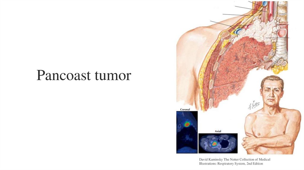
Pancoast tumor
Idiopathic pulmonary fibrosis
Khoroshaev Oleg
Infiltrative form of the secondary tuberculosis
Alveolar echinococcosis
Pulmonary hamartoma
Alveolar adenoma
Sclerosing hemangioma
Syphiliswith Pulmonary Involvement
2.
Pancoast tumorDavid Kaminsky The Netter Collection of Medical
Illustrations: Respiratory System, 2nd Edition
3.
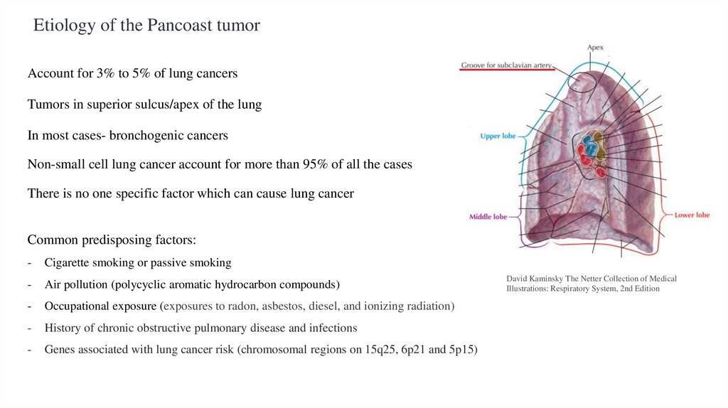
Etiology of the Pancoast tumorAccount for 3% to 5% of lung cancers
Tumors in superior sulcus/apex of the lung
In most cases- bronchogenic cancers
Non-small cell lung cancer account for more than 95% of all the cases
There is no one specific factor which can cause lung cancer
Common predisposing factors:
-
Cigarette smoking or passive smoking
-
Air pollution (polycyclic aromatic hydrocarbon compounds)
-
Occupational exposure (exposures to radon, asbestos, diesel, and ionizing radiation)
-
History of chronic obstructive pulmonary disease and infections
-
Genes associated with lung cancer risk (chromosomal regions on 15q25, 6p21 and 5p15)
David Kaminsky The Netter Collection of Medical
Illustrations: Respiratory System, 2nd Edition
4.
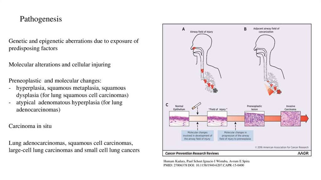
PathogenesisGenetic and epigenetic aberrations due to exposure of
predisposing factors
Molecular alterations and cellular injuring
Preneoplastic and molecular changes:
- hyperplasia, squamous metaplasia, squamous
dysplasia (for lung squamous cell carcinomas)
- atypical adenomatous hyperplasia (for lung
adenocarcinomas)
Carcinoma in situ
Lung adenocarcinomas, squamous cell carcinomas,
large-cell lung carcinomas and small cell lung cancers
Humam Kadara, Paul Scheet Ignacio I Wistuba, Avrum E Spira
PMID: 27006378 DOI: 10.1158/1940-6207.CAPR-15-0400
5.
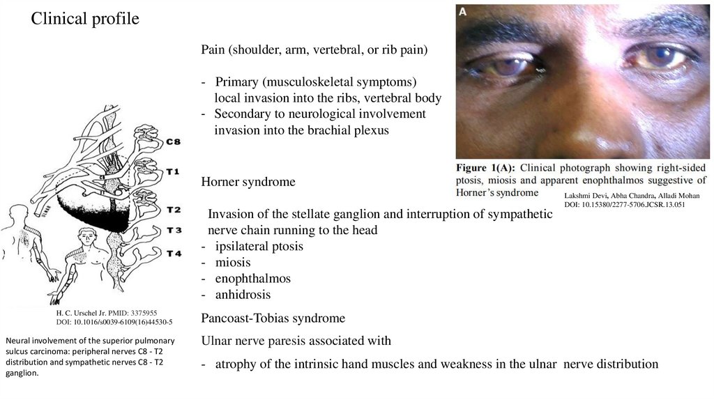
Сlinical profilePain (shoulder, arm, vertebral, or rib pain)
- Primary (musculoskeletal symptoms)
local invasion into the ribs, vertebral body
- Secondary to neurological involvement
invasion into the brachial plexus
Horner syndrome
Lakshmi Devi, Abha Chandra, Alladi Mohan
DOI: 10.15380/2277-5706.JCSR.13.051
Invasion of the stellate ganglion and interruption of sympathetic
nerve chain running to the head
- ipsilateral ptosis
- miosis
- enophthalmos
- anhidrosis
H. C. Urschel Jr. PMID: 3375955
DOI: 10.1016/s0039-6109(16)44530-5
Neural involvement of the superior pulmonary
sulcus carcinoma: peripheral nerves C8 - T2
distribution and sympathetic nerves C8 - T2
ganglion.
Pancoast-Tobias syndrome
Ulnar nerve paresis associated with
- atrophy of the intrinsic hand muscles and weakness in the ulnar nerve distribution
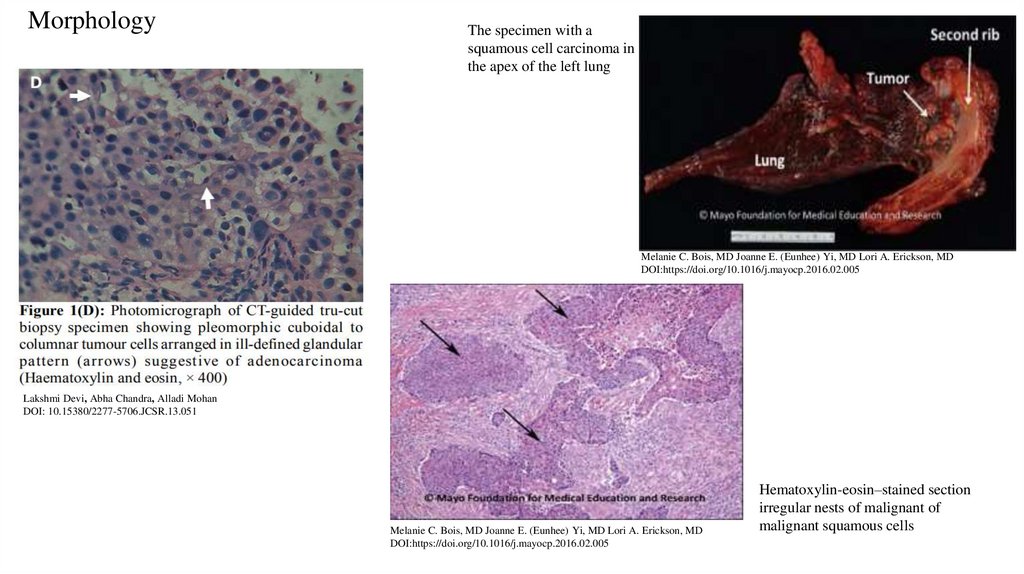
6.
MorphologyThe specimen with a
squamous cell carcinoma in
the apex of the left lung
Melanie C. Bois, MD Joanne E. (Eunhee) Yi, MD Lori A. Erickson, MD
DOI:https://doi.org/10.1016/j.mayocp.2016.02.005
Lakshmi Devi, Abha Chandra, Alladi Mohan
DOI: 10.15380/2277-5706.JCSR.13.051
Melanie C. Bois, MD Joanne E. (Eunhee) Yi, MD Lori A. Erickson, MD
DOI:https://doi.org/10.1016/j.mayocp.2016.02.005
Hematoxylin-eosin–stained section
irregular nests of malignant of
malignant squamous cells
7.
References8.
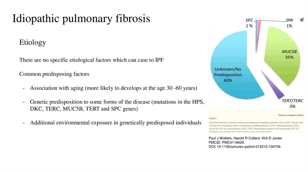
Idiopathic pulmonary fibrosisEtiology
There are no specific etiological factors which can case to IPF
Common predisposing factors
- Association with aging (more likely to develops at the age 30 -60 years)
- Genetic predisposition to some forms of the disease (mutations in the HPS,
DKC, TERC, MUC5B, TERT and SPC genes)
- Additional environmental exposure in genetically predisposed individuals
Paul J Wolters, Harold R Collard, Kirk D Jones
PMCID: PMC4116429
DOI: 10.1146/annurev-pathol-012513-104706
9.
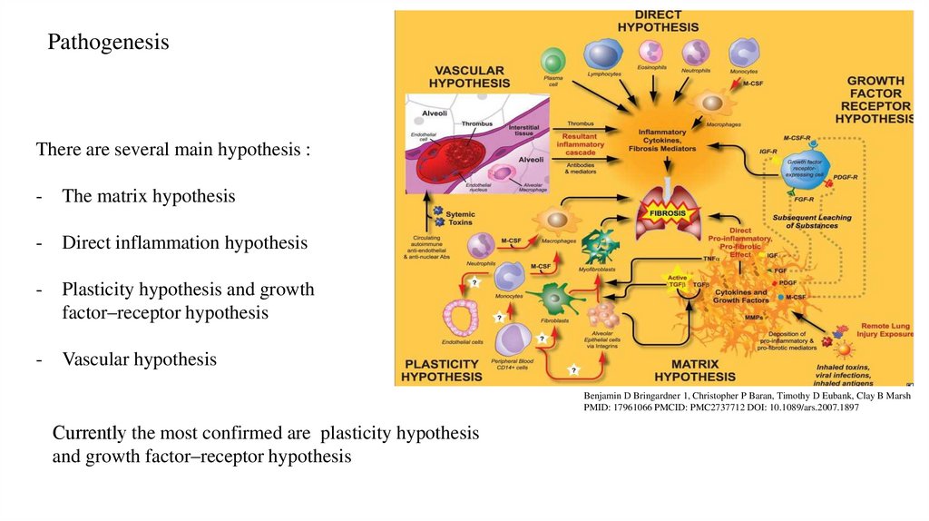
PathogenesisThere are several main hypothesis :
-
The matrix hypothesis
-
Direct inflammation hypothesis
-
Plasticity hypothesis and growth
factor–receptor hypothesis
-
Vascular hypothesis
Benjamin D Bringardner 1, Christopher P Baran, Timothy D Eubank, Clay B Marsh
PMID: 17961066 PMCID: PMC2737712 DOI: 10.1089/ars.2007.1897
Сurrently the most confirmed are plasticity hypothesis
and growth factor–receptor hypothesis
10.
The main mechanisms of plasticity hypothesis and growthfactor-receptor hypothesis
- Endoplasmic reticulum stress
- Transforming growth factor activation (TGF-β activation)
Paul J Wolters, Harold R Collard, Kirk D Jones PMCID: PMC4116429
DOI: 10.1146/annurev-pathol-012513-104706
11.
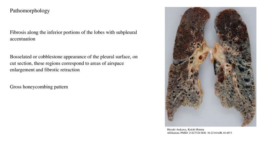
PathomorphologyFibrosis along the inferior portions of the lobes with subpleural
accentuation
Bosselated or cobblestone appearance of the pleural surface, on
cut section, these regions correspond to areas of airspace
enlargement and fibrotic retraction
Gross honeycombing pattern
Hiroaki Arakawa, Koichi Honma
Affiliations PMID: 21427324 DOI: 10.2214/AJR.10.4873
12.
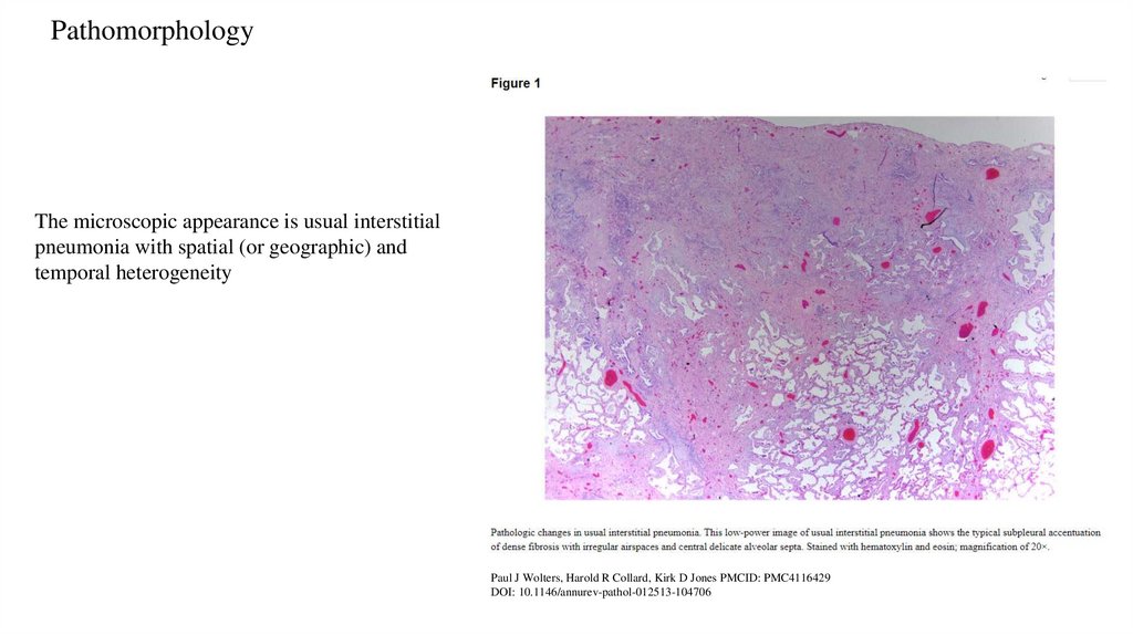
PathomorphologyThe microscopic appearance is usual interstitial
pneumonia with spatial (or geographic) and
temporal heterogeneity
Paul J Wolters, Harold R Collard, Kirk D Jones PMCID: PMC4116429
DOI: 10.1146/annurev-pathol-012513-104706
13.
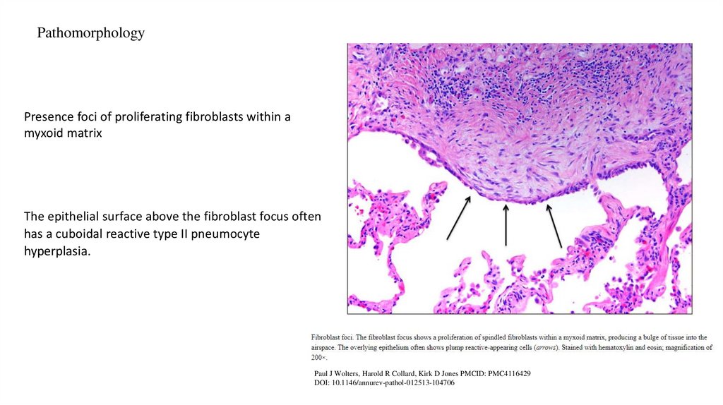
PathomorphologyPresence foci of proliferating fibroblasts within a
myxoid matrix
The epithelial surface above the fibroblast focus often
has a cuboidal reactive type II pneumocyte
hyperplasia.
Paul J Wolters, Harold R Collard, Kirk D Jones PMCID: PMC4116429
DOI: 10.1146/annurev-pathol-012513-104706
14.
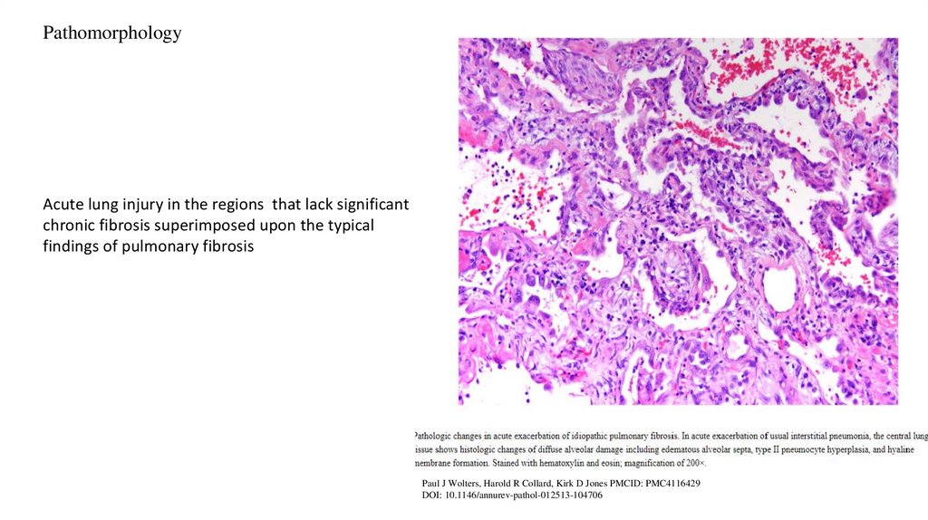
PathomorphologyAcute lung injury in the regions that lack significant
chronic fibrosis superimposed upon the typical
findings of pulmonary fibrosis
Paul J Wolters, Harold R Collard, Kirk D Jones PMCID: PMC4116429
DOI: 10.1146/annurev-pathol-012513-104706
15.
References16.
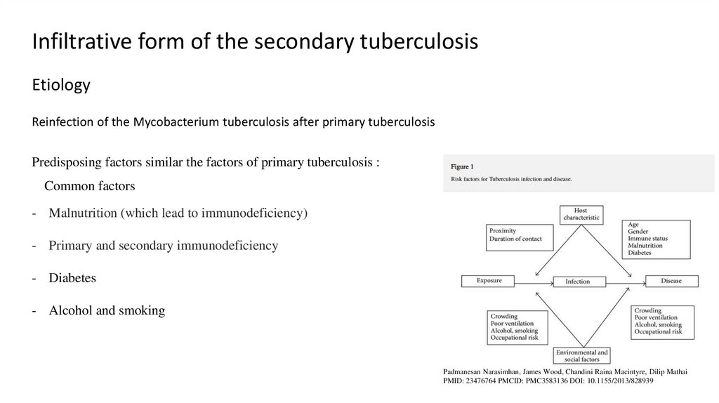
Infiltrative form of the secondary tuberculosisEtiology
Reinfection of the Mycobacterium tuberculosis after primary tuberculosis
Predisposing factors similar the factors of primary tuberculosis :
Common factors
- Malnutrition (which lead to immunodeficiency)
- Primary and secondary immunodeficiency
- Diabetes
- Alcohol and smoking
Padmanesan Narasimhan, James Wood, Chandini Raina Macintyre, Dilip Mathai
PMID: 23476764 PMCID: PMC3583136 DOI: 10.1155/2013/828939
17.
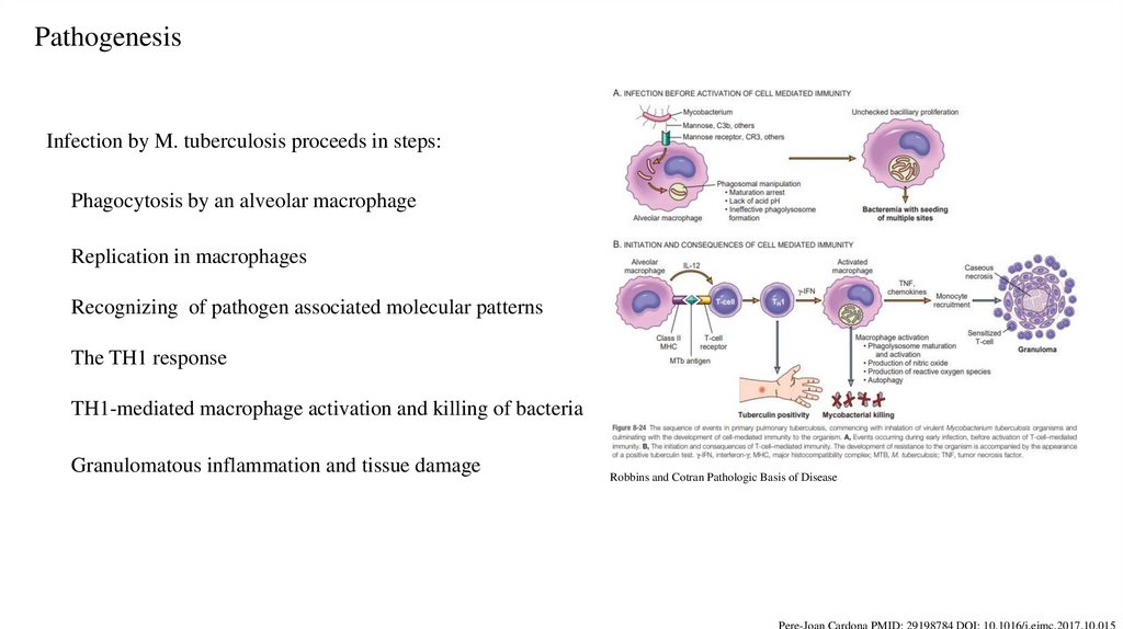
PathogenesisInfection by M. tuberculosis proceeds in steps:
Phagocytosis by an alveolar macrophage
Replication in macrophages
Recognizing of pathogen associated molecular patterns
The TH1 response
TH1-mediated macrophage activation and killing of bacteria
Granulomatous inflammation and tissue damage
Robbins and Cotran Pathologic Basis of Disease
Pere-Joan Cardona PMID: 29198784 DOI: 10.1016/j.eimc.2017.10.015
18.
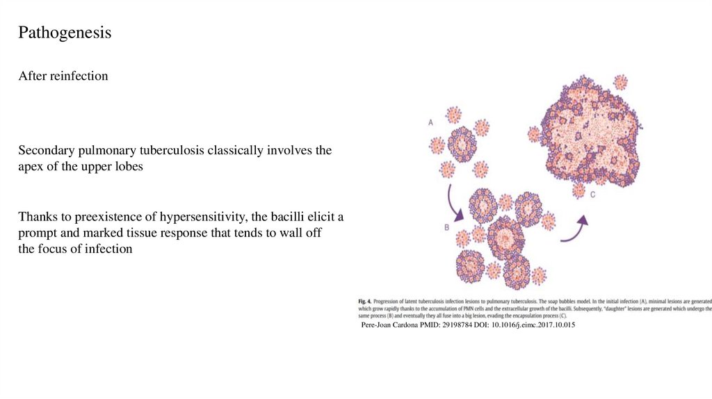
PathogenesisAfter reinfection
Secondary pulmonary tuberculosis classically involves the
apex of the upper lobes
Thanks to preexistence of hypersensitivity, the bacilli elicit a
prompt and marked tissue response that tends to wall off
the focus of infection
Pere-Joan Cardona PMID: 29198784 DOI: 10.1016/j.eimc.2017.10.015
19.
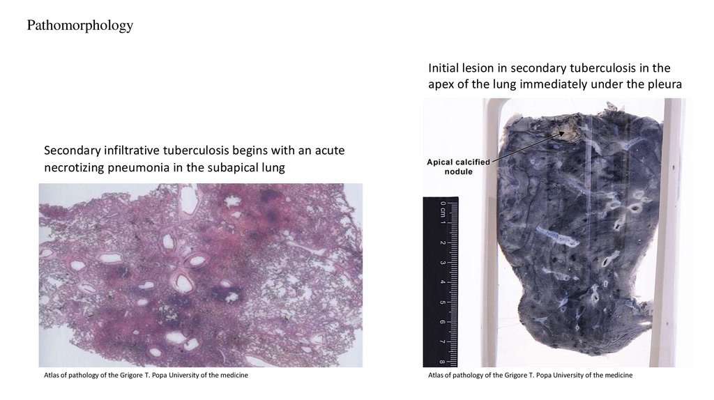
PathomorphologyInitial lesion in secondary tuberculosis in the
apex of the lung immediately under the pleura
Secondary infiltrative tuberculosis begins with an acute
necrotizing pneumonia in the subapical lung
Atlas of pathology of the Grigore T. Popa University of the medicine
Atlas of pathology of the Grigore T. Popa University of the medicine
20.
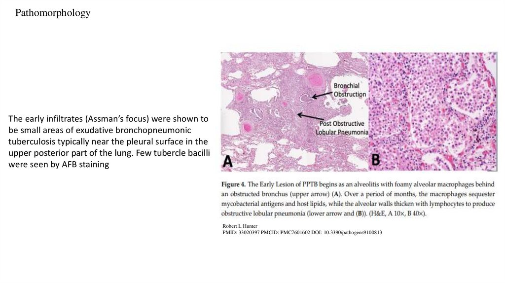
PathomorphologyThe early infiltrates (Assman’s focus) were shown to
be small areas of exudative bronchopneumonic
tuberculosis typically near the pleural surface in the
upper posterior part of the lung. Few tubercle bacilli
were seen by AFB staining
Robert L Hunter
PMID: 33020397 PMCID: PMC7601602 DOI: 10.3390/pathogens9100813
21.
References22.
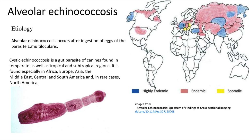
Alveolar echinococcosisEtiology
Alveolar echinococcosis occurs after ingestion of eggs of the
parasite E.multilocularis.
Cystic echinococcosis is a gut parasite of canines found in
temperate as well as tropical and subtropical regions. It is
found especially in Africa, Europe, Asia, the
Middle East, Central and South America and, in rare cases,
North America
images from
Alveolar Echinococcosis: Spectrum of Findings at Cross-sectional Imaging
doi.org/10.1148/rg.327125708
23.
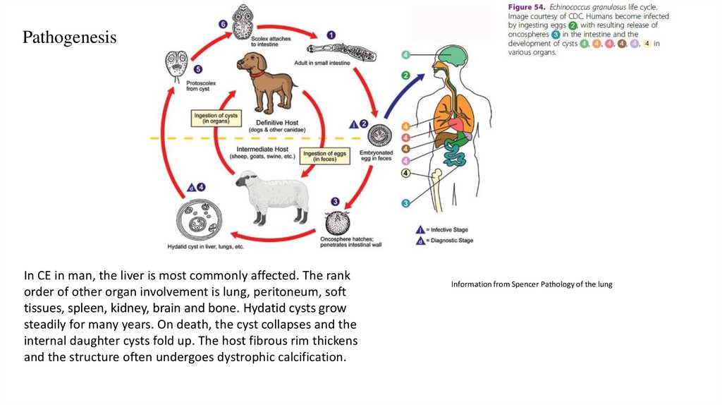
PathogenesisIn CE in man, the liver is most commonly affected. The rank
order of other organ involvement is lung, peritoneum, soft
tissues, spleen, kidney, brain and bone. Hydatid cysts grow
steadily for many years. On death, the cyst collapses and the
internal daughter cysts fold up. The host fibrous rim thickens
and the structure often undergoes dystrophic calcification.
Information from Spencer Pathology of the lung
24.
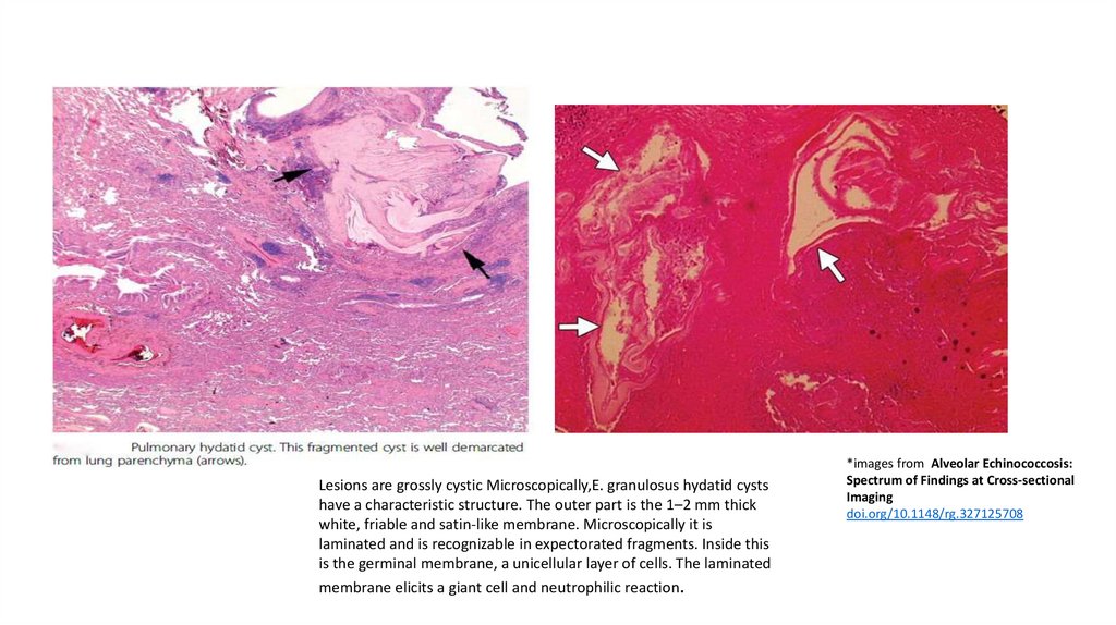
Lesions are grossly cystic Microscopically,E. granulosus hydatid cystshave a characteristic structure. The outer part is the 1–2 mm thick
white, friable and satin-like membrane. Microscopically it is
laminated and is recognizable in expectorated fragments. Inside this
is the germinal membrane, a unicellular layer of cells. The laminated
membrane elicits a giant cell and neutrophilic reaction.
*images from Alveolar Echinococcosis:
Spectrum of Findings at Cross-sectional
Imaging
doi.org/10.1148/rg.327125708
25.
26.
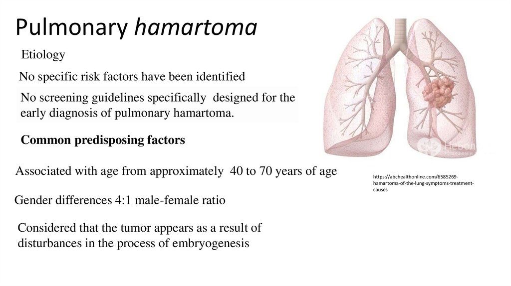
Pulmonary hamartomaEtiology
No specific risk factors have been identified
No screening guidelines specifically designed for the
early diagnosis of pulmonary hamartoma.
Common predisposing factors
Associated with age from approximately 40 to 70 years of age
Gender differences 4:1 male-female ratio
Considered that the tumor appears as a result of
disturbances in the process of embryogenesis
https://abchealthonline.com/6585269hamartoma-of-the-lung-symptoms-treatmentcauses
27.
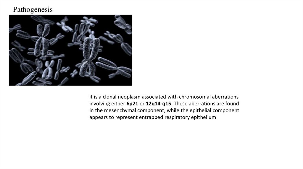
Pathogenesisit is a clonal neoplasm associated with chromosomal aberrations
involving either 6p21 or 12q14-q15. These aberrations are found
in the mesenchymal component, while the epithelial component
appears to represent entrapped respiratory epithelium
28.
PathomorphologyImages from:
Hamartoma
Updated: Oct 09, 2019
Author: Rohit Seth, MD,
PhD, MRCS(Edin); Chief
Editor: Harris Gellman
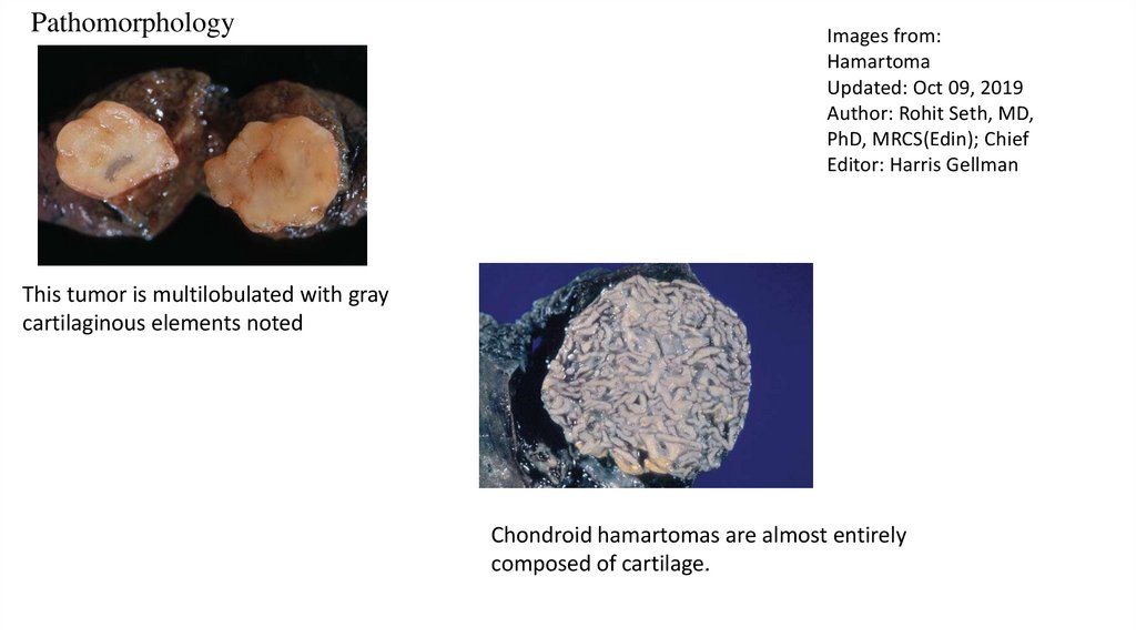
This tumor is multilobulated with gray
cartilaginous elements noted
Chondroid hamartomas are almost entirely
composed of cartilage.
29.
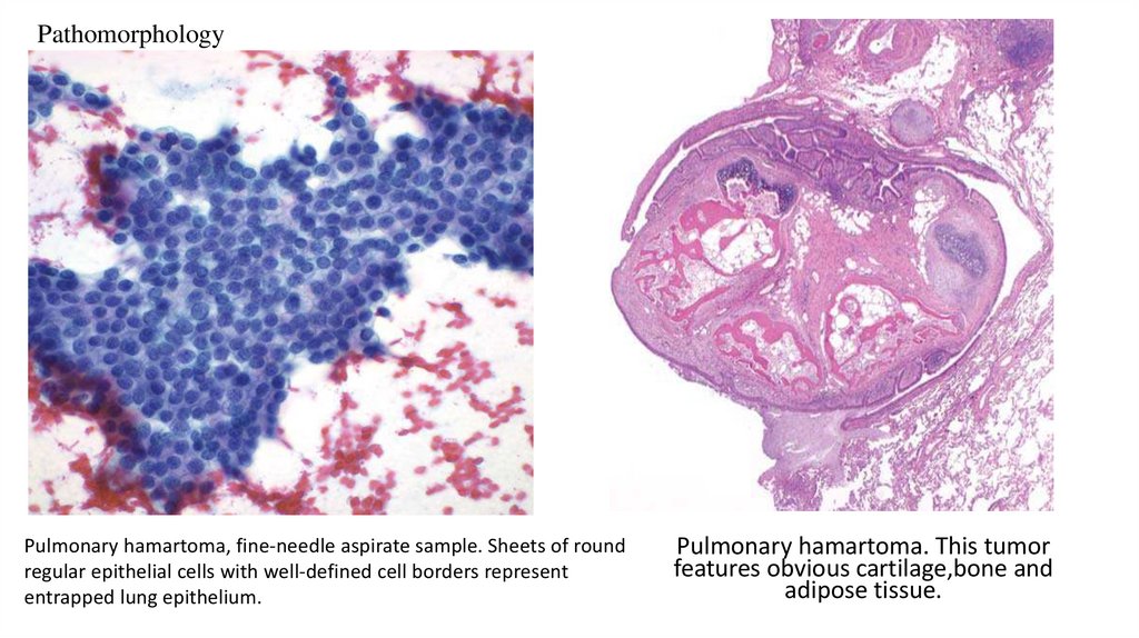
PathomorphologyPulmonary hamartoma, fine-needle aspirate sample. Sheets of round
regular epithelial cells with well-defined cell borders represent
entrapped lung epithelium.
Pulmonary hamartoma. This tumor
features obvious cartilage,bone and
adipose tissue.
30.
31.
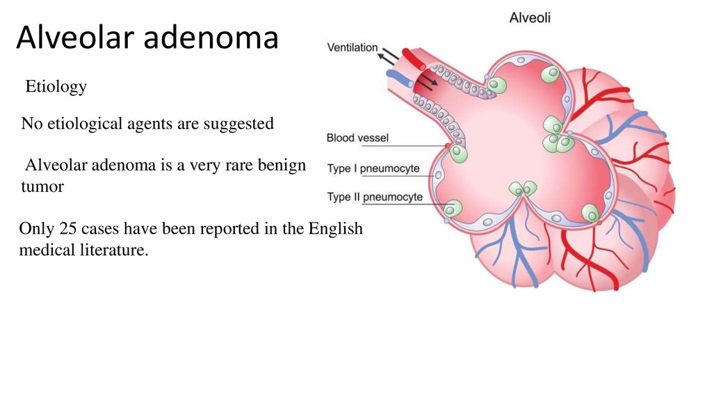
Alveolar adenomaEtiology
No etiological agents are suggested
Alveolar adenoma is a very rare benign
tumor
Only 25 cases have been reported in the English
medical literature.
32.
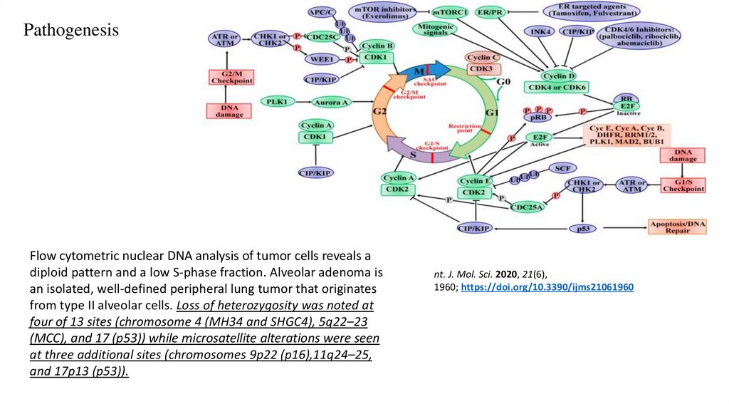
PathogenesisFlow cytometric nuclear DNA analysis of tumor cells reveals a
diploid pattern and a low S-phase fraction. Alveolar adenoma is
an isolated, well-defined peripheral lung tumor that originates
from type II alveolar cells. Loss of heterozygosity was noted at
four of 13 sites (chromosome 4 (MH34 and SHGC4), 5q22–23
(MCC), and 17 (p53)) while microsatellite alterations were seen
at three additional sites (chromosomes 9p22 (p16),11q24–25,
and 17p13 (p53)).
nt. J. Mol. Sci. 2020, 21(6),
1960; https://doi.org/10.3390/ijms21061960
33.
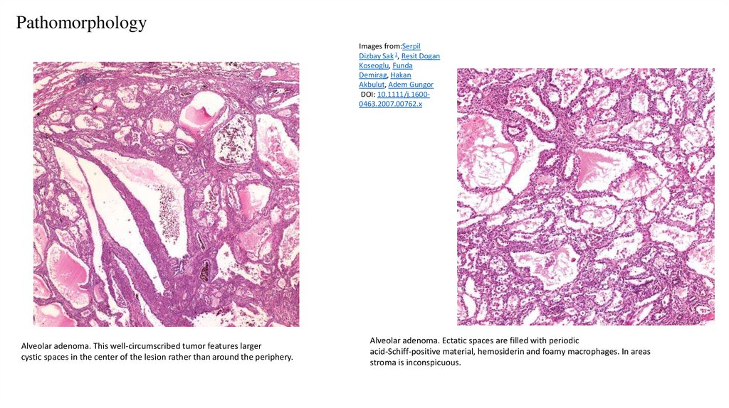
PathomorphologyImages from:Serpil
Dizbay Sak 1, Resit Dogan
Koseoglu, Funda
Demirag, Hakan
Akbulut, Adem Gungor
DOI: 10.1111/j.16000463.2007.00762.x
Alveolar adenoma. This well-circumscribed tumor features larger
cystic spaces in the center of the lesion rather than around the periphery.
Alveolar adenoma. Ectatic spaces are filled with periodic
acid-Schiff-positive material, hemosiderin and foamy macrophages. In areas
stroma is inconspicuous.
34.
35.
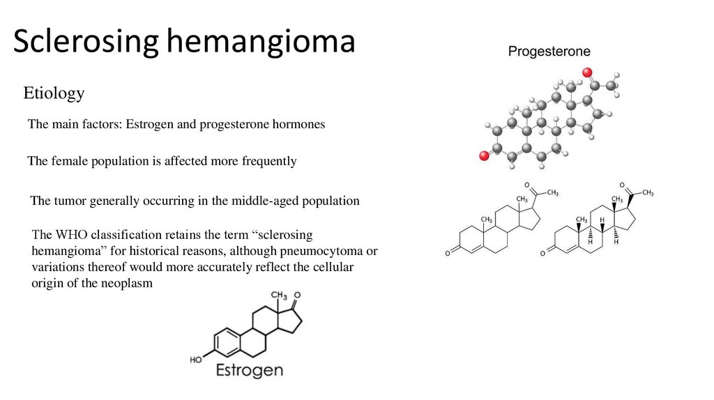
Sclerosing hemangiomaEtiology
The main factors: Estrogen and progesterone hormones
The female population is affected more frequently
The tumor generally occurring in the middle-aged population
The WHO classification retains the term “sclerosing
hemangioma” for historical reasons, although pneumocytoma or
variations thereof would more accurately reflect the cellular
origin of the neoplasm
36.
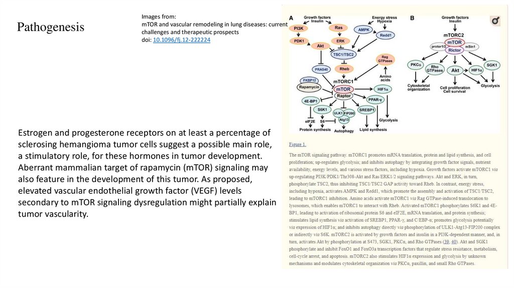
PathogenesisImages from:
mTOR and vascular remodeling in lung diseases: current
challenges and therapeutic prospects
doi: 10.1096/fj.12-222224
Estrogen and progesterone receptors on at least a percentage of
sclerosing hemangioma tumor cells suggest a possible main role,
a stimulatory role, for these hormones in tumor development.
Aberrant mammalian target of rapamycin (mTOR) signaling may
also feature in the development of this tumor. As proposed,
elevated vascular endothelial growth factor (VEGF) levels
secondary to mTOR signaling dysregulation might partially explain
tumor vascularity.
37.
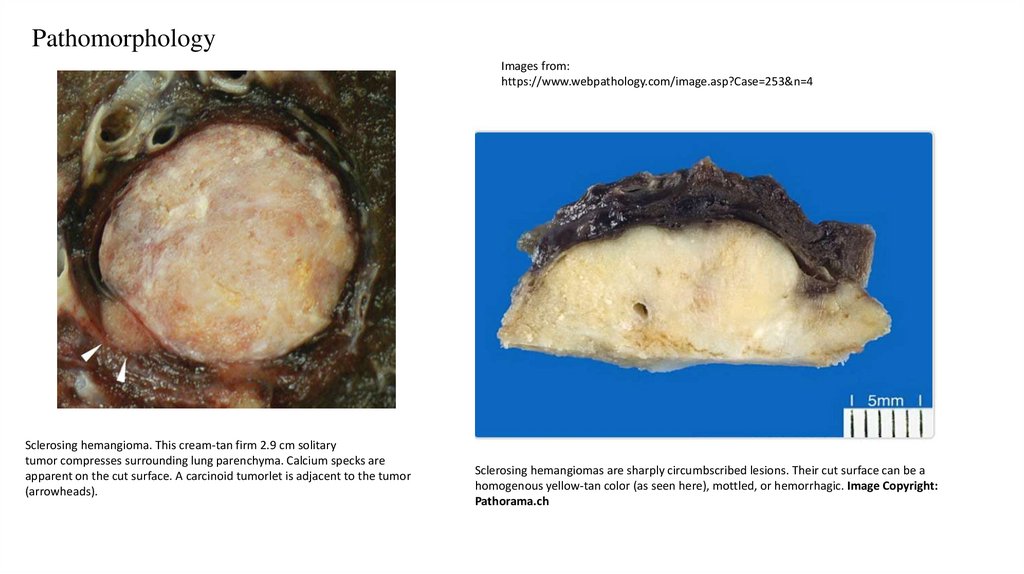
PathomorphologyImages from:
https://www.webpathology.com/image.asp?Case=253&n=4
Sclerosing hemangioma. This cream-tan firm 2.9 cm solitary
tumor compresses surrounding lung parenchyma. Calcium specks are
apparent on the cut surface. A carcinoid tumorlet is adjacent to the tumor
(arrowheads).
Sclerosing hemangiomas are sharply circumbscribed lesions. Their cut surface can be a
homogenous yellow-tan color (as seen here), mottled, or hemorrhagic. Image Copyright:
Pathorama.ch
38.
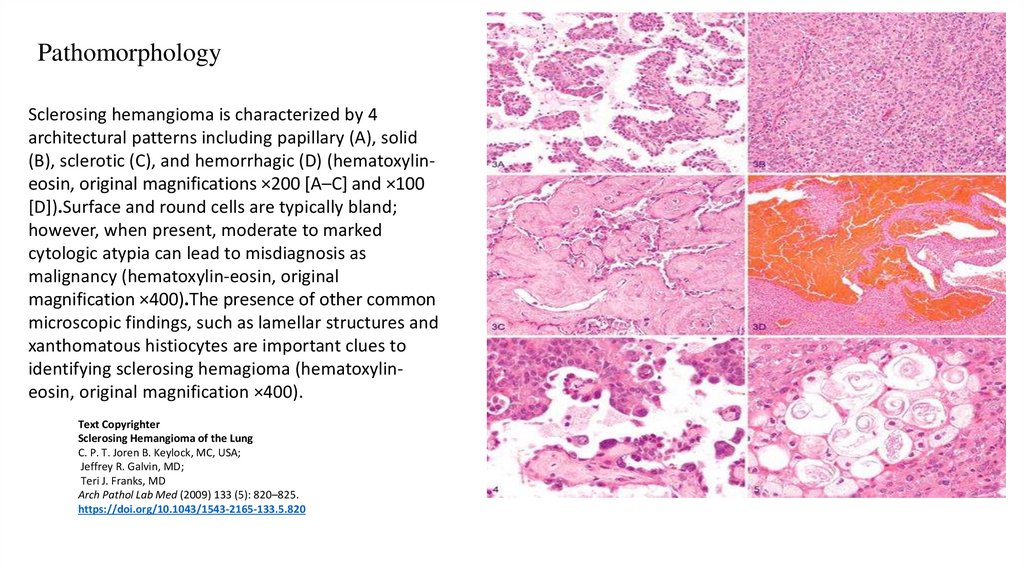
PathomorphologySclerosing hemangioma is characterized by 4
architectural patterns including papillary (A), solid
(B), sclerotic (C), and hemorrhagic (D) (hematoxylineosin, original magnifications ×200 [A–C] and ×100
[D]).Surface and round cells are typically bland;
however, when present, moderate to marked
cytologic atypia can lead to misdiagnosis as
malignancy (hematoxylin-eosin, original
magnification ×400).The presence of other common
microscopic findings, such as lamellar structures and
xanthomatous histiocytes are important clues to
identifying sclerosing hemagioma (hematoxylineosin, original magnification ×400).
Text Copyrighter
Sclerosing Hemangioma of the Lung
C. P. T. Joren B. Keylock, MC, USA;
Jeffrey R. Galvin, MD;
Teri J. Franks, MD
Arch Pathol Lab Med (2009) 133 (5): 820–825.
https://doi.org/10.1043/1543-2165-133.5.820
39.
40.
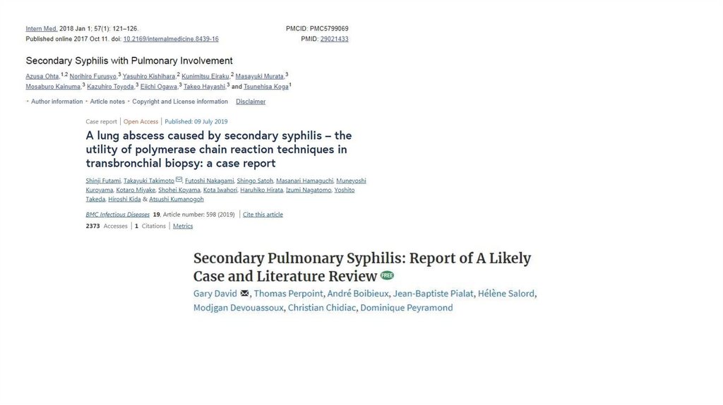
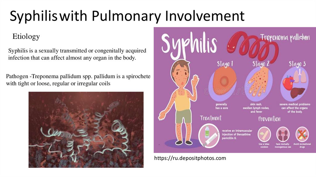
Syphiliswith Pulmonary InvolvementEtiology
Syphilis is a sexually transmitted or congenitally acquired
infection that can affect almost any organ in the body.
Pathogen -Treponema pallidum spp. pallidum is a spirochete
with tight or loose, regular or irregular coils
https://ru.depositphotos.com
41.
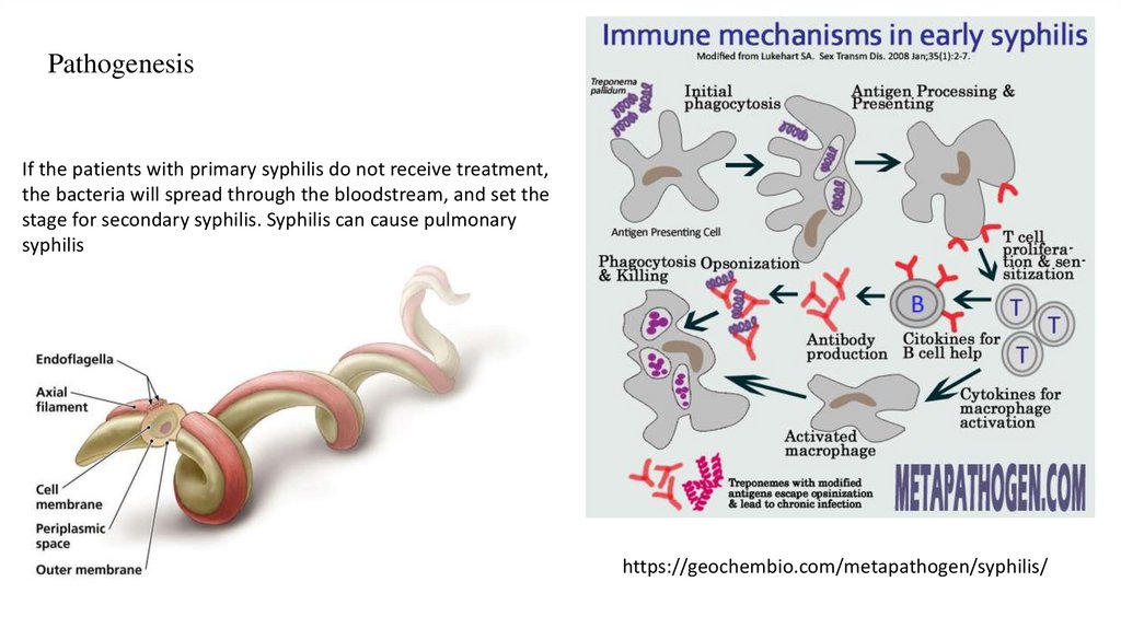
PathogenesisIf the patients with primary syphilis do not receive treatment,
the bacteria will spread through the bloodstream, and set the
stage for secondary syphilis. Syphilis can cause pulmonary
syphilis
https://geochembio.com/metapathogen/syphilis/
42.
PathomorphologyMicroscopic pathology showed granuloma formation by
epithelioid histiocytes and Langhans giant cells (arrow), in addition
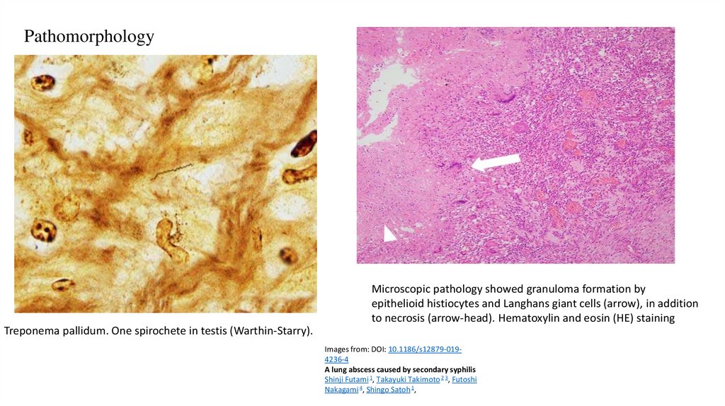
to necrosis (arrow-head). Hematoxylin and eosin (HE) staining
Treponema pallidum. One spirochete in testis (Warthin-Starry).
Images from: DOI: 10.1186/s12879-0194236-4
A lung abscess caused by secondary syphilis
Shinji Futami 1, Takayuki Takimoto 2 3, Futoshi
Nakagami 4, Shingo Satoh 1,