 e-mail: yarn85@mail.ru")


")


 TB")


 spinal cord, caused by MBT. Specific lesion of cerebral membranes and substances is tuberculous meningoencephalitis.")


 are discriminated in the clinical picture of tuberculous meningitis:")


 medicine
Similar presentations:

")

")

")
")


")
Primary and secondary tuberculosis. (Lecture 5)
1. Zaporizhzhia State Medical University Department of phthisiology and pulmonology R.N. Yasinskiy (PhD, assistant of department) e-mail: yarn85@mail.ru
Primary and secondarytuberculosis
2. CLINICAL FORMS
TB of respiratory organsPrimary tuberculous complex.
Disseminated lung tuberculosis.
Nidus lung tuberculosis.
Infiltrative lung tuberculosis.
Caseous pneumonia.
Lung tuberculoma.
Lung fibrous-cavernous tuberculosis.
Lung cirrhotic tuberculosis.
3. CLINICAL FORMS
• TB of exstrarespiratory organsTB of bronchi, trachea and upper respiratory tract.
TB of intrathoracic lymphatic nodes.
TB pleurisy (including empyema).
TB of brain tunics and the central nervous system.
TB of bones and joints.
TB of urinary and sexual organs.
TB of intestine, peritoneum, mesenteric lymphatic
nodes.
Miliary tuberculosis.
TB of other organs and systems.
4. CHARACTERISTIC OF TUBERCULOUS PROCESS
Localization and spreading: Localization ofdefects in lungs according to the numbers
(names) of segments, names of lung sections,
and in other organs and systems - according to
anatomical names of localization of a wound.
Presence of destruction: Destr+ Destr Facultative it is necessary to specify phase of
process:
- infiltration, decay, sowing;
- suction, condensation, scarring, calcination
5. ETIOLOGIC METHOD OF CONFIRMATION:
(MBT +) – it is confirmed by results ofbacteriological analysis (cipher code A
15), in this case to specify:
M+ positive result of sputum analysis
on acid-resisting bacteria (ARB)
C0 – cultural analysis wasn’t done
C- negative result of cultural analysis
C+ positive result of cultural analysis,
in this case to specify:
6.
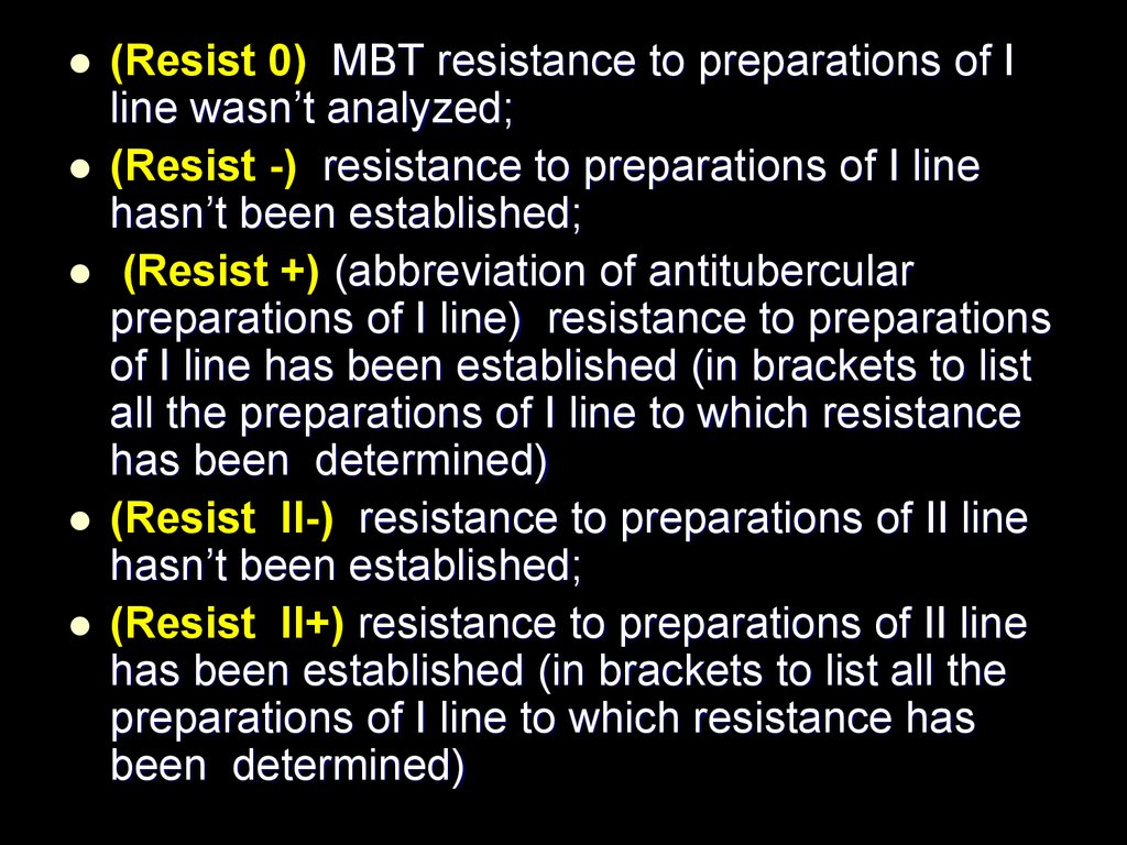
(Resist 0) MBT resistance to preparations of Iline wasn’t analyzed;
(Resist -) resistance to preparations of I line
hasn’t been established;
(Resist +) (abbreviation of antitubercular
preparations of I line) resistance to preparations
of I line has been established (in brackets to list
all the preparations of I line to which resistance
has been determined)
(Resist II-) resistance to preparations of II line
hasn’t been established;
(Resist II+) resistance to preparations of II line
has been established (in brackets to list all the
preparations of I line to which resistance has
been determined)
7. Types of TB cases
• New case of TB – A patient who has never beentreated for TB or has taken anti-TB drugs for less than
one month.
• Previously treated case of TB – A patient who has
been treated for one month or more with anti-TB drugs
in the past. Retreatment cases are further classified by
the outcome of their most recent course of treatment
into four categories.
8. Previously treated case of TB
1. Relapse patients have previously been treated for TB, were declared cured or
treatment completed at the end of their most recent course of treatment, and are now
diagnosed with a recurrent episode of TB (either a true relapse or a new episode of TB
caused by reinfection).
2. Treatment after failure patients have previously been treated for TB and their most
recent course of treatment failed i.e. they had a positive sputum smear or culture result at
month 5 or later during treatment.
3. Treatment after loss to follow-up patients have previously been treated for TB and
were declared ‘lost to follow-up’ at the end of their most recent course of treatment.
4. Other previously treated patients are those who have previously been treated for
TB but whose outcome after their most recent course of treatment is unknown or
undocumented.
9.
Case of multidrug-resistant TB (MDR-TB) – TB that is resistant to
two first-line drugs: isoniazid and rifampicin.
Case of rifampicin-resistant TB (RifTB) – A patient with TB that is
resistant to rifampicin detected using phenotypic or genotypic methods,
with or without resistance to other anti-TB drugs. It includes any
resistance to rifampicin, whether mono-resistance, multidrug resistance,
polydrug resistance or extensive drug resistance.
Case of extremaly drug-resistant TB (XDR-TB) – TB, that is resistant
to isoniazid, rifampicin, at least one fluoroquinolone and aminoglycosid.
Case of risk of MDR (RMDR) – TB in cases, while patient has
contact with MDR patient, but hasn’t results of bacteriological
investigation yet, or has negative bacteriological result.
10. Clinical forms of pulmonary tuberculosis
• There such clinical forms of pulmonaryTB,
as
milliary,
disseminated,
focal,
infiltrative, tuberculoma, caseous pneumonia,
fibrous-cavernous,
cirrhotic
primary tuberculosis complex.
tuberculosis,
11. Clinical forms of extra-pulmonary tuberculosis
• It depends on the affected organ. Miliarytuberculosis, tuberculosis of intrathoracic
lymph nodes, bronchial TB, pleural effusion
considers as pulmonary process in lung
lesions cases.
12. Phases of TB
• There are such TB process phases: infiltration, decay(corresponding
Destruction
+),
contamination,
resorption, seals, scarring and calcification. Infiltration,
decay and contamination characterize tubercular activity
changes in patients. Resorption, seals, scarring and
calcification (calcination) means dicreasing of active
tuberculosis process in dynamics with a tendency to
stabilization.
13. Diagnosis examples
1) New case of TB (01.02.2016) upper lobe of right lung (infiltrative),contamination phase, Destr +, MBT+, M+, MG+, Rif-, C+, Resist-,
Hist0, Cat 1, Coh 1 (2016).
2) Relapse of TB (01.04.2016) lungs (disseminative), infiltration phase,
Destr-, MBT+, M-, MG+, Rif-, C0, Resist0, Hist0, Cat 2, Coh 2 (2016).
3) MDR-TB (05.12.2015) left lung (caseous pneumonia), contamination
phase, Destr+, MBT+, M+, MG0, Rif0, C+, Resist+ (HRES), Resist 2+
(EtOfx), infiltrative TB of B1B2B6 (bronchi) of right lungs with 2 stage
of B2B6 stenosis, Hist0, Cat 4 (New case of TB), Coh 4 (2015).
14. RADIOLOGICAL SYNDROMS
• To explain radiological features of tuberculosisclinical form we must understand radiological
syndroms.
There
are
10
syndroms:
abnormal
pulmonary pattern, lung roots pathology, focal shadow,
infiltrative shadow, disseminative syndrome, rounded
shadow, ring-like shadow, increased enlightenment of
the lung fields, mediastinal pathology and free fluid in
the pleural cavity.
15. Abnormal pulmonary pattern syndrome
- Increased and enriching the lung picture (at inflammatoryprocesses,
collagenous
diseases,
tumor,
pneumoconiosis,
sarcoidosis, vascular lesions with symptoms of congestion and
interstitial edema);
- deformation of lung pattern (at formation of inflammatory
infiltrates, peri-bronchial inflammation, cicatricial due to wrinkling
certain segments, the interparticle pathology and partial internal
connective tissues, lung`s fibrosis with chronic venous stasis, the
appearance of a thin mesh picture at hemosiderosis, the formation of
numerous small ring shadows at scleroderma);
16. Abnormal pulmonary pattern syndrome
- weakeningpattern
of
(in
lung`s
diffuse
pulmonary dissemination,
development of numerous
small cavities);
17. Abnormal pulmonary pattern syndrome
- depletion of the picture (at inflating the lungs,lung arterial nets hypoplasia);
• unusual elements of the picture
18. Lung`s roots pathology
• Manifested with increase,deformation, increase the intensity and
root of the lung shade structures
violation, associated with vascular or
bronchial lymph nodes disorders.
Changes root of the lung occur when
tuberculosis internal thoracic lymph
nodes, sarcoidosis, lymphosarcoma,
central cancer, lymphogranulomatosis,
nonspecific inflammation (basal
pneumonia), aortic aneurysm,
expanding the trunk of pulmonary
blood vessels in heart diseases with
hypertension in the pulmonary
circulation (mitral stenosis) and at
primary pulmonary hypertension,
benign tumors (thymoma), retrosternal
goiter, acute childhood diseases
(measles, scarlet fever) and others.
19. Focal shadow
Characterized by one or more shades (up to
10), round or irregular shapes, up to 1 cm in
diameter, which can have a different intensity and
are usually placed in a limited space in one or
both lungs. Symptom “focal shadow” is a
manifestation of many diseases, occurring with
lesions of the lung parenchyma. Inflammation
(bacterial
and
tuberculosis,
viral
fungal
acute
lesions),
pneumonia,
benign
and
malignant tumors, vascular disorders, collagen
diseases, blood diseases, reticular and lymphoid
tissues are the main pathological processes that
accompanied the emergence of focal shadows, the
formation
of
which
results
in
disappearance of air from the alveoli.
the
local
20. Infiltrative shadow
This syndrome characterized by shadow areas
of more than 1 cm, round or irregular shape, which
has no clear contours. Depending on the severity
there are syndrome “limited infiltrative shadow” in
size from lobules up to lobe, and the syndrome of
“total infiltrative syndrome”, which is characterized
by the size of the shadow over 1 share for total
blackout of all lung fields. Infiltrative changes in
the lungs are the most widespread (50 %) among
other pulmonary diseases. Causes of this syndrome
may be inflammation, tumor process, atelectasis,
pulmonary infarction, hematoma, accompanied by
hypoventilation. This syndrome can develop at
congenital defects – lobe hypoplasia and aplasia.
21. Disseminative syndrome
Characterized by multiple focal and retinal
shadows of varying intensity to 1 cm in diameter,
that are placed on a large lung`s length the and are
usually bilateral. There are more than 200 diseases of
different aetiology and genesis, accompanied by
disseminative syndrome in the lungs. Depending on
the etiology and pathogenesis all diseases are divided
into: 1) infectious-inflammatory (bacterial, viral,
mycobacterial, fungal), 2) the tumor; 3) parasitic; 4)
pneumoconiosis; 5) allergic; 6) collagenoses; 7) an
inhaled and aspiration; 8) congenital constitutional;
9) metabolic-toxic; 10) reticulo-endothelial and
hematopoietic; 11) cardiovascular; 12) traumatic; 13)
unknown etiology.
22. Rounded shadow
Characterized
by
volume
spherical or oval formation of
correct,
incorrect
or
polycyclic
forms with clear or blurred contours
more than 1 cm in diameter. It may
be tuberculoma, nonspecific round
pneumonia, eosinophilic rounded
infiltrate, cancer, benign tumors
(neurinoma,
hemangioma,
arteriovenous aneurysm, adenoma,
veins varicose), tumors of bronchi,
asperhiloma,
Echinococcus.
retention
cyst,
23. Ring-like shadow
It
characterized
by
rounded
enlightenment, which is surrounded by a
ring-liked shadow. Enlightenment in the
lung may be due to lack of lung tissue
and replacement of air with restriction
tissue defect from the surrounding areas
of wall or capsule. The cavity of the lungs
may have primary (congenital cysts air,
emphysematous bullae, bronchiectasis) or
secondary
(decay
or
inflammatory
infiltrate tumors, cleaning parasitic cysts,
tumors traumatic penetration in the
interstitial tissue) native.
24. Increased enlightenment of the lung fields
It
includes
varying
prevalence
of
enlightenment, not limited by ring-like shadow
and is located in the lungs or in the pleural
cavity. Symptom can be caused by: defects in
lung
tissue
(pneumothorax);
degenerative-
dystrophic changes of intrapulmonary bronchial
artery branches with presence of capillary or
venous stasis; violation of bronchial patency as
a result of chronic inflammation in them,
increased viscosity of bronchial secretions,
bronchial
compression
for
inflammatory,
neoplastic and sclerotic processes in the lung
parenchyma; congenital bronchial pathology;
adaptive reactions after lung resection.
25. Mediastinal pathology syndrome
It
manifested
by
changing
the
mediastinum form or position. This
syndrome may be present at fibrosis,
cirrhosis, after lung`s resection, lung`s
agenesia, lung atelectasis, in the presence
of air or fluid in the pleural cavity, at
diaphragmatic hernia, sometimes in large
lung tumors or giant lungs cyst cases, at
the bronchial tumors and enthetic bodies.
Mediastinum forms changing (extension)
may be at mediastinal cysts and tumors,
inflammatory processes (acute, chronic,
mediastinit encysted abscess of the
mediastinum).
26. Free fluid in the pleural cavity
It characterized by a one- or two-way
shadow areas of different sizes, with the
predominant
divisions,
placement
with
oblique
in
the
lower
upper
limit.
Depending on the position of the body
section blackout can change the location.
Pleural effusions are divided into exudate
and transudate. Transudate resulting in
increase of capillary pressure, or in decrease
of oncotic pressure of blood plasma. The
transudate nature pleural fluid has in
congestive heart failure, liver cirrhosis,
hydrothorax,
myxedema cases.
glomerulonephritis,
27. Free fluid in the pleural cavity
The most frequent cause of exudative pleurisy of different etiology is
increased permeability of the pleural surfaces for protein and decreased oncotic
pressure gradient. The second reason is the lymphatic outflow reduction from the
pleural cavity. A third reason could be a pressure reduction in the pleural cavity.
Pleural effusion may develop in lung cancer, breast cancer, lymphoma,
lymphogranulematosis, benign and malignant pleural mesothelioma, bacterial
pneumonia, tuberculosis, fungal infections (aspergillosis, cryptococcosis,
actinomycosis) and parasitic diseases (amebiasis, echinococcosis) at viral,
infections, pulmonary embolism, pancreatitis, hepatic and subdiaphragmatic
abscess, at collagen diseases (rheumatism, systemic lupus erythematosus,
Wegener's granulomatosis), rupture of thoracic lymphatic duct.
28. Depending on pathogenesis the tuberculosis is divided primary and secondary. Primary tuberculosis develops after the first contact of macroorganism with MBT. Secondary forms of a tuberculosis arise at people which have been earlier infected, and after pri
Depending on pathogenesis the tuberculosisis divided primary and secondary.
Primary tuberculosis develops after the first
contact of macroorganism with MBT.
Secondary forms of a tuberculosis arise at people
which have been earlier infected, and after
primary infestation should pass not less than year.
It passes some years more often.
29. PRIMARY TUBERCULOSIS
Primary is considered tuberculosis thatdevelops in firstly infected persons.
The period from the moment of the
intensity of tuberculin reaction during
one year without signs of intoxication is
called the period of early tuberculous
infection
30. Para-specific reactions (tuberculosis “masks”)
• In primary tuberculosis there are situationswhere the disease occurs more on the type of
therapeutic, hematological, rheumatologic
disease. This is due to the fact that the body is
infected TB patient responsible development
of vasculitis and allergic reactions.
31. “Flu-like” TB mask
• The most frequentlytuberculosis in active phase
occurs in such frequent,
long, unusual flu-like illness
without clearly expressed
inflammation of the upper
respiratory tract and causes
the patient's family
outbreaks of influenza states
– its a “flu-like”
tuberculosis mask
32. “pneumonic” mask
The second frequency is “pneumonic” mask. Thisis repeated recurrent pneumonia, especially in the
same lungs place with torpent course, having
atypical clinic and course, difficult to treated,
slowly resolved with the formation of small focal
and fibrotic changes.
33. Poncet`s disease
Tuberculosis can begin on type “rheumatic”mask,
called
“Poncet`s
disease”.
It
manifested a long course articular syndrome
with pain, swelling, breach of mobility in the
joints with deformation, ankylosis. When Xrays there are typical signs of rheumatoid
arthritis.
There
no
efficiency
after
antirheumatic therapy in “Poncet`s disease”
cases, no complications such as endocarditis.
Only TB positive tuberculin tests, specific
X-ray changes and the effect of specific
therapy allows to confirm the diagnosis of
tuberculosis.
34. “Neurological” TB mask
“Neurological” TB mask manifests as long,persistent neuralgia, which can not be usually
treated, especially for intercostal and sciatic
nerves, but without signs of compression
(osteochondrosis) or inflammatory lesions nerve
(a radiculitis).
35. “Lupus-like” TB mask
“Lupus-like” mask manifeststypical erythema on the face
in the form of "butterfly",
trophic disorders, arthralgia,
leukopenia, sharply increased
ESR,
sometimes
specific
blood cells and antibodies to
DNA are finding.
36. “Hematological” mask
“Hematological” mask of tuberculosis occurs withbone marrow hypoplasia, leukopenia, anemia,
thrombocytopenia,
sometimes
with
reactions.
manifests
lymphadenopathy,
Often
splenomegaly,
B12-deficiency
hypoplastic anemia.
leukemoid
anemia
and
37. Keratoconjunctivitis phlyctenular
• Keratoconjunctivitisphlyctenular. Most often its
tubercular-allergic process in
children with broncho-adenitis
and tuberculosis of the lymph
nodes, and other allergic
reactions. On the bulbar
conjunctiva and cornea near the
limbus there are single or
multiple inflammatory nodules
of yellowish-pink color with a
bunch of the blood vessels that
are often completely resolve,
but sometimes disintegrate with
the formation of ulcers followed
by replacement with connective
tissue
38. CHARACTERISTIC SIGNS OF PRIMARY TUBERCULOSIS:
the intensity of tuberculin reactionsorganism hypersensibilization to MBT
injury of lymphatic system (lymphatic nodes) with the
susceptibility to caseous necrosis
susceptibility to lymphogenous and haematogenous
dissemination, possibility of spontaneous recovery
availability of paraspecific reactions
The main forms of primary tubercular process:
1.Tubercular intoxication at children and teenagers.
2.Primary - tubercular complex.
3.Tuberculosis of intrathoracic lymph nodes.
39. PRIMARY TUBERCULOUS COMPLEX
1.PATHOGENESIS
After the penetration of MBT into the lungs, primary
lesion (primary affect), of the size from a millet grain to a
section of a lung, is predominantly localized subpleurally
in the II, III, VIII, IX segments. From the primary affect the
infection spreads along lymphatic vessels to
intrathoracic lymphatic nodes.
2.
PATHOLOGICAL ANATOMY OF THE PRIMARY
TUBERCULOSIS
In the primary lung focus, alveolitis develops, which is quickly
replaced by the typical development of caseosis necrosis. In
the centre of primary focus, caseosis forms but in the
periphery – elements of non specific inflammation occur.
The primary lung affect localizes more often just under pleura,
therefore frequently pleura is involved in the inflammation
process. The lymphatic vessels expand, their walls becoming
infiltrated and tubercles appear. In the regional lymphatic
nodes, there are elements of inflammations converting into
specific caseous changes with necrosis
40. PRIMARY TUBERCULOUS COMPLEX
• The dynamicstudy of primary
pulmonary
processes
among children
has allowed to
allot 4 phases
of the primary
tuberculosis:
41. PRIMARY TUBERCULOUS COMPLEX
1) PNEUMONIC;In the first phase
(pneumatic) the focus of
broncho-lobular pneumonia
(3) is determined with a
size of 1,5-2 till 5 cm.
The form of the lung focus
(3) is round or irregular,
with heterogenous
character and dim contours.
Enlarged regional lymphatic
nodes (1) are determined
simultaneously (the picture
of infiltrative
bronchoadenitis) and there
is an amplification of
bronchial vessels picture
– lymphangitis (2)
between the focus and the
lung root.
42. PRIMARY TUBERCULOUS COMPLEX
• 2)PHASE
DISSOLVING;
OF
In the second phase of
dissolving (bipolarity) the
reduction of the perifocal
zone of inflammation (3) is
observed.
The
centrally
located
caseous focus becomes
more prominent. The signs
of inflammation in regional
lymphatic nodes (1) and in
the
zone
of
bronchopulmonary vessels
are decreaseding (2).
43. PRIMARY TUBERCULOUS COMPLEX
• 3)PHASE
OF
CONDENSATION;
In the third phase, the
phase of condensation:
the primary focus is well
outlined (3), its contours
are cleared, on periphery
of the focus there is the
beginning
of
calcification
as
fine
pieces; at peripheral
regions
of
lung
bronchial
lymphatic
nodes calcification is
also present (1).
44. PRIMARY TUBERCULOUS COMPLEX
4) FORMATION OF GOHN’SFOCUS.
• In the fourth phase, in
the place of broncholobular pneumonia (3)
calcification become
compact, the focus is
round with regular
precise contours, its
size does not exceed
3-5 mm. This
formation is called
Gohn’s focus.
45. TUBERCULOSIS OF INTRATHORACIC LYMPHATIC NODES
Bronchoadenitis is a disease of the lymph nodes of thelungs roots and the mediastinum. In this form of primary
tuberculosis, intrathoracic lymph nodes are mainly
involved in the process of inflammation
Pathogenesis. Infestation generally takes place by the
droplet-dust way, through the mucous membrane of
tonsils and bronchi MBT penetrate into lymphatic
vessels, nodes, where a specific process develops.
Depending on the state of micro- and macroorganism,
infiltrative-inflammatory
or
necrotic
changes
in
lymphatic nodes prevail.
Pathomorphism. One or some groups of lymphatic nodes
may be in jured at tuberculosis. Paratracheal,
tracheobronchial, bronchopulmonary, bifurcating and
оther lymphatic nodes are hurt. The process may be unior bilateral, predominantly asymmetric.
46. Clinical pattern of tuberculous bronchoadenitis
acute intoxicationspecific clinical symptoms: subfebrile temperature,
deterioration of general condition, loss of appetite,
weight loss, adynamia or excitation of nervous system
in progression, and especially in small children,
appears bitonal cough
among adults, attacks of dry, «hoarse» tickling cough
take place. It is caused by irritation of the mucous of
bronchi or formation of the broncho-pulmonary fistula
Blood analyses are without any features
Detection of MBT. In gastric lavage, it is possible to
find out MBT, especially often it is discovered in the
sputum and in bronchial lavage during the rupture of
the caseous mass into the bronchus.
47. Clinico-roentgenologically variants of intrathoracic lymphatic nodes TB
tumour like (tumoursimilar)form
Left side tumorous lymphadenitis.
Massive enlargement of
broncho-pulmonic lymphatic nodes
infiltrative form
On chest x-ray the shadow of
the right lung root is extended
the outside contour is dim,
the structure is blurred and
intensity is increased.
48. Miliary tuberculosis
Generalized TB clinical form with the hematogenictype of dissemination and acute course
49. Sub-acute disseminated tuberculosis
Bilateral lung injurywith predominant
affecting upper
lobes and trend
for destructive
progressive
course
50. Chronic disseminated tuberculosis
• Disseminatedlung injury with
the wavy course
and progressive
alveoli
substitution by
connective tissue
51. Focal (nidus) TB
• The mildest limitedlung TB clinical form
which is characterized
by the appearance of
single or several nidi
(opacity with the
diameter up to 1 cm in
diameter) in one or
more segments of one
or both lungs with
predominantly
productive type of the
inflammation and slow
torpid progression.
52. Infiltrative TB
The expansive lung TB clinical formwhich is characterized by the
appearance of different size and shape
lesions in one or more segments of one
or both lungs with predominantly
exudative type of the inflammation and
strong trend to the rapid progression
and decay.
53. Infiltrative TB
54. Infiltrative TB
55. Infiltrative TB
56. Infiltrative TB
57. Caseous pneumonia
An acute TBform with the
decay
predominance
and severe
progressive
course
58. Tuberculoma
Theinflammative
focus limited
by connective
tissue
capsule
59. Fibrous-cavernous tuberculosis
The chronicdestructive
clinical TB form
characterized by
the presence of
the old fibrous
cavity (cavern) in
the lung tissue
surrounded by
extended fibrosis
and satellite nidi
60. Cirrhotic lung tuberculosis
The chronicdestructive
clinical TB form
characterized by
the predominant
cirrhosis in the
lung tissue with
the embedded
cavitations inside
61. Tuberculous meningitis is the inflammation of the membranes of cerebrum and (or) spinal cord, caused by MBT. Specific lesion of cerebral membranes and substances is tuberculous meningoencephalitis.
Causes, incidence, and riskfactors
Tuberculous meningitis is
caused by the spread of
Mycobacterium tuberculosis
to the brain, from another
site in the body. The
symptoms usually begin
gradually. Risk factors
include a history of
pulmonary tuberculosis,
excessive alcohol use,
AIDS, or other disorders that
compromise the immune
system.
62.
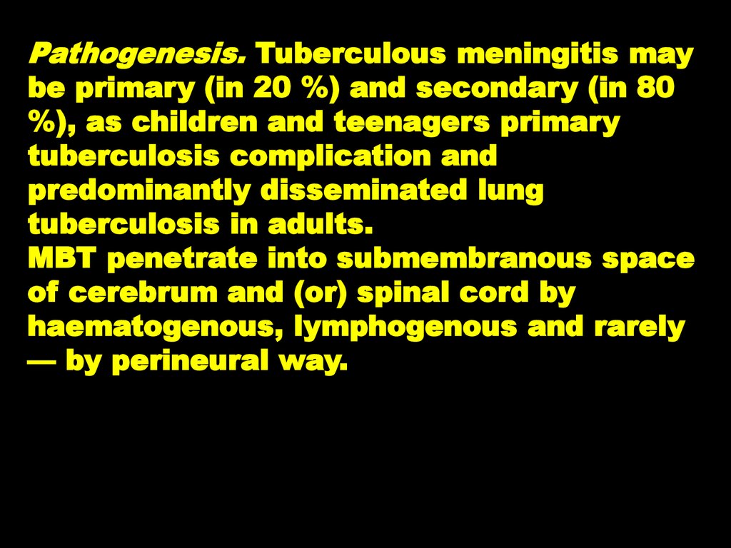
Pathogenesis. Tuberculous meningitis maybe primary (in 20 %) and secondary (in 80
%), as children and teenagers primary
tuberculosis complication and
predominantly disseminated lung
tuberculosis in adults.
MBT penetrate into submembranous space
of cerebrum and (or) spinal cord by
haematogenous, lymphogenous and rarely
— by perineural way.
63.
Pathological anatomy. The specificprocess is predominantly localized in
the soft membrane of cerebral base,
in which connection cranial nerves,
located here, are injured. The illness
may progress as meningoencephalitis
(64-70 %) - inflammation of cerebral
membranes and substance, as basal
meningitis (20-30 %) - inflammation of
cerebral membranes and as spinal
meningitis (4-6%) - inflammation of
spinal cord membranes.
Exudate, tubercles, tender fibrin
threads appear on soft cerebral
membrane at tuberculous meningitis.
Pathologic changes are also abserved
on the vessel membrane of cerebral
ventricles.
64. Clinic of Tuberculous meningitis
I. A prodromic periodthe duration is from 1 to 4 weeks: general weakness,
irritability, sleeplessness, lability, unstable headache, often
subfebrile body temperature
II. obvious clinical manifestations
the body temperature rises to febrile, the headache of sharp
intensity, joined by vomiting
meningeal syptoms develop gradually: rigidity of cervical
muscles, Kernig's and Brudzinsky's symptoms
eye motional and drain cranial nerves are injured
pareses according to the central type of the VII, IX, X, XII
pairs of cranial nerves
vegetovascular disturbances in the form of vasomotor
reactions develop, stable red dermographism, Trusso spots,
relative bradycardia, disturbance of sleep and appetite
III. The period of pareses and paralyses
expressed adynamy, apathy to all surrounding, later on
soporose state develops, coma
65. 5 components (syndromes) are discriminated in the clinical picture of tuberculous meningitis:
Intoxicating syndromeMeningeal syndrome: headache,
vomoting, hyperestasis
Symptoms of cranial nerves
lesions (III, VI, VII, XII) and the
spinal cord roots
Changes of spinal fluid
Symptoms of irritation and
prolapse of functions owing to
cerebral tissue lesion
66. Meningeal symptoms
• rigidity cervical muscles67. Characteristics of spinal fluid
Liquor is usually transparent oropalescent
flows under increased pressure
greater number of cells (100 –
300 in 1 ml, at norm - up to 10)
increased protein content
(0,6 –1,0 g/L and more,
at norm – 0,2-0,4 g/L)
lowered sugar concentration 1,5 mmol/L and less, norm2,22-3,88 mmol/L ) and chlorides concentration (110
mol/L and less, norm -120 -130 mmol/L )
Pandi and None-Apelta’s positive reactions
in liquor a weblike film is formed in 24 hours, in which in
10-20 % of cases MBT are revealed