


 medicine
Similar presentations:

")
")


")

Tuberculosis
1.
TUBERCULOSIS1
2.
Tuberculosis is a chroniccommunicable disease with
specific
granulomatous
inflammation caused by a
variety of tubercle bacilli,
especially
Micobacterium
tuberculosis hominis and M. t.
bovis.
3.
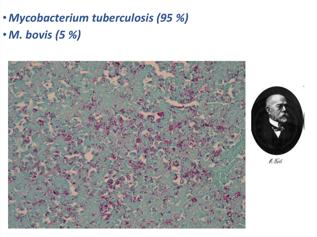
• Mycobacterium tuberculosis (95 %)• M. bоvis (5 %)
4. Mode of transmission
• By inhalation into the respiratory tract.• Ingestion. Through ingestion into GI tract leads to
development to tonsillar or intestinal tuberculosis.
• Inoculation. Through mucous membranes of mouth
and throat, skin.
• Transplacental route results in development of
congenital tuberculosis in fetus from infected
mother.
5.
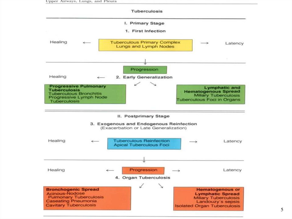
56. Features of Primary Tuberculosis
• Development of disease at the first getting of the activatorinto the organism.
• Sensibilization and allergy of HIT (Hypersensitivity of
Immediate Type) .
• Prevalence of the exudative - necrotic changes.
• Tendency
to
hematogenous
and
lymphogenous
generalization and also to chronic duration.
• Paraspecific reactions such as: vasculitis, nodous erythema,
arthritis.
• Primary Tuberculosis used to be found most often in young
children, but in industrialized countries it has become more
common in the elderly and debilitated, in alcoholics, and in
high-risk racial groups.
7.
PRIMARY COMPLEX OF TUBERCULOSIS"Ghon complex“ consists of
I. Pulmonary component
so called Primary affect
or primary focus or
Ghon’s focus.
II. Lymphatic vessel
component occurs by
Tuberculous
lymphangitis.
III
III. Lymph node component
occurs by Tuberculous
lymphadenitis.
II
I
11
8.
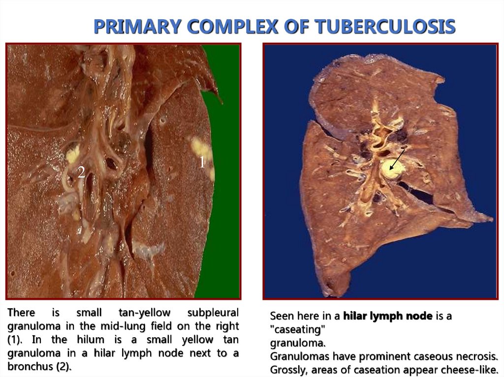
PRIMARY COMPLEX OF TUBERCULOSIS2
1
There is
small tan-yellow
subpleural
granuloma in the mid-lung field on the right
(1). In the hilum is a small yellow tan
granuloma in a hilar lymph node next to a
bronchus (2).
Seen here in a hilar lymph node is a
"caseating"
granuloma.
Granulomas have prominent caseous necrosis.
Grossly, areas of caseation appear cheese-like.
9.
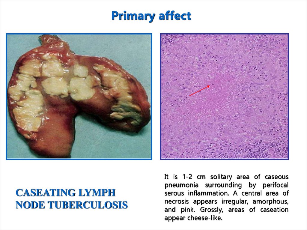
Primary affectCASEATING LYMPH
NODE TUBERCULOSIS
It is 1-2 cm solitary area of caseous
pneumonia surrounding by perifocal
serous inflammation. A central area of
necrosis appears irregular, amorphous,
and pink. Grossly, areas of caseation
appear cheese-like.
10.
Primary tuberculosis of alimentary tractTuberculous
mesenterial
11.
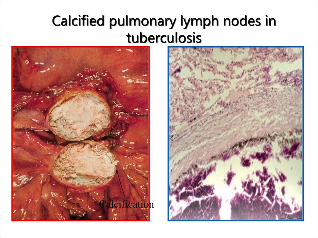
Calcified pulmonary lymph nodes intuberculosis
Calcification
12.
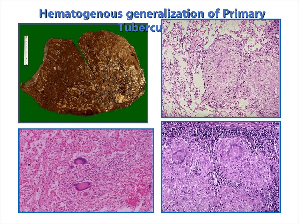
Hematogenous generalization of PrimaryTuberculosis
13. Classifications of hematogenous tuberculosis
• Generalized hematogenous tuberculosis:а) The most acute tubercular sepsis.
b) Acute general miliary tuberculosis.
c) Acute general large-focal tuberculosis.
d) Chronic miliary tuberculosis.
• Hematogenous pulmonary tuberculosis:
а) Acute miliary tuberculosis.
б) Chronic miliary tuberculosis.
в) Chronic
large-focal tuberculosis or hematogenousdisseminative.
• Hematogenous tuberculosis with unpulmonary
lesions or organic tuberculosis:
Tuberculosis of the kidneys, of urinary- genital tract, of skin,
of bone- articular, of endocrine organs and others .
32
14.
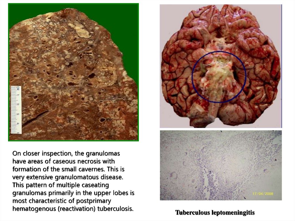
On closer inspection, the granulomashave areas of caseous necrosis with
formation of the small cavernes. This is
very extensive granulomatous disease.
This pattern of multiple caseating
granulomas primarily in the upper lobes is
most characteristic of postprimary
hematogenous (reactivation) tuberculosis.
Tuberculous leptomeningitis
15.
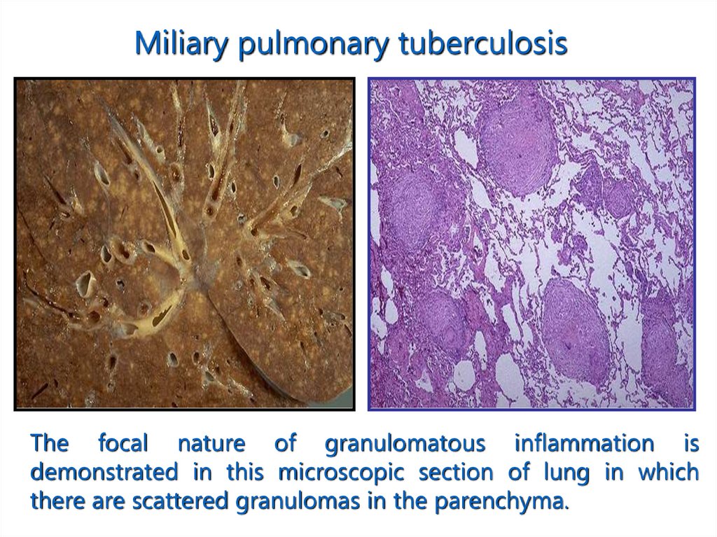
Miliary pulmonary tuberculosisThe focal nature of granulomatous inflammation is
demonstrated in this microscopic section of lung in which
there are scattered granulomas in the parenchyma.
16.
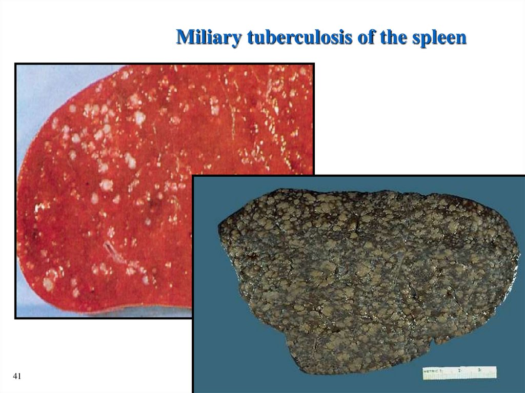
Miliary tuberculosis of the spleen41
17.
RENAL TUBERCULOSIS39
18.
MILIARY TUBERCULOSIS IN LIVER40
19. Forms or stages of the secondary tuberculosis:
1.Acute local tuberculosis.2.Fibrous-local tuberculosis.
3.Infiltrative tuberculosis.
4.Tuberculoma.
5.Caseous pneumonia.
6.Acute cavernous tuberculosis.
7.Fibrous – cavernous tuberculosis.
8.Cirrhotic tuberculosis.
43
20.
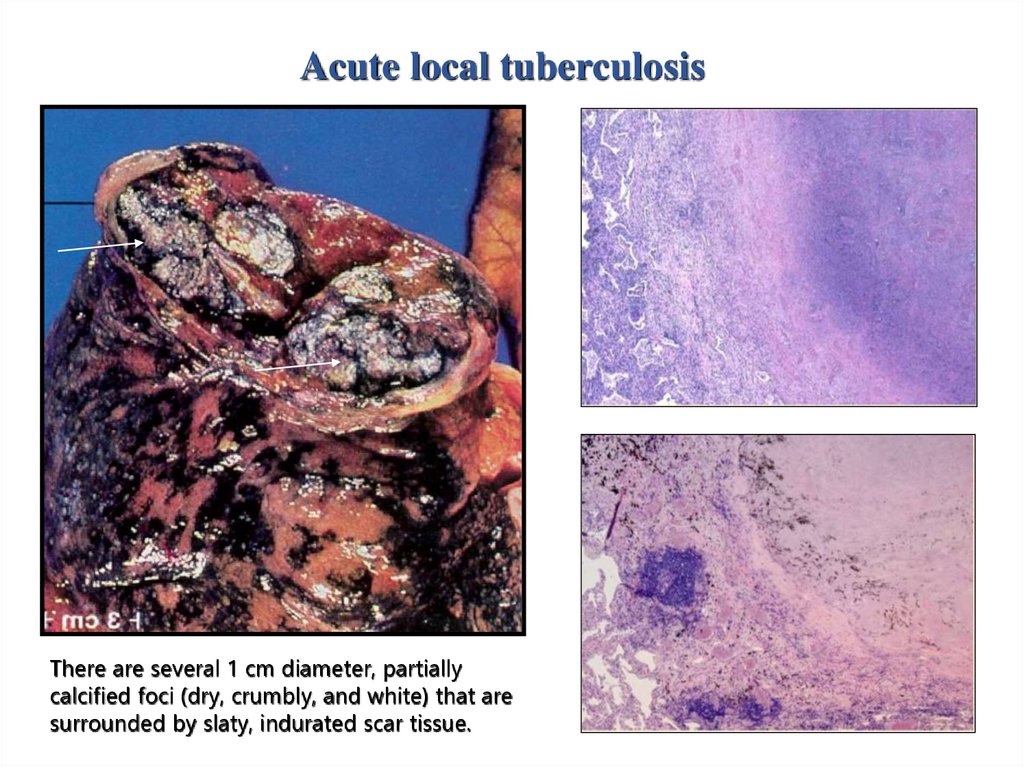
Acute local tuberculosisThere are several 1 cm diameter, partially
calcified foci (dry, crumbly, and white) that are
surrounded by slaty, indurated scar tissue.
21.
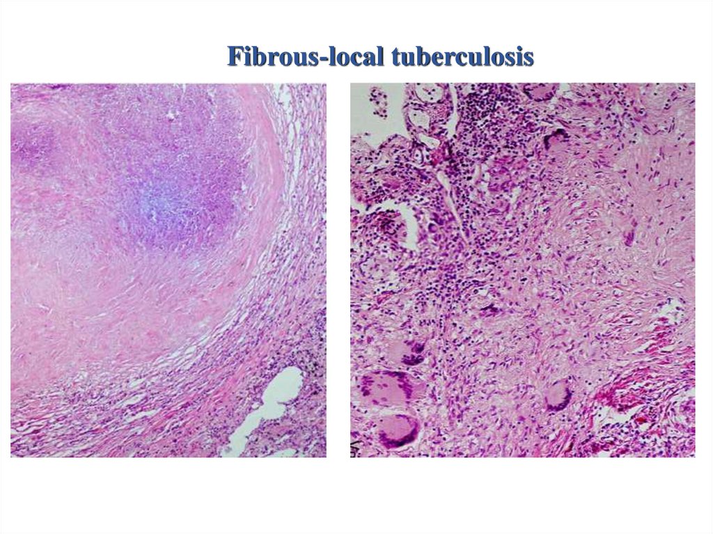
Fibrous-local tuberculosis22.
Infiltrative tuberculosis23.
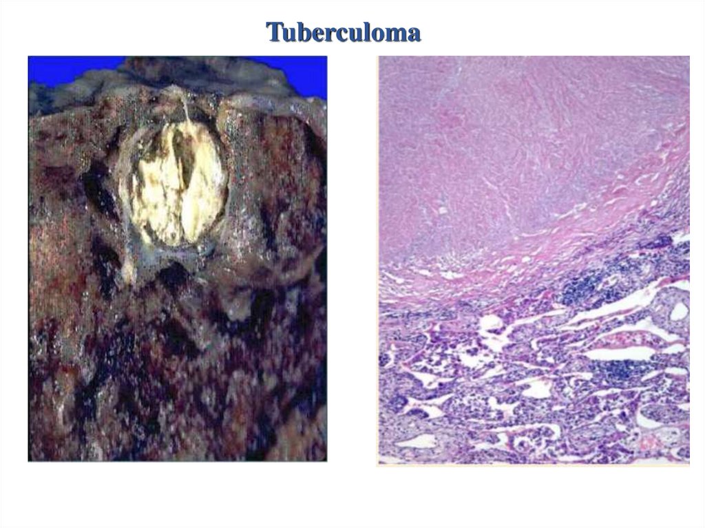
Tuberculoma24.
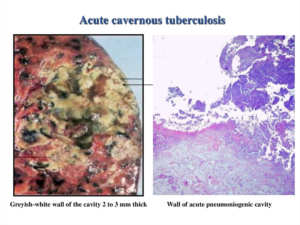
Caseous pneumonia25.
Acute cavernous tuberculosisGreyish-white wall of the cavity 2 to 3 mm thick
Wall of acute pneumoniogenic cavity
26.
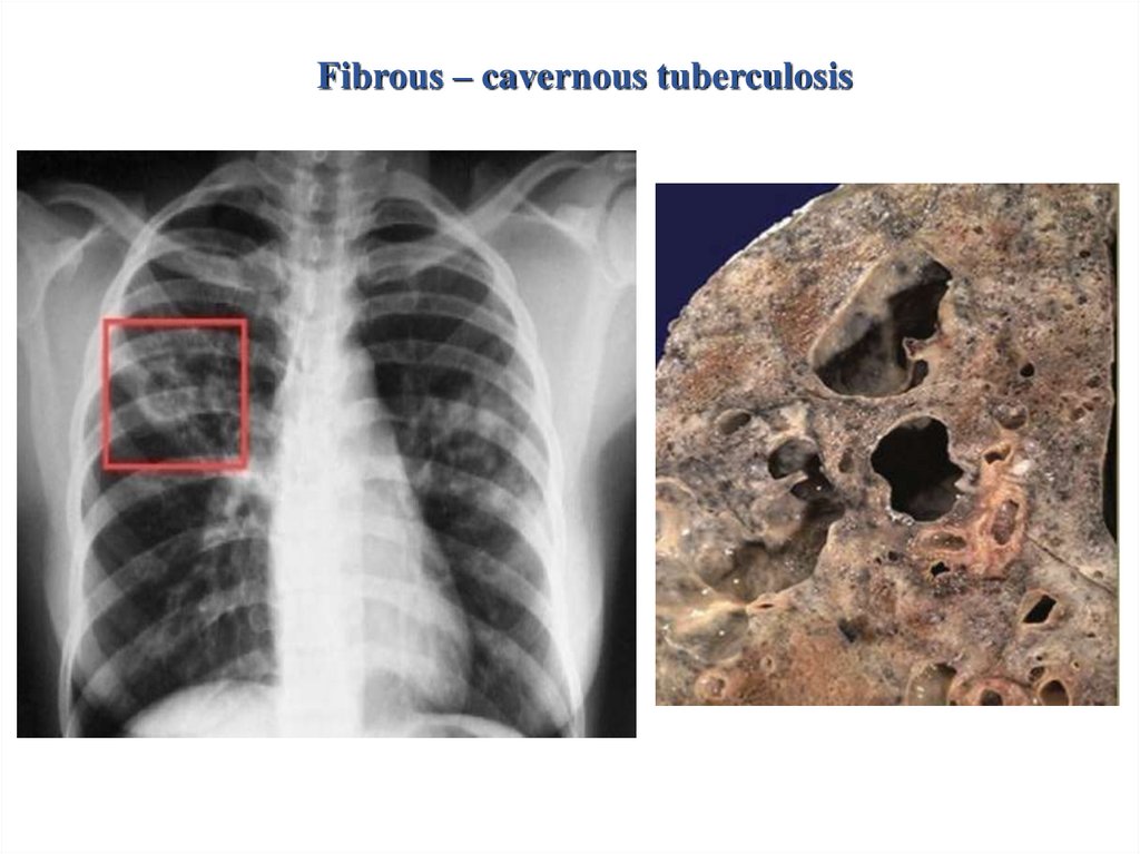
Fibrous – cavernous tuberculosis27.
Fibrotic scar in the wall oftuberculous cavity consists of
fibroblast, collagen, and scattered
Langerhans giant cells
The wall of tuberculous cavity
contains foci of calcification
replacing the caseating
granulomas
47
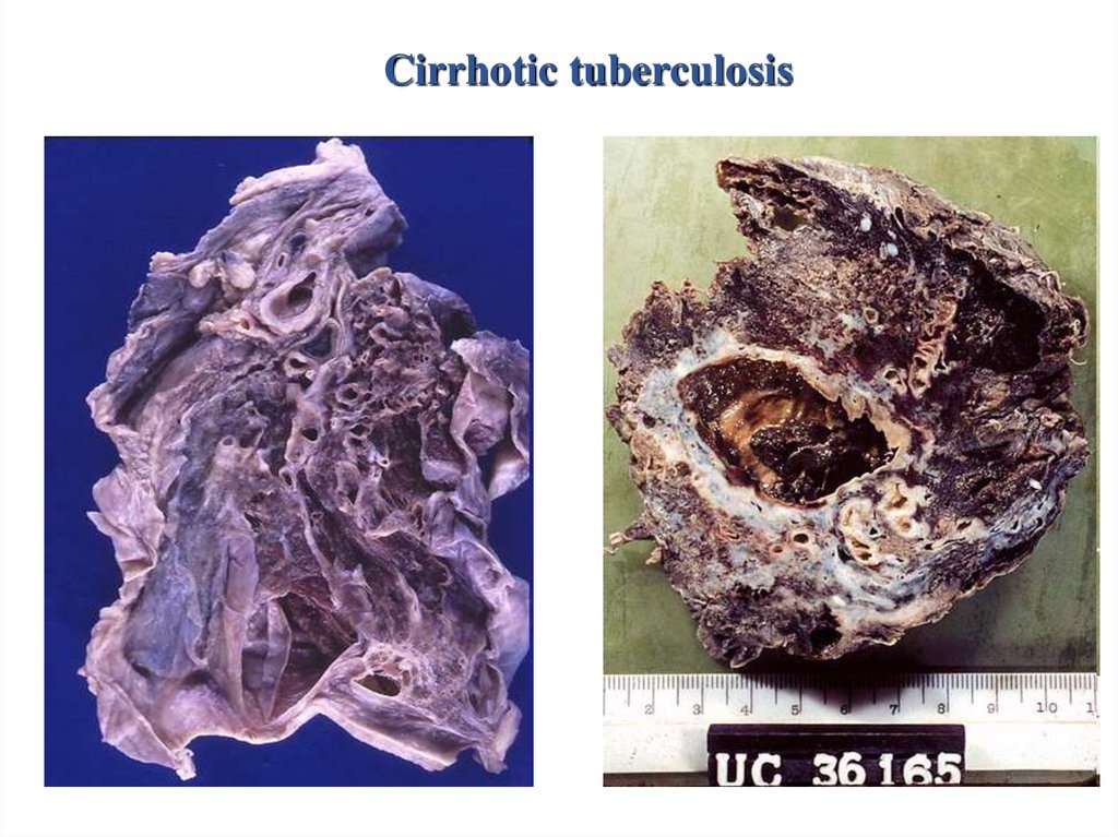
28.
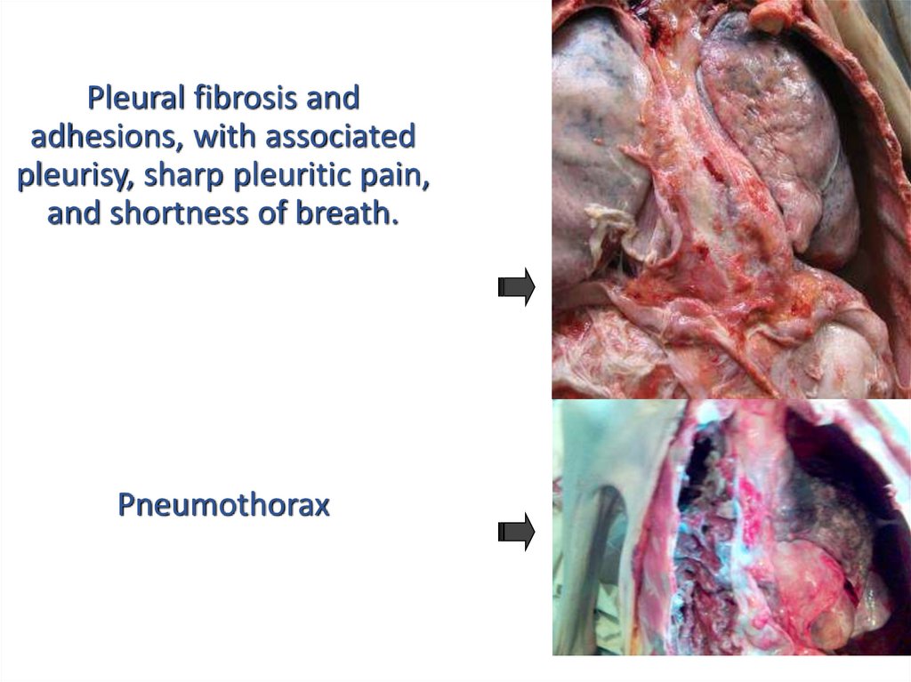
Cirrhotic tuberculosis29. Complications and causes of death
• Scarring and calcification.• Pneumothorax.
• Empyema.
• Pleural fibrosis and adhesions, with associated pleurisy,
sharp pleuritic pain, and shortness of breath.
• Chronic respiratory-cardiac insufficiency due to
development “cor pulmonale”.
• Acute hemorrhage due to erosion of vessels.
• Chronic renal insufficiency due to development of
amiloidosis of kidneys.
• Intoxication.
48
30.
Pleural fibrosis andadhesions, with associated
pleurisy, sharp pleuritic pain,
and shortness of breath.
Pneumothorax
31.
Acute hemorrhage due toerosion of vessels.