


 medicine
Similar presentations:

")


")


")
Definition of sarcoidosis
1.
Definition of Sarcoidosis• Sarcoidosis (Behnier-Beck-Shauman's disease) is a systemic
disease characterized by the development of productive
inflammation with the formation of epithelioid-cell
granulomas without necrosis with the outcome of resorption
or fibrosis.
• Sarcoidosis is characterized by the formation of
noncaseating granulomas in one or more organs and tissues;
the etiology is unknown. Most often the lungs and
lymphatic system are affected, but sarcoidosis can affect
any organ. Symptoms of sarcoidosis of the lungs vary from
total absence (limited disease) to shortness of breath when
exercising and, rarely, respiratory or other organ failure (a
common disease).
2.
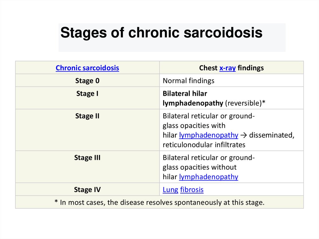
Stages of chronic sarcoidosisChronic sarcoidosis
Chest x-ray findings
Stage 0
Normal findings
Stage I
Bilateral hilar
lymphadenopathy (reversible)*
Stage II
Bilateral reticular or groundglass opacities with
hilar lymphadenopathy → disseminated,
reticulonodular infiltrates
Stage III
Bilateral reticular or groundglass opacities without
hilar lymphadenopathy
Stage IV
Lung fibrosis
* In most cases, the disease resolves spontaneously at this stage.
3.
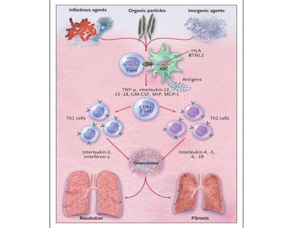
Etiology of Sarcoidosis1. Genetics
Studies have shown that a mutation of the gene BTNL2, as well as the HLA-DQB1 variant of
the gene HLA, are associated with an increased risk for the disease.
2. Infectious agents
The major implicated infectious agents include: mycobacteria, fungi, borrelia, and rickettsia.
Mycobacterium tuberculosis infection.
M.Tuberculosis catalase – peroxidase has been identified as a possible antigen catalyst of
sarcoidosis.
The disease has also been reported by transmission via organ transplants.
3. Autoimmune
Association of autoimmune disorders has been frequently observed. The exact mechanism
of this relation is not known, but some evidence supports the hypothesis that this is a
consequence of Th1 lymphokine prevalence
4.
Risk factors• While anyone can develop sarcoidosis, factors that may
increase your risk include:
• Age. Sarcoidosis can occur at any age, but often occurs
between the ages of 20 and 60 years. Women are
slightly more likely to develop the disease.
• Race. People of African descent and those of Northern
European descent have a higher incidence of
sarcoidosis. African-Americans are more likely to have
involvement of other organs along with the lungs.
• Family history. If someone in your family has had
sarcoidosis, you're more likely to develop the disease.
5.
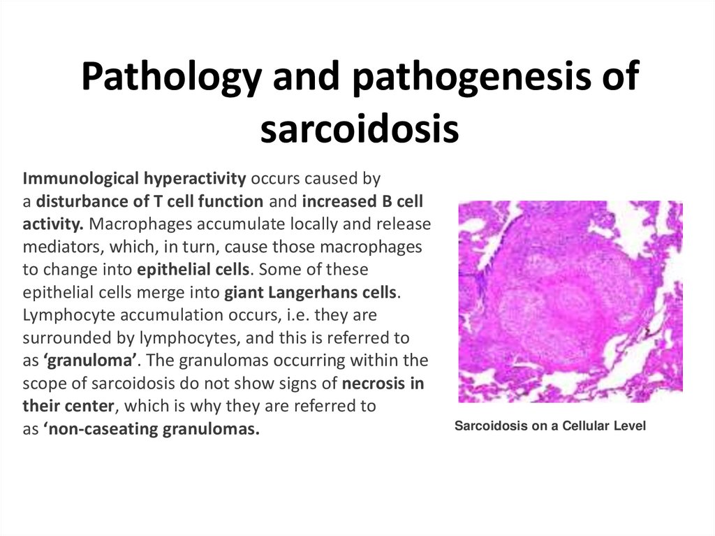
Pathology and pathogenesis ofsarcoidosis
Immunological hyperactivity occurs caused by
a disturbance of T cell function and increased B cell
activity. Macrophages accumulate locally and release
mediators, which, in turn, cause those macrophages
to change into epithelial cells. Some of these
epithelial cells merge into giant Langerhans cells.
Lymphocyte accumulation occurs, i.e. they are
surrounded by lymphocytes, and this is referred to
as ‘granuloma’. The granulomas occurring within the
scope of sarcoidosis do not show signs of necrosis in
their center, which is why they are referred to
as ‘non-caseating granulomas.
Sarcoidosis on a Cellular Level
6.
. While this information may not be as relevant for the exam, one may come across thisin clinical practice where a patient may want to know about this in more detail. Within
the aforementioned giant cells, shell-shaped calcified inclusions can be found here and
there, the so-called ‘Schaumann bodies’. These were named after J. N. Schaumann,
who was the first to recognize that sarcoidosis is a systemic disease that can attack
several organs and not only the skin, which had been the theory until that time.
Note: Histologically, in cases of sarcoidosis, non-caseating granulomas are present,
while in cases of tuberculosis, the granulomas are caseating
Sarcoidosis is a multisystem disease that involves the lungs in 90 percent of cases. It has
a predilection for the upper lobes of the lung and bronchovascular bundles more than
other lung compartments, although it can affect any area. Lung involvement is often
associated with hilar and mediastinal lymphadenopathy.
On histopathology, classic sarcoid granulomas are non-necrotizing with a tightly packed
central area composed of macrophages, epithelioid cells, multinucleated giant cells, and
T lymphocytes that are CD4 positive. The central areas are surrounded by CD8 and CD4
positive T lymphocytes, B lymphocytes, monocytes, mast cells, and fibroblasts, which in
turn are surrounded by lamellar rings of hyaline collagen. The proportions of
lymphocytic infiltrate and fibrosis surrounding the granulomas vary depending on the
patient and disease duration. Additional histopathologic features of sarcoid granulomas
that may be present include asteroid bodies, Schaumann bodies, and birefringent
crystalline particles (calcium oxalate and other calcium salts.
7.
8.
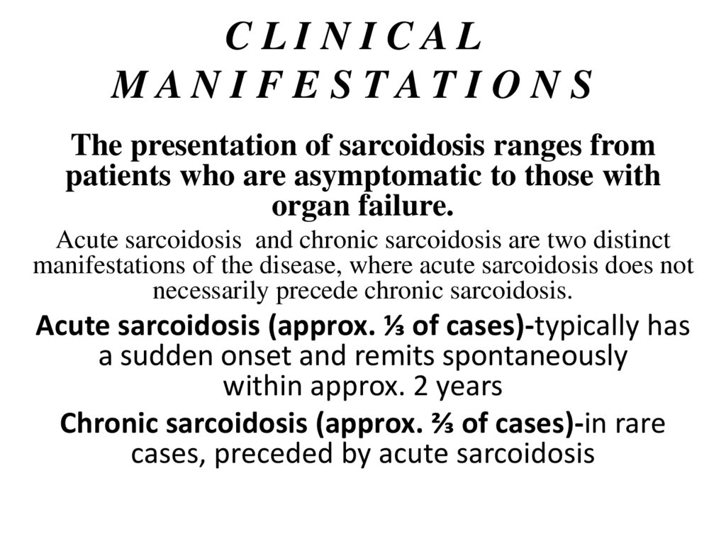
CLINICALMANIFESTATIONS
The presentation of sarcoidosis ranges from
patients who are asymptomatic to those with
organ failure.
Acute sarcoidosis and chronic sarcoidosis are two distinct
manifestations of the disease, where acute sarcoidosis does not
necessarily precede chronic sarcoidosis.
Acute sarcoidosis (approx. ⅓ of cases)-typically has
a sudden onset and remits spontaneously
within approx. 2 years
Chronic sarcoidosis (approx. ⅔ of cases)-in rare
cases, preceded by acute sarcoidosis
9.
Respiratory complaints including
cough and dyspnea are the most common presenting
symptoms. In many cases, the patient presents with
a 2- to 4-week history of these symptoms.
• Symptoms related to cutaneous and ocular
disease are the next two most common complaints.
Skin lesions are often nonspecific.
• Nonspecific constitutional symptoms include
fatigue, fever, night sweats, and weight loss. Fatigue
is perhaps the most common constitutional
symptom that affects these patients.
10.
Lung symptomsLung involvement occurs in >90 % of sarcoidosis patients
and may cause lung problems, such as:
• Persistent dry cough is a very common symptom. Airway
hyperreactivity, as determined by methacholine challenge, will
be positive in some of these patients.
• Shortness of breath
• Wheezing
• Chest pain
• Pulmonary arterial hypertension is reported in at least 5%
of sarcoidosis patients. Either direct vascular involvement or
the consequence of fibrotic changes in the lung can lead to
pulmonary arterial hypertension.
11.
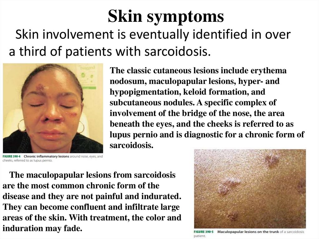
Skin symptomsSkin involvement is eventually identified in over
a third of patients with sarcoidosis.
The classic cutaneous lesions include erythema
nodosum, maculopapular lesions, hyper- and
hypopigmentation, keloid formation, and
subcutaneous nodules. A specific complex of
involvement of the bridge of the nose, the area
beneath the eyes, and the cheeks is referred to as
lupus pernio and is diagnostic for a chronic form of
sarcoidosis.
The maculopapular lesions from sarcoidosis
are the most common chronic form of the
disease and they are not painful and indurated.
They can become confluent and infiltrate large
areas of the skin. With treatment, the color and
induration may fade.
12.
Eye symptoms• Blurred vision
• Eye pain
• Burning, itching
or dry eyes
• Severe redness
• Sensitivity to
light
Heart symptoms
Chest pain
Shortness of breath (dyspnea)
Fainting (syncope)
Fatigue
Irregular heartbeats (arrhythmias)
Rapid or fluttering heart beat
(palpitations)
• Swelling caused by excess fluid
(edema)
Sarcoidosis can also affect calcium metabolism, the
nervous system, the liver and spleen, muscles, bones and
joints, the kidneys, lymph nodes, or any other organ.
13.
Laboratory diagnosticsIn case of suspicion, the patient is prescribed a General
and biochemical blood test, a urine test.
Preparation for laboratory diagnostics:
*Alcohol and smoking are excluded 24
hours before the study;
*blood and urine sampling is performed
in the morning before meals;
*some medications are canceled within a
few days.
14.
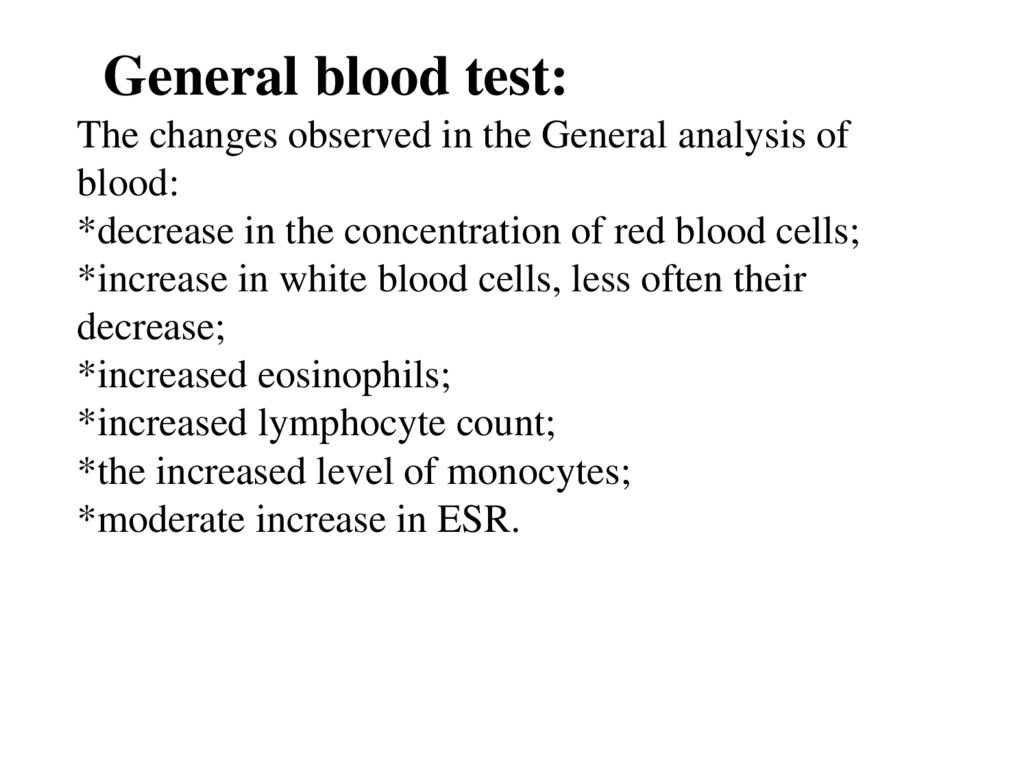
General blood test:The changes observed in the General analysis of
blood:
*decrease in the concentration of red blood cells;
*increase in white blood cells, less often their
decrease;
*increased eosinophils;
*increased lymphocyte count;
*the increased level of monocytes;
*moderate increase in ESR.
15.
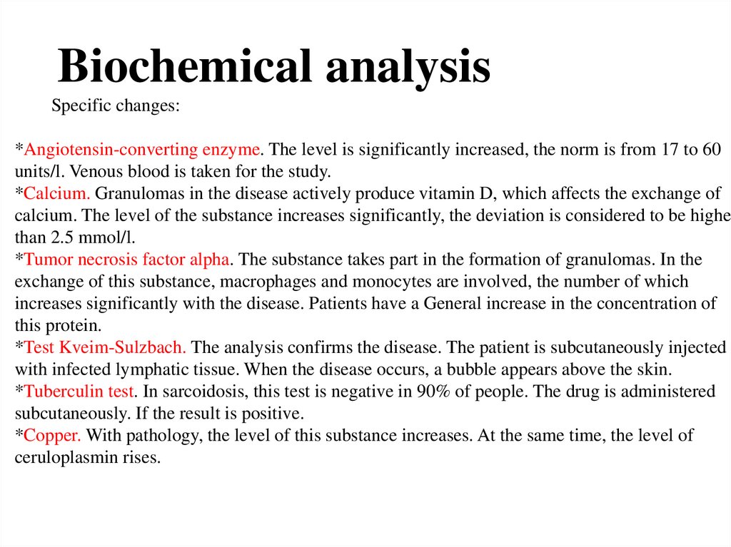
Biochemical analysisSpecific changes:
*Angiotensin-converting enzyme. The level is significantly increased, the norm is from 17 to 60
units/l. Venous blood is taken for the study.
*Calcium. Granulomas in the disease actively produce vitamin D, which affects the exchange of
calcium. The level of the substance increases significantly, the deviation is considered to be higher
than 2.5 mmol/l.
*Tumor necrosis factor alpha. The substance takes part in the formation of granulomas. In the
exchange of this substance, macrophages and monocytes are involved, the number of which
increases significantly with the disease. Patients have a General increase in the concentration of
this protein.
*Test Kveim-Sulzbach. The analysis confirms the disease. The patient is subcutaneously injected
with infected lymphatic tissue. When the disease occurs, a bubble appears above the skin.
*Tuberculin test. In sarcoidosis, this test is negative in 90% of people. The drug is administered
subcutaneously. If the result is positive.
*Copper. With pathology, the level of this substance increases. At the same time, the level of
ceruloplasmin rises.
16.
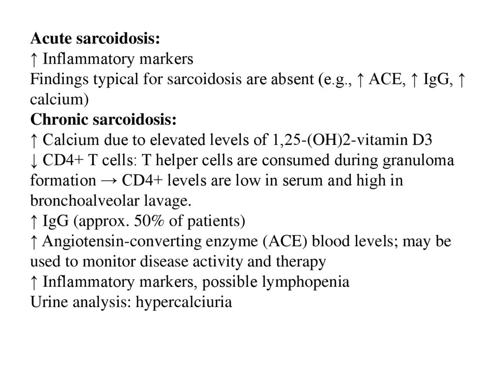
Acute sarcoidosis:↑ Inflammatory markers
Findings typical for sarcoidosis are absent (e.g., ↑ ACE, ↑ IgG, ↑
calcium)
Chronic sarcoidosis:
↑ Calcium due to elevated levels of 1,25-(OH)2-vitamin D3
↓ CD4+ T cells: T helper cells are consumed during granuloma
formation → CD4+ levels are low in serum and high in
bronchoalveolar lavage.
↑ IgG (approx. 50% of patients)
↑ Angiotensin-converting enzyme (ACE) blood levels; may be
used to monitor disease activity and therapy
↑ Inflammatory markers, possible lymphopenia
Urine analysis: hypercalciuria
17.
Instrumental investigations:Chest x-ray
CT
Biopsy
Endoscopic examination: bronchoscopy and
thoracoscopy
Pulmonary function tests (to assess the
severity of the disease)
18.
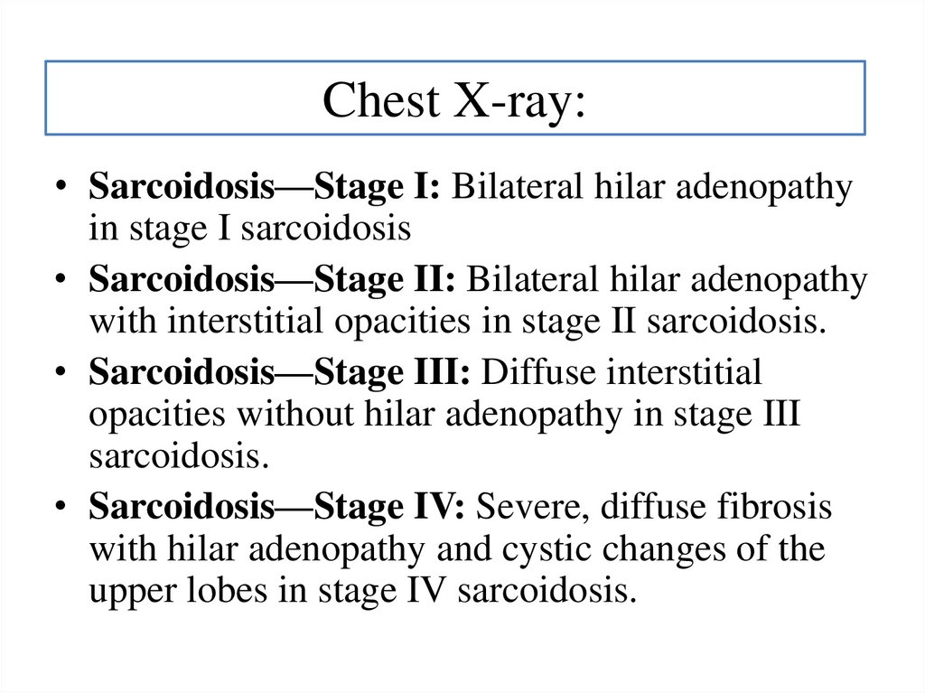
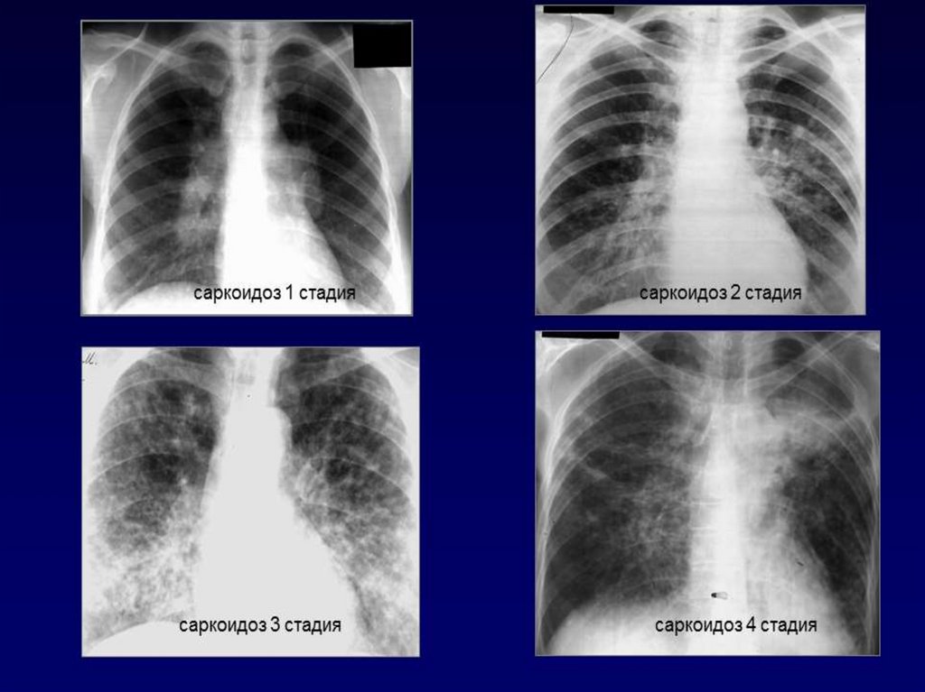
Chest X-ray:• Sarcoidosis—Stage I: Bilateral hilar adenopathy
in stage I sarcoidosis
• Sarcoidosis—Stage II: Bilateral hilar adenopathy
with interstitial opacities in stage II sarcoidosis.
• Sarcoidosis—Stage III: Diffuse interstitial
opacities without hilar adenopathy in stage III
sarcoidosis.
• Sarcoidosis—Stage IV: Severe, diffuse fibrosis
with hilar adenopathy and cystic changes of the
upper lobes in stage IV sarcoidosis.
19.
20.
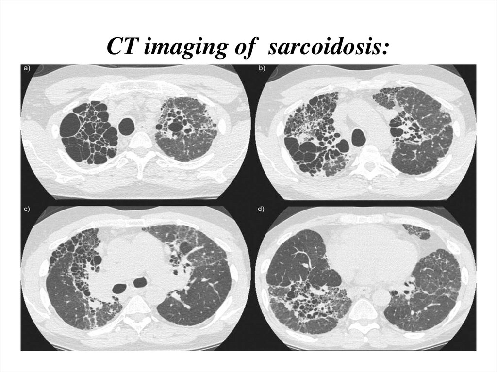
CT findings in more advanced stages (II to IV)include:
Thickening of the bronchovascular bundles
and bronchial walls
Beading of the interlobular septa
Ground-glass opacification
Parenchymal nodules, cysts, or cavities
Traction bronchiectasis
21.
CT imaging of sarcoidosis:22.
When imaging suggests sarcoidosis, thediagnosis is confirmed by demonstration of
noncaseating granulomas on biopsy and
exclusion
of
alternative
causes
of
granulomatous disease .
Biopsy sites:
peripheral lymph nodes;
skin lesions;
conjunctiva;
23.
Biopsy methods:Bronchoscopic:
Transbronchial lung biopsy (PLL).
Classical transbronchial needle biopsy of intrathoracic lymph nodes
Endoscopic fine-needle puncture of mediastinal lymph nodes
under the control of endosonography.
Direct biopsy of the bronchial mucosa (direct biopsy).
Brush biopsy of the bronchial mucosa (brush biopsy).
Bronchoalveolar lavage (BAL).
Surgical biopsy techniques:
Thoracotomy with biopsy of the lung and intrathoracic lymph
nodes.
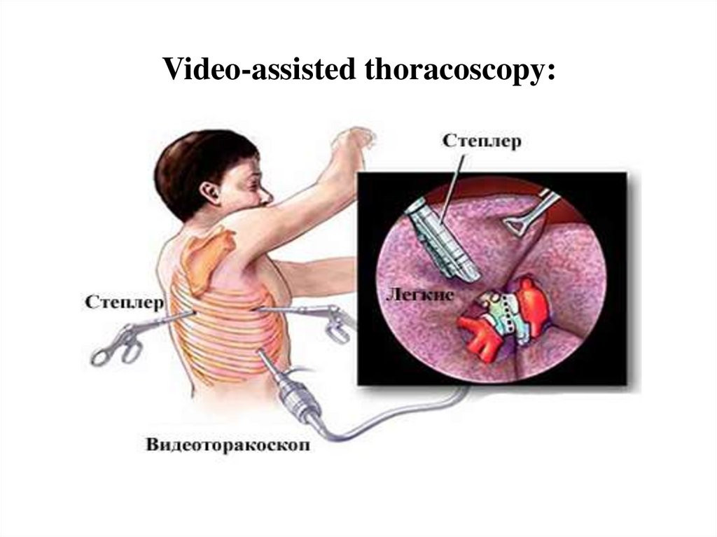
Video-assisted thoracoscopy
Mediastinoscopy.
24.
Video-assisted thoracoscopy:25.
Bronchoscopy:changes in the vessels of the bronchial mucosa
(expansion)
lumpy eruptions (sarcoid granulomas) in the form of
plaques of various sizes (from millet grains to a pea);
on the mucous membrane of the bronchi, ischemic
spots are visible - pale areas devoid of blood vessels.
Thoracoscopy:
Whitish-yellowish sarcoid granulomas are visible on
the pleural surface.
26.
Pulmonary function test:• Pulmonary function test results are often
normal in early stages but demonstrate
restriction and reduced diffusing capacity for
carbon monoxide (DLCO) in advanced disease.
27.
Differential diagnosissarcoidosis
28.
DiseaseClinical manifestations
X-ray picture
Sarcoidosis
More often asymptomatic onset, with
progression of subfebrile fever,
weakness, aching chest pain
An increase in hilar lymph nodes, less
often parabronchial, tracheobronchial.
The apperance of a large-spotted
pattern in the basal and small-spotted in
the meddle zones, as well as small focal
shadows
Silicosis
Shortness of breath, cough, chest pain,
lymph nodes are not enlarged. Slowly
progressive course.
Diffuse interstitial fibrosis, nodular
process. Monomorphic shadows.
Disseminated tuberculosis
Intoxication syndrome. There may be a
cough, excretion of micobacterium
tuberculosis in the macrobacterium,
hemoptysis, chest pain.
Shadows are polymorphic. There may
be interstitial changes and enlargement
of the LN.
Exogenous allergic alveolits
Chills, fever, shortness of breath,
cough, pain in the chest, muscles,
joints.
Strengthening the pulmonary pattern
due to the intertial component, the
summation of these shadows creates a
picture of miliary foci.
Idiopathic fibrosing alveolits
Dyspnoe with acute progressive course,
fever, weight loss, chest pain, muscles,
joints
Strengthening and deformation of the
pulmonary pattern, interstitial fibrosis.
Lymphogranulomatosis
General malaise, fever
An increase in the mediastinal lumen,
more often with the formation of
conglomerates. In the lung tissue,
interstitial and infiltrative changes
29.
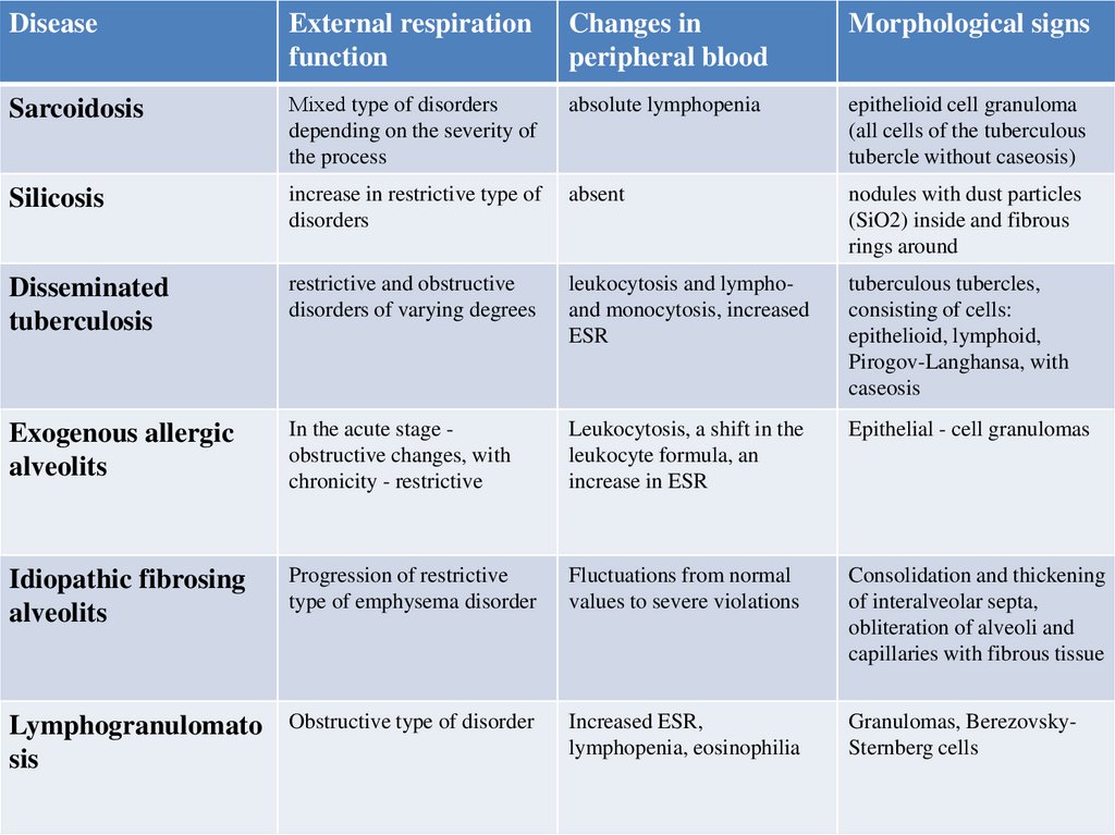
DiseaseExternal respiration
function
Changes in
peripheral blood
Morphological signs
Sarcoidosis
Мixed type of disorders
depending on the severity of
the process
absolute lymphopenia
epithelioid cell granuloma
(all cells of the tuberculous
tubercle without caseosis)
Silicosis
increase in restrictive type of
disorders
absent
nodules with dust particles
(SiO2) inside and fibrous
rings around
Disseminated
tuberculosis
restrictive and obstructive
disorders of varying degrees
leukocytosis and lymphoand monocytosis, increased
ESR
tuberculous tubercles,
consisting of cells:
epithelioid, lymphoid,
Pirogov-Langhansa, with
caseosis
Exogenous allergic
alveolits
In the acute stage obstructive changes, with
chronicity - restrictive
Leukocytosis, a shift in the
leukocyte formula, an
increase in ESR
Epithelial - cell granulomas
Idiopathic fibrosing
alveolits
Progression of restrictive
type of emphysema disorder
Fluctuations from normal
values to severe violations
Consolidation and thickening
of interalveolar septa,
obliteration of alveoli and
capillaries with fibrous tissue
Lymphogranulomato
sis
Obstructive type of disorder
Increased ESR,
lymphopenia, eosinophilia
Granulomas, BerezovskySternberg cells
30.
Differential diagnosis of granulomatous diseaseRisk factors
Sarcoidosis
Tuberculosis (TB)
Hodgkin lymphoma
Clinical presentation
Biopsy
•African American
females in the US
•Dry cough
•Noncaseating granulomas
•Erythema nodosum
•Lupus pernio
•Giant cells
•Anterior (and
possibly posterior) uvei
tis
•Immunocompromised i
ndividuals
•Previous TB and/or
recent TB exposure
•Fever, weight loss, and
night sweats
•Productive cough that
does not respond to
conventional antibiotic
therapy
•Hemoptysis
•History of infectious
mononucleosis
•Pel-Ebstein fever
•Non•Alcohol-induced pain caseating granulomas
•Pruritus
•Reed-Sternberg cells
•Inflammatory cell
infiltrate
(e.g., eosinophils, fibro
blasts, plasma cells)
Non-Hodgkin lymphoma •Infections (e.g., EBV •Indolent lymph
infection or Helicobacte node enlargement
Other laboratory
findings
+
+
•↑ CD4 /CD8 ratio in
bronchoalveolar lavage
•Caseating granulomas •M. tuberculosis or
its DNA
•Langhans giant
cells, epithelioid
macrophages,
and lymphocytes
•Acid-fast M.
tuberculosis
•Single or
combined cytopenias (i.
e., anemia, leukopenia,
and/or thrombocytopeni
a)
•Non•Single or
caseating granulomas w combined cytopenias
31.
examination isunremarkable.
•In symptomatic
patients
• Progressive e
xertional
dyspnea
• Chronic coug
h (possibly
with sputum)
• Auscultatory
findings
(e.g., rales, cr
ackles)
• Signs of
respiratory
failure
(e.g., digital
clubbing)
Granulomatosis with •Caucasian individuals
polyangiitis
aged 65–74 years
Histoplasmosis
•Chronic rhinitis/sinusit •Nonis with
caseating granulomas
thick purulent/bloody
discharge
•Treatmentresistant pneumonia
•Glomerulonephritis
•AIDS
•Pulmonary
•Exposure to bird or bat (e.g., dry cough,
excrement
oral ulcers) or
extrapulmonary
(e.g., splenomegaly)
•Positive cytoplasmic
ANCA
•Caseating granulomas •Positive polysaccharid
e urine and serum
•Identification of H.
capsulatum yeast with s antigen test
ilver stain
32.
Treatment• NSAIDs
• Corticosteroids
• Sometimes used immunosuppressants
33.
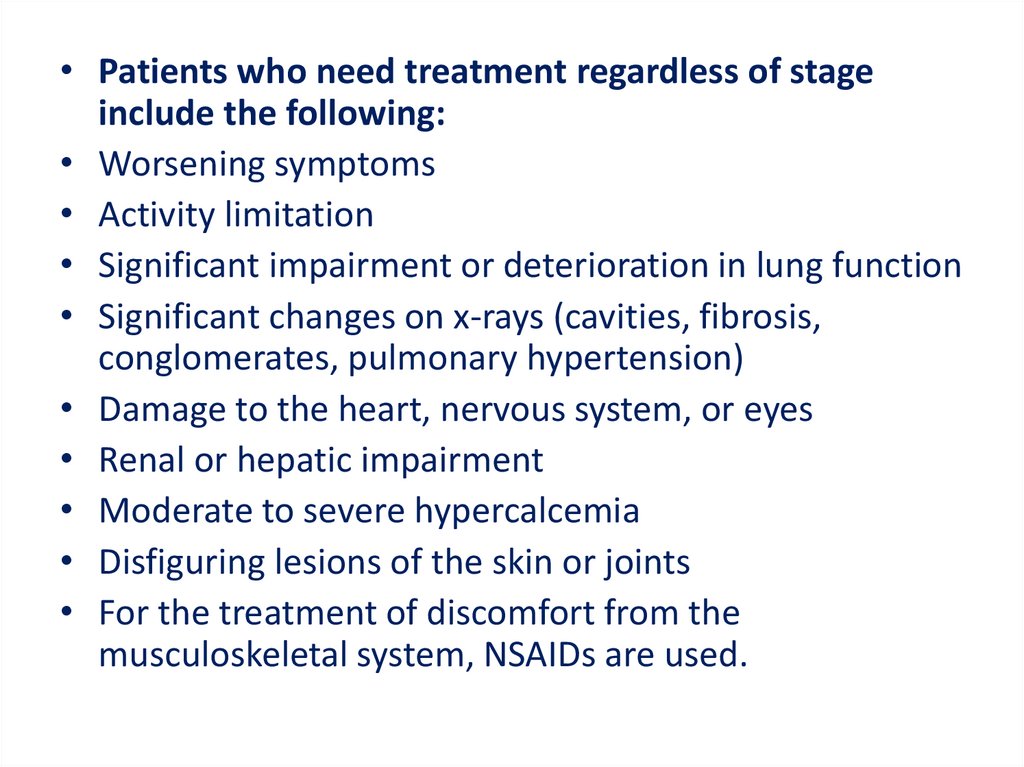
• Patients who need treatment regardless of stageinclude the following:
• Worsening symptoms
• Activity limitation
• Significant impairment or deterioration in lung function
• Significant changes on x-rays (cavities, fibrosis,
conglomerates, pulmonary hypertension)
• Damage to the heart, nervous system, or eyes
• Renal or hepatic impairment
• Moderate to severe hypercalcemia
• Disfiguring lesions of the skin or joints
• For the treatment of discomfort from the
musculoskeletal system, NSAIDs are used.
34.
Corticosteroids• Symptom management begins with corticosteroids.
• ! The presence of abnormalities on chest scans without significant
symptoms or evidence of decreased organ function is not an
indication for treatment.
• The standard protocol is prednisone 20–40 mg orally once a day,
depending on symptoms and severity of the disease. Alternatively,
you can use an every other day regimen: for example, prednisone
40 mg orally once every other day.
• Although patients rarely need a dose> 40 mg / day, higher doses
may be required to reduce complications of neurological disease.
Response usually occurs within 6-12 weeks, so symptoms and
pulmonary function tests can be re-evaluated between 6 and 12
weeks. In chronic and latent cases, the reaction may be delayed. If
there is an effect, the dose of corticosteroids is gradually reduced to
maintenance (for example, prednisone 10-15 mg / day); with
improvement, therapy is continued for at least 6-12 months.
35.
• The optimal duration of treatment is unknown. Apremature dose reduction may lead to relapse. In case of a
doubtful reaction or ineffectiveness of treatment, the use
of the drug is gradually discontinued. Ultimately,
corticosteroids can be discontinued in most patients, but
since relapse occurs in 50% of cases, follow-up
examinations should be performed, usually every 3–6
months.
• Treatment with corticosteroids should be resumed if
complaints and symptoms recur, including dyspnea,
arthralgia, fever, liver failure, cardiac arrhythmias, CNS
symptoms, hypercalcemia, eye damage, lack of topical drug
control, and disfiguring skin lesions. Because low doses of
corticosteroids suppress ACE production, it may be useful to
monitor serum ACE levels over time when assessing
adherence to corticosteroid treatment in the presence of
elevated ACE levels.
36.
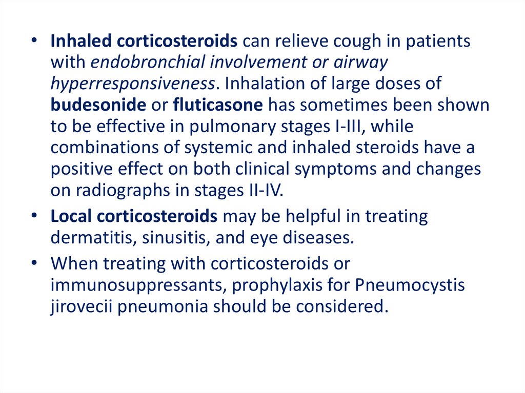
• Inhaled corticosteroids can relieve cough in patientswith endobronchial involvement or airway
hyperresponsiveness. Inhalation of large doses of
budesonide or fluticasone has sometimes been shown
to be effective in pulmonary stages I-III, while
combinations of systemic and inhaled steroids have a
positive effect on both clinical symptoms and changes
on radiographs in stages II-IV.
• Local corticosteroids may be helpful in treating
dermatitis, sinusitis, and eye diseases.
• When treating with corticosteroids or
immunosuppressants, prophylaxis for Pneumocystis
jirovecii pneumonia should be considered.
37.
Immunosuppressants• ! Treatment with immunosuppressants is carried out in case of
intolerance to moderate doses of corticosteroids, refractoriness of
sarcoidosis to corticosteroids, or if treatment with corticosteroids is
required for a long time.
• In about 10% of cases when therapy is necessary, tolerated doses of
corticosteroids are ineffective, and a 6-month trial of methotrexate
therapy at a dose of 10-15 mg / week should be carried out.
Methotrexate and corticosteroids are given initially; after 6-8
weeks, the dose of corticosteroids can be gradually reduced, and in
many cases their use can be discontinued. However, the maximum
effect of methotrexate can be observed after 6-12 months. In such
cases, the dose of prednisolone should be decreased more slowly.
Determination of blood corpuscles and liver enzymes should be
performed first every 1–2 weeks, then every 4–6 weeks, as soon as
a stable dose is reached. In patients receiving methotrexate, folate
(1 mg orally per day) is recommended.
38.
• Other drugs that have been effective in a small number of patientswho do not respond to corticosteroid treatment or who experience
complicating side effects include azathioprine, mycophenolate
mofetil, cyclophosphamide, chloroquine or hydroxychloroquine,
and infliximab. Immunosuppressants are often more effective in
refractory cases, and relapse is common after stopping treatment.
The TNF inhibitor infliximab may be effective in treating chronic
steroid-dependent pulmonary sarcoidosis, refractory lupus fever,
and neurosarcoidosis. It is administered intravenously at a dose of
3-5 mg / kg once, repeated in 2 weeks, and then administered 1
time / month.
• Hydroxychloroquine 400 mg orally once a day or 200 mg orally
twice a day may be as effective for treating hypercalcemia, sarcoid
skin lesions, or enlarged patient-discomfortable or disfiguring
peripheral lymph nodes.
39.
Oxygen therapyThe administration of oxygen to patients with LH on
the background of sarcoidosis is indicated for
chronic hypoxemia (Rao2 < 55 mm Hg), while the
dose is titrated to reach SpO2 >90% when breathing
through an oxygen concentrator
Specific LH therapy for sarcoidosis
Currently, there are effective drugs for the treatment of
pulmonary arterial hypertension (PAH), such forms as
idiopathic (primary) PAH, PAH in systemic scleroderma, etc. It is
possible that these same drugs are also the most effective
therapy for LH associated with sarcoidosis.
40.
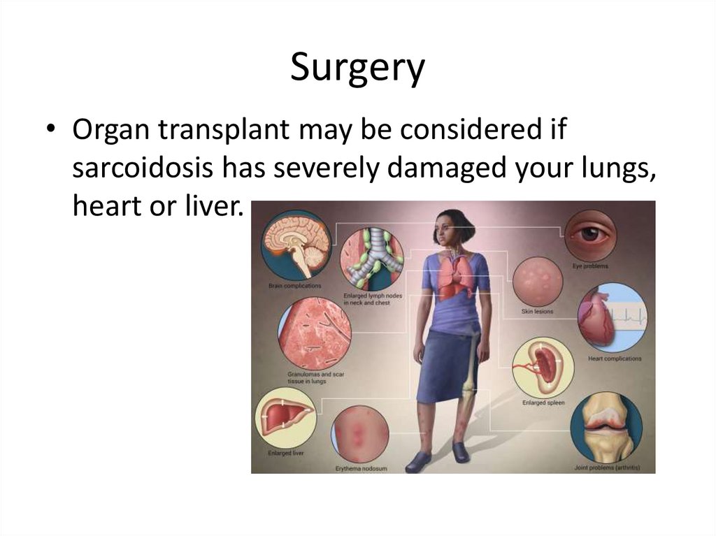
Surgery• Organ transplant may be considered if
sarcoidosis has severely damaged your lungs,
heart or liver.
41.
Complication of sarcoidosis:Complications list for Sarcoidosis:
Lung damage - about 90% of cases
Collapsed lung
Lung granulomas
Lung fibrosis
Frequent pneumonia
Bleedings
Eye complications:
Cataracts
Glaucoma
Blindness
Heart damage
Nervous system - only about 1-5% of cases.
Liver damage
Death
Kidney damage
Arthritis
Psychological problem
42.
Collapsed lunginflammation or the growth of
granulomas
rupture of pleura
the pressure is equalized with
atmospheric pressure
the lungs begin to shrink
43.
Pulmonary fibrosisPulmonary fibrosis is the end stage of
pulmonary sarcoidosis. This process begins at
stages 2 - 3 of the disease, when symptoms are
just beginning to appear
44.
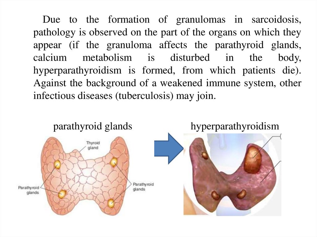
Due to the formation of granulomas in sarcoidosis,pathology is observed on the part of the organs on which they
appear (if the granuloma affects the parathyroid glands,
calcium metabolism is disturbed in the body,
hyperparathyroidism is formed, from which patients die).
Against the background of a weakened immune system, other
infectious diseases (tuberculosis) may join.
parathyroid glands
hyperparathyroidism