 e-mail: yarn85@mail.ru")


 medicine
Similar presentations:
")

")
")


")

")

Importance of tuberculosis as scientific and practical problem. Tuberculosis epidemiology in the world. (Lecture 1)
1. Zaporizhzhia State Medical University Department of phthisiology and pulmonology R.N. Yasinskiy (assistant of department) e-mail: yarn85@mail.ru
Importance of tuberculosis as scientificand practical problem.
Tuberculosis epidemiology in the
world. Etiology and pathogenesis of
tuberculosis.
Immunity at tuberculosis
2.
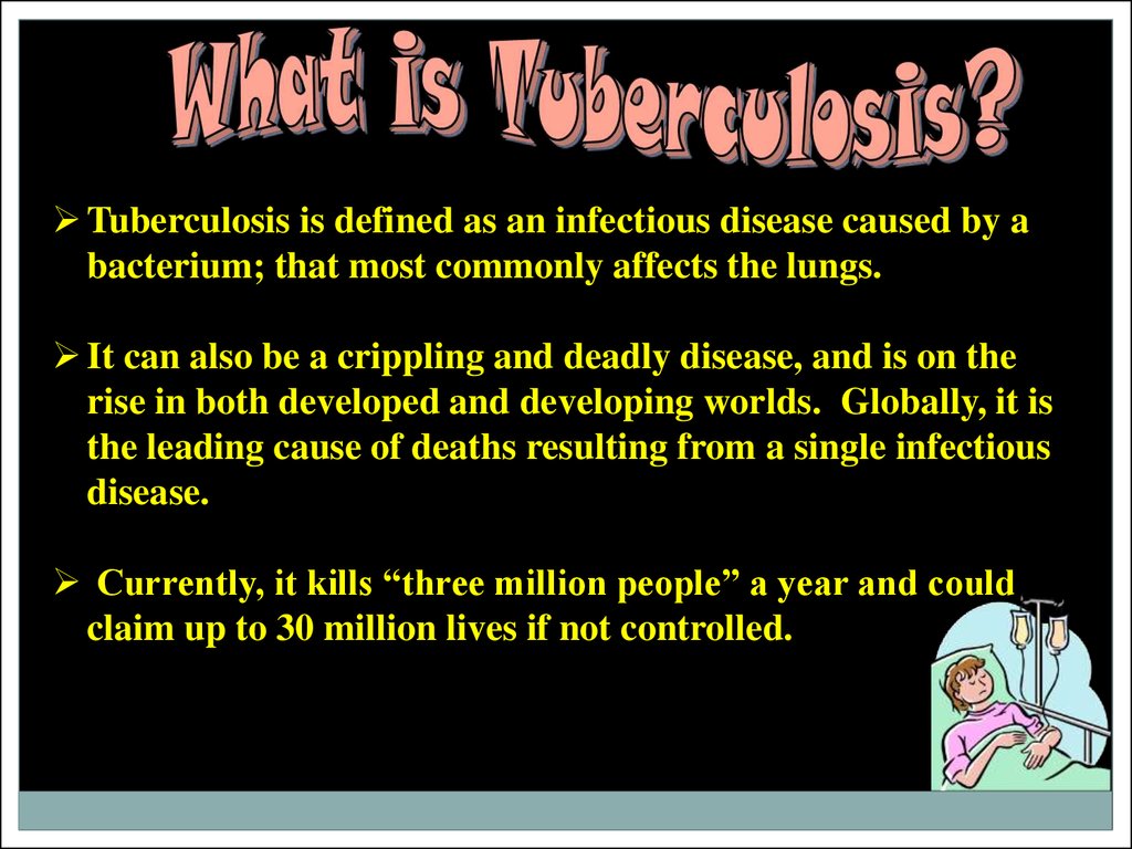
Tuberculosis is defined as an infectious disease caused by abacterium; that most commonly affects the lungs.
It can also be a crippling and deadly disease, and is on the
rise in both developed and developing worlds. Globally, it is
the leading cause of deaths resulting from a single infectious
disease.
Currently, it kills “three million people” a year and could
claim up to 30 million lives if not controlled.
3.
These numbers are expected to increase inthe coming years because of the acquired
immune deficiency syndrome (AIDS)
pandemic – a high percentage of the patients
with human immunodeficiency virus (HIV)
are co-infected with MBТ, and the
emergence of drug-resistant strains of the
TB organisms. This alarming increase in
morbidity and mortality highlights the need
to strengthen control measures.
Accurate and rapid diagnosis is essential for controlling the
disease, yet the traditional tests for TB produce results that are
either inaccurate or take too long to be definitive.
4.
People who have healthy immune systems can often fight off atuberculosis infection after breathing in MBT.
These people have no symptoms and are not sick, because the
immune system is able to prevent the MBT from growing and
multiplying.
This is called latent tuberculosis. People with latent tuberculosis
are not contagious and cannot spread the disease to others.
However, anything that stresses the immune system, such as the
development of a chronic disease, can allow the bacteria to
become active and begin to multiply in the body.
5. The magnitude of the problem:
Tuberculosis kills more than 3 million peopleper year
Tuberculosis produces 25% of all avoidable
deaths in developing countries
Tuberculosis produces more death than any
other single infectious disease , so its the
deadliest one
About the 1/3 of the world population are
infected by tuberculosis
6. Tuberculosis uniqueness:
the most ancient among known infectionthe most ubiquitous infection
infection which can coexist with the
human being without producing the
disease
infection from which human being isn’t
able to deliver
7. Main reasons for tuberculosis reappearance as a global challenge:
Drug-resistanceHuman immunodeficiency virus
(HIV)
Social disturbances
8. EPIDEMIOLOGY
9.
10. 5 PRIORITIES TO ELIMINATE TB
Reaching the “missed” cases (3 million not in thesystem)
Address MDR-TB as crisis
Accelerate response to TB/HIV
Increase financing to close resource gaps
Intensify research and ensure rapid uptake of
innovations
11. Reaching the "missed" cases early means cutting transmission
Reaching the "missed" cases early means cuttingtransmission
Share of total missed cases
12.
Addressing MDR-TB as a crisisPercentage of new TB cases with MDR-TB
13.
Five priority actions to address the globalMDR-TB crisis
14.
Accelerating response to TB/HIV means cuttingtransmission and mortality
Estimated HIV prevalence in new TB cases, 2013
15.
67th World Health Assembly, Geneva, May 201416.
The End TB Strategy – Components1. INTEGRATED, PATIENT-CENTRED CARE AND
PREVENTION
A. Early diagnosis of tuberculosis including universal drugsusceptibility testing, and systematic screening of contacts and
high-risk groups
B. Treatment of all people with tuberculosis including drugresistant tuberculosis, and patient support
C. Collaborative tuberculosis/HIV activities, and management of
co-morbidities
D. Preventive treatment of persons at high risk, and vaccination
against tuberculosis
17.
2. BOLD POLICIES AND SUPPORTIVE SYSTEMSA. Political commitment with adequate resources for tuberculosis care and prevention
B. Engagement of communities, civil society organizations, and public and private care
providers
C. Universal health coverage policy, and regulatory frameworks for case notification,
vital registration, quality and rational
use of medicines, and infection control
D. Social protection, poverty alleviation and actions on other determinants of
tuberculosis
3. INTENSIFIED RESEARCH AND INNOVATION
A. Discovery, development and rapid uptake of new tools, interventions and strategies
B. Research to optimize implementation and impact, and promote innovations
18. Mycobacteria types
1.M. causing tuberculosis
2.
Non-patogenous M.
3.
M. tuberculosis (human)
M. bovis.
M. Africans.
M.avium-intracellulare.
M.smegmaticus.
M. xenopi
M. scrofulaceum.
M. causing leprosy
M. leprae
19. Important Mycobacterium
Mycobacterium tuberculosis, along with M.bovis, M. africanum, and M. microti all cause
the disease known as tuberculosis (TB) and are
members of the tuberculosis species complex.
Each member of the TB complex is pathogenic,
but M. tuberculosis is pathogenic for humans
while M. bovis is usually pathogenic for animals
20.
The Causative agent of tuberculosis is opened byR. Kokh on March, 24, 1892
21. Morphology of Mycobacterium tuberculosis
Straight, slightly curvedRod shaped 3 x
0.3microns
May be single, in pairs
or in small clumps
On conditions in growth
appears as filamentous,
club shaped, or in
Branched forms.
22. Atypical Mycobacterium
PhotochromogensScotochromogens
Non Photochromogens
Rapid growers
22
23.
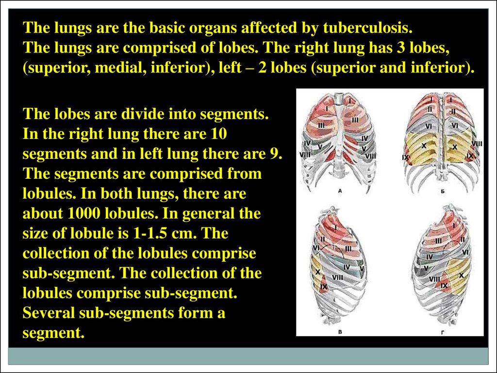
The lungs are the basic organs affected by tuberculosis.The lungs are comprised of lobes. The right lung has 3 lobes,
(superior, medial, inferior), left – 2 lobes (superior and inferior).
The lobes are divide into segments.
In the right lung there are 10
segments and in left lung there are 9.
The segments are comprised from
lobules. In both lungs, there are
about 1000 lobules. In general the
size of lobule is 1-1.5 cm. The
collection of the lobules comprise
sub-segment. The collection of the
lobules comprise sub-segment.
Several sub-segments form a
segment.
24.
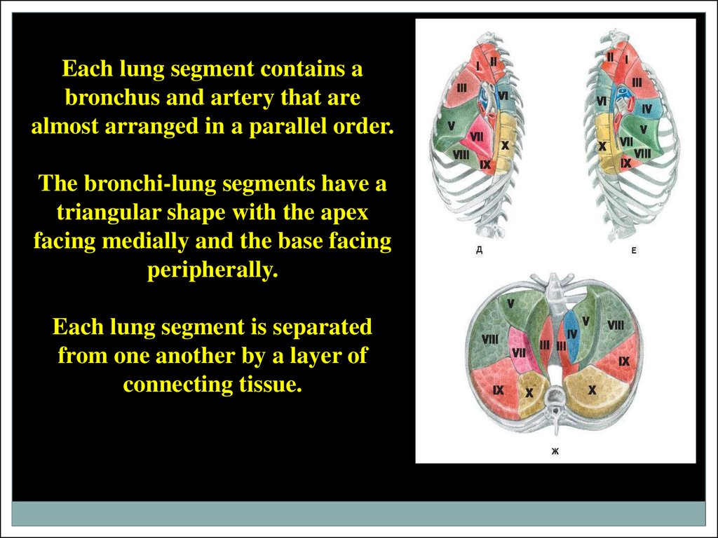
Each lung segment contains abronchus and artery that are
almost arranged in a parallel order.
The bronchi-lung segments have a
triangular shape with the apex
facing medially and the base facing
peripherally.
Each lung segment is separated
from one another by a layer of
connecting tissue.
25.
Bronchial AirwaysThe two bronchi proceed from the
bifurcation of the trachea opposite to
the 4-th thoracic vertebra to their
corresponding lungs.
Upon entering the lungs, the bronchi
divide into branches in which each of
these branches divide and subdivide
dichotomously to their ultimate
termination (smallest bronchi).
26.
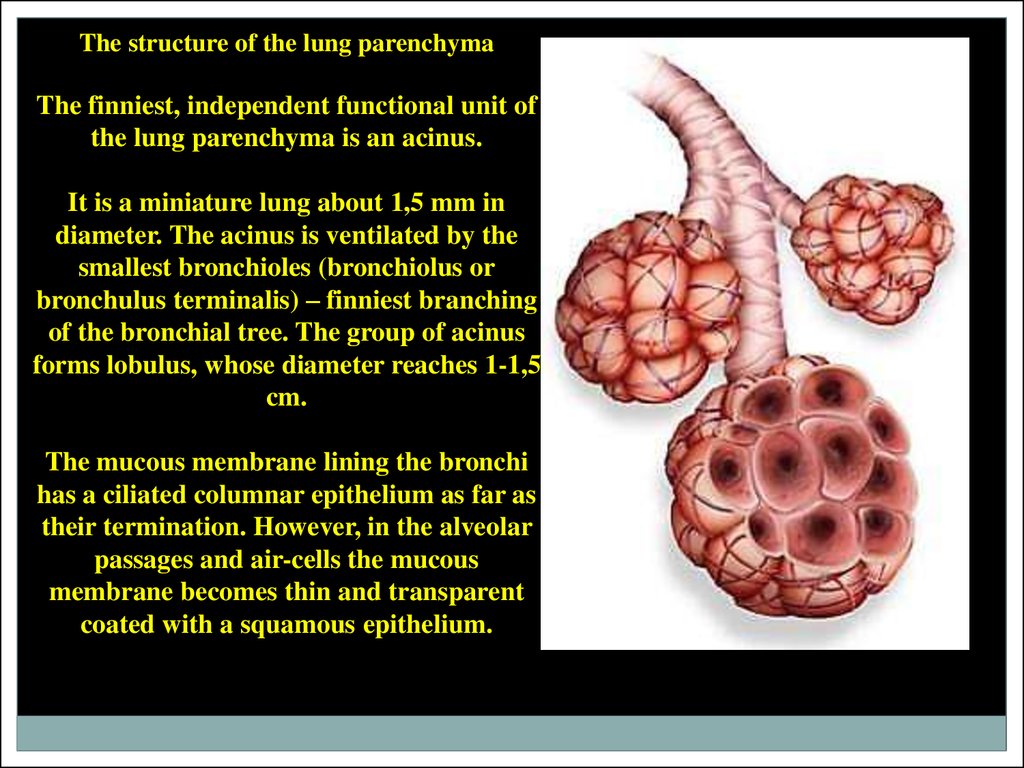
The structure of the lung parenchymaThe finniest, independent functional unit of
the lung parenchyma is an acinus.
It is a miniature lung about 1,5 mm in
diameter. The acinus is ventilated by the
smallest bronchioles (bronchiolus or
bronchulus terminalis) – finniest branching
of the bronchial tree. The group of acinus
forms lobulus, whose diameter reaches 1-1,5
cm.
The mucous membrane lining the bronchi
has a ciliated columnar epithelium as far as
their termination. However, in the alveolar
passages and air-cells the mucous
membrane becomes thin and transparent
coated with a squamous epithelium.
27.
PleuraEach lung is enclosed and its structure
supported by a serous membrane, the
pleura, which invests it as far as the
root, and is then reflected on the
parietals of the chest.
That portion of the membrane which is
in relation with the lung is called (pleura
visceralis s. pleura pulmonalis), and that
in contact with the parietes, pleura
costalis, pleura diaphragmatica and
(pleura mediastinalis).
The pulmonary pleura is very thin,
elastic, and inseparably connected with
the structure of the lung; the costal
pleura is thick and strong, has very little
elasticity, and can be readily stripped off
the ribs and intercostal muscles which it
covers.
pleura visceralis
parietal pleura
28.
The lymphatic lung systemThe lung surface is formed of a thin subpleural network of lymphatic vessels that
communicate with the pleural cavity by a
system of pores.
The lung parenchyma consists of 2 types of
lymphatic structures.
The 1st type forms an elaborate network
located beneath the bronchi’s mucous
membrane.
The 2nd type originates in the capillaries between alveolar ducts and alveolar sacs.
Lymphatic vessels of both types terminate in the broncho-pulmonary nodes in the
hilus of the lung. These numerous and large nodes are located around the bronchi and
within the tracheal bifurcation.
29. Transmission of tuberculosis
TB is spread from person to person through the air. The dots in the airrepresent droplet.
The dots in the air represent droplet nuclei containing tubercle bacilli.
30. The ways of the transmission:
Inhalation (about 90%)Dusty
o Droplet
Alimentary
Contact
Vertical
o
31.
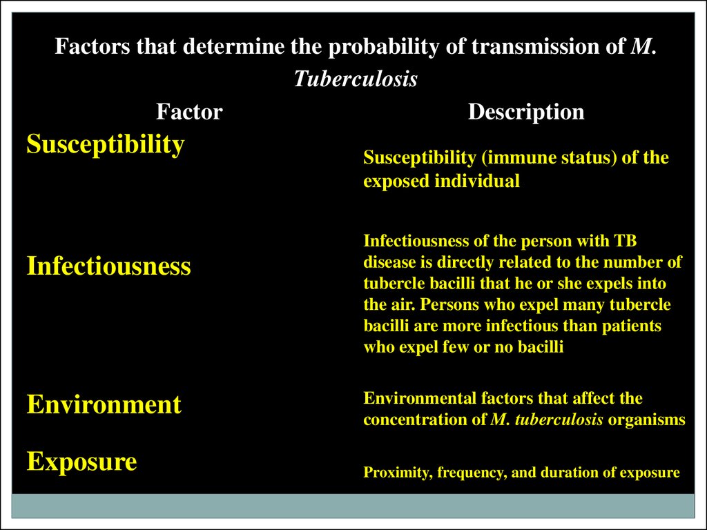
Factors that determine the probability of transmission of M.Tuberculosis
Factor
Description
Susceptibility
Infectiousness
Environment
Exposure
Susceptibility (immune status) of the
exposed individual
Infectiousness of the person with TB
disease is directly related to the number of
tubercle bacilli that he or she expels into
the air. Persons who expel many tubercle
bacilli are more infectious than patients
who expel few or no bacilli
Environmental factors that affect the
concentration of M. tuberculosis organisms
Proximity, frequency, and duration of exposure
32.
Characteristics of a patient with TB disease that areassociated with Infectiousness
Factor
Clinical
Procedure
Radiographic and laboratory
Description
Presence of cough, especially lasting 3 weeks or longer
1. Respiratory tract disease, especially with
involvement of the larynx (highly infectious)
2. Failure to cover the mouth and nose when
coughing
3. Inappropriate or inadequate treatment (drugs,
duration)
1.
Undergoing cough-inducing or aerosol-generating
procedures (e.g., bronchoscopy, sputum induction,
administration of aerosolized medications)
1.
2.
3.
Cavitation on chest radiograph
Positive culture for M. tuberculosis
Positive AFB sputum smear result
33.
Proximity and length of exposure factors that can affecttransmission of M. Tuberculosis
Factor
Description
Duration of exposure to a
person with infectious TB
The longer the duration of
exposure, the higher the risk for
transmission
Frequency of exposure to
infectious person
The more frequent the
exposure, the higher the risk for
transmission
Physical proximity to infectious The closer the proximity, the
person
higher the risk for transmission
34.
Pathogenesis of TBDroplet nuclei
containing
tubercle bacilli
are inhaled, enter
the lungs, and
travel to the
alveoli.
35.
Tuberclebacilli
multiply in the
alveoli.
36.
A small number of tuberclebacilli enter the bloodstream
and spread throughout the
body. The tubercle bacilli
may reach any part of the
body, including areas where
TB disease is more likely to
develop (such as the brain,
larynx, lymph node, lung,
spine, bone, or kidney).
37.
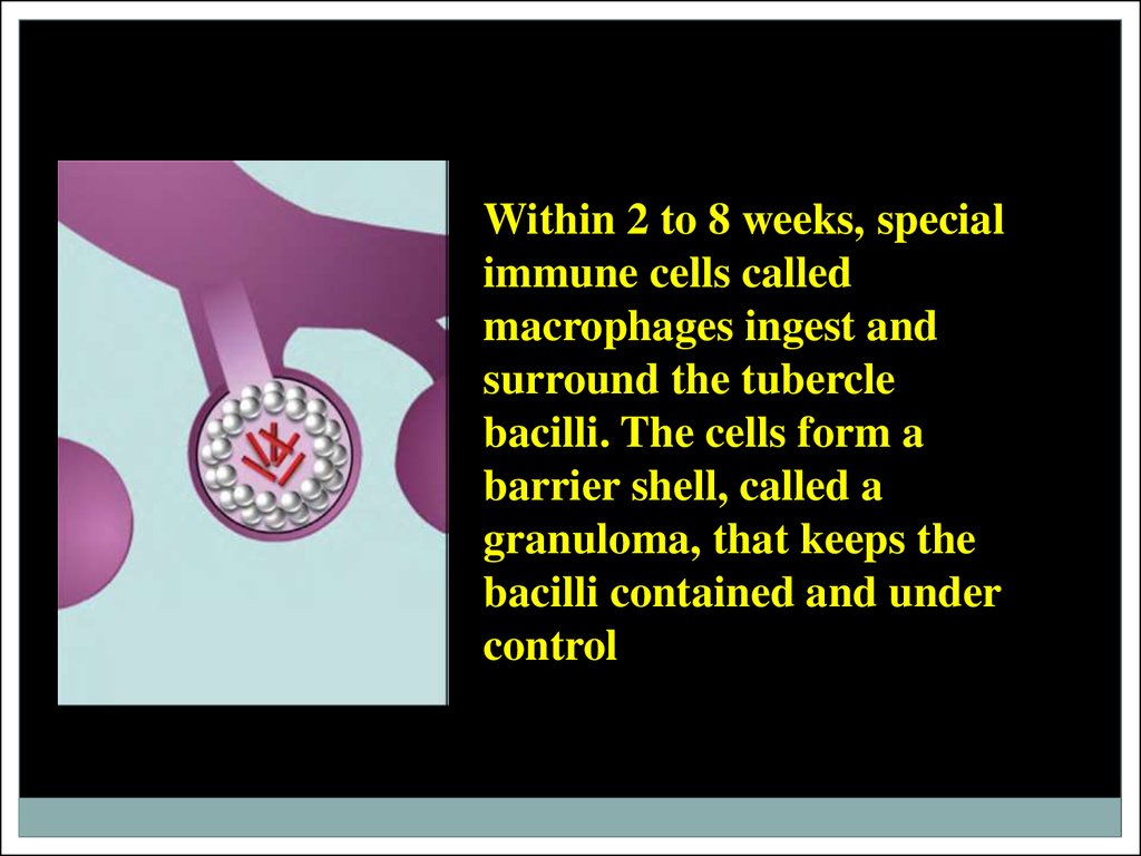
Within 2 to 8 weeks, specialimmune cells called
macrophages ingest and
surround the tubercle
bacilli. The cells form a
barrier shell, called a
granuloma, that keeps the
bacilli contained and under
control (LTBI).
38.
If the immune systemcannot keep the tubercle
bacilli under control, the
bacilli begin to multiply
rapidly (TB disease).
This process can occur in
different areas in the
body, such as the lungs,
kidneys, brain, or bone.
39.
Latent Tuberculosis Infection (LTBI)Persons with LTBI have M. tuberculosis in their bodies, but do not have TB disease and
cannot spread the infection to other people. A person with LTBI is not regarded as
having a case of TB. The process of LTBI begins when extracellular bacilli are ingested
by macrophages and presented to other white blood cells. This triggers the immune
response in which white blood cells kill or encapsulate most of the bacilli, leading to the
formation of a granuloma. At this point, LTBI has been established. LTBI may be
detected by using the tuberculin skin test (TST) or an interferon-gamma release assay
(IGRA) . It can take 2 to 8 weeks after the initial TB infection for the body’s immune
system to be able to react to tuberculin and for the infection to be detected by the TST
or IGRA. Within weeks after infection, the immune system is usually able to halt the
multiplication of the tubercle bacilli, preventing further progression.
Persons with LTBI have M. tuberculosis in their bodies, but
do not have TB disease and cannot spread the infection to
other people.
40.
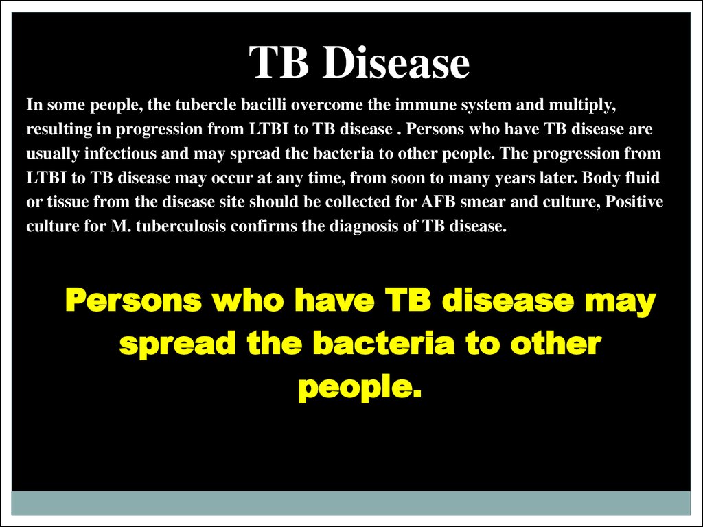
TB DiseaseIn some people, the tubercle bacilli overcome the immune system and multiply,
resulting in progression from LTBI to TB disease . Persons who have TB disease are
usually infectious and may spread the bacteria to other people. The progression from
LTBI to TB disease may occur at any time, from soon to many years later. Body fluid
or tissue from the disease site should be collected for AFB smear and culture, Positive
culture for M. tuberculosis confirms the diagnosis of TB disease.
Persons who have TB disease may
spread the bacteria to other
people.
41.
Risk of developing TB disease over alifetime
Without treatment, approximately 5% of persons who
have been infected with M. tuberculosis will develop
disease in the first year or 2 after infection, and another
5% will develop disease sometime later in life. Thus,
without treatment, approximately 10% of persons with
normal immune systems who are infected with M.
tuberculosis will develop TB disease at some point in
their lives.
42.
Risk of LTBI Progressing to TB DiseaseAnyone who has LTBI can develop TB disease, but some people
are at higher risk than others. HIV infection is the greatest risk
factor for the development of TB disease in persons with LTBI,
due to a weakened immune system. The risk of developing TB
disease is 7% to 10% each year for persons who are infected with
both M. tuberculosis and HIV and who are not receiving highly
active treatment for HIV; it is 10% over a lifetime for persons
infected only with M. tuberculosis. Children younger than 5 years
of age are also at increased risk for progression of LTBI to TB
disease.
43.
Persons at Increased Risk for Progression ofLTBI to TB Disease
Risk Factor
Risk of Developing TB
TB infection and no risk
factors
About 10% over a lifetime
TB infection and diabetes
About 30% over a lifetime
TB infection and HIV
infection
About 7% to 10% PER
YEAR
Description
For people with TB infection,
no risk factors, and no
treatment, the risk is about 5%
in the first 2 years after
infection and about 10% over a
lifetime.
For people with TB infection
and diabetes, and with no
treatment, the risk is three
times as high, or about 30%
over a lifetime.
For people with TB infection
and untreated HIV infection
and with no LTBI treatment,
the risk is about 7% to 10%
PER YEAR, a very high risk
over a lifetime.
44.
The tubercular inflammationThe tubercular inflammation, like any other inflammation is a
manifestation of alteration, exudation, proliferation, leading to
the formation of tubercular granuloma
(Тuberculum, tubercular tumor).
The term granuloma is derived from the diminutive of the Latin
term for a grain, granulum, which was first used by Rudolf
Virchov [1818] to describe tumors that may ulcerate and give
rise to granulation tissue.
The tubercular granuloma is not a mere collection of
inflammatory cells but is an active site of action of numerous
enzymes and cytokines in the very complex process of removing
the causative agent MBT.
45.
Participate in the formation of tubercular granuloma- hematogenic elements (lymphocytes, monocytes,
polymorphonuclear leucocytes),
- histiogenic elements (histocytes, macrophages, fibroblasts,
reticular cells, endothelium of blood vessels, plasmatic and
mast cells),.
46.
The tubercular granuloma has the following structure:- The center consists of amorphous tissue detritus (due to alteration
and necrosis), the peripheral region contains several layers of
epithelial cells.
- Lymphoid and
plasma cells are
present in the
external layers of
the tuberculum.
- Giant
multinucleated
Pirogov – Langhans
cells can be seen
among the epithelial
cells.
47.
Diagram of aGranuloma
NOTE: ultimately a fibrin
layer develops around
granuloma (fibrosis), further
“walling off” the lesion.
Typical progression in
pulmonary TB involves
caseation, calcification and
cavity formation.
48.
Tubercle bacilliLymphocyte
Giant cells
Fully activated
macrophage
Partially activated
macrophage
49.
The tuberculum histogenesis depends on the development of theinflammation process, which is either progressive or regressive.
When there is a decreased host resistance to tuberculosis,
progression of the tubercular inflammation takes place.
The tissue exudative reaction develops with the formation of
cheesy necrosis which might develop within the tuberculum and
surrounding tissues.
These tissues will generally be impregnated with serousfibrinous exudates.
50.
Various foci of different sizes of cheesy necrosis arise during thefurther progression of specific tubercular inflammation.
Foci of cheesy necrosis can spread and merge into bigger foci
from which foci with sites of caseation (infiltrates) are formed.
Caseation is diluted under the action of proteolytic enzymes and
is coughed out through the bronchi.
Cavities of disintegration appear in these sites of the lungs but
ulcers appear on the mucous membrane and skin.
The cavity formed during the disintegration of caseation will be
the source of dissemination of MBT in other parts of the lungs
and formation of new foci and cavities.
51.
The particular danger is represented by vascular blood erosionsupplying sites of lungs where caseous degeneration occurred.
During cavity formation, blood from the damaged vessels
penetrates the bronchi and from there, either penetrates other
parts of lungs or is expectorated externally.
Reversible development of process (regression) occurs during
high resistance of the organism the tuberculum will be
substituted by fibrosis and calcification. (Chronic development
of tuberculous inflammation).
52.
The morphological and biochemical components of microbialcells cause various reactions in the host.
The basic biochemical components of МВТ are:
– proteins;
– carbohydrates;
– lipids.
Proteins (tuberculoproteids) is the basic carrier of МВТ
antigenic properties.
53.
Delayed-type hypersensitivity (DTH)The substances, which are included in the MBT
wall structure, induce tissue specific
inflammation reaction and granuloma
formation, with the development of the
delayed-type hypersensitivity (DTH), which
could be detected by a positive tuberculin test
reaction, and a weak antibody formation.
54.
In general, term DTH is used forcharacteristics of a type IV immune response
(induration at the site of intradermal
injection of tuberculin develops after 48
hours) among individuals who are infected
with Mycobacterium tuberculosis.
DTH is to be concerned as an immune
response from the damaged tissue factors.
55.
The cycle of tuberculosis development from MBTcontamination till the occurrence of its clinical manifestations
and distribution of MBT in envi-ronment conditionally is
classified into 5 stages.
Stages:
1. Spreading of infection (contamination).
2. Beginning of infection, proliferation and dissemination in an
infected host.
3. Formation of immune reaction in the host.
4. Formation of caseous necrosis, and proliferation of bacteria.
5. Secondary spreading of infection (ability to infect).
56.
Primary tuberculosisPrimary tuberculosis develops after the first contact of
macroorganism with MBT.
MBT fill in the peripheral parts of the lungs when tiny particles
containing MBT are inhaled through the superior respiratory
tract.
The mycobacterium remains there and reproduces slowly
forming the primary pulmonary affection (focus).
In this way, mycobacterium falling into the lymph through
which they are transported to the lymph nodes.
The classical form of morphological manifestation of primary
tuberculosis is the primary tuberculosis complex.
57.
In the primary lung focus, alveolitis develops, which is quicklyreplaced by the typical development of caseosis necrosis.
In the centre of primary focus, caseosis forms but in the
periphe- ry-elements of non specific inflammation occur.
The primary lung affect localizes more often just under pleura,
therefore frequently pleura is involved in the inflammation
process.
The lymphatic vessels expand, their walls becoming infiltrated
and tubercles appear.
In the regional lymphatic nodes, there are elements of
inflammations converting into specific caseous changes with
necrosis
58.
Perifocal inflammation around the lymph nodes willspread in the mediastinum and surround the lung
tissues.
The inflammation process within the lymph nodes is
most intense in the primary affection area.
Therefore, reparative changes in the lymph nodes will
be slower.
59.
The dynamic study of primary pulmonaryprocesses among children has allowed to allot 4
phases of the primary tuberculosis course:
1) pneumonic;
2) phase of dissolving;
3) phase of condensation;
4) formation of Gohn’s focus.
60.
In the first phase (pneumatic) the focus ofbroncho-lobular pneumonia (3) is determined
with a size of 1,5-2 till 5 cm.
The form of the lung focus (3) is round or
irregular, with heterogenous character and dim
contours.
phase 1 (pneumonic)
Enlarged regional lymphatic nodes (1) are
determined simultaneously (the picture of
infiltrative bronchoadenitis) and there is an
amplification of bronchial vessels picture –
lymphangitis (2) between the focus and the lung
root.
61.
In the second phase of dissolving (bipolarity) the reduction ofthe perifocal zone of inflammation (3) is observed.
The centrally located caseous focus becomes more prominent.
The signs of inflammation in
regional lymphatic nodes (1) and in
the zone of bronchopulmonary
vessels are decreaseding (2).
phase 2 of dissolving (bipolarity)
62.
In the third phase, the phase ofcondensation: the primary focus
is well outlined (3), its contours
are cleared, on periphery of the
focus there is the beginning of
calcification as fine pieces; at
peripheral regions of lung
bronchial lymphatic nodes
calcification is also present (1).
the phase 3 – condensation
63.
In the fourth phase, in the placeof broncho-lobular pneumonia
(3) calcification become
compact, the focus is round
with regular precise contours,
its size does not exceed 3-5 mm.
This formation is called Gohn’s
focus.
phase 4 formation of Gohn’s focus
64.
Outcomes of the primary tubercular complex may be in thefollowing way:
1) healing with encapsulation, calcification or ossification;
2) progression and generalization of the inflammation process.
It may be accompanied with additional complications such as
atelectasis, pneumosclerosis, etc.
65.
There are 2 types of generalization of thetubercular complex progression:
1) hematogenic;
2) lymphogenic
3) bronchogenic.
66.
At progression of hematogenous disseminated tuberculosis the cavities areformed.
The formation of cavities is the result of cheesy disintegration and
dissolution of necrotic masses.
The cavities are usually thin-walled, multiple and settled down
symmetrically in both lungs.
In an origin of such cavities, important role plays damage of blood vessels,
their thrombosis and obliteration.
The blood supply of these focuses is disturbed in lungs and destruction is
formed resembling trophic ulcers.
During the formation of the cavities, the possibility of bronchogenic
dissemination of healthy regions of lungs can appear.
67.
Immunity at tuberculosisNatural resistance to tuberculosis is inherited. It involves non-specific
antimicrobial humoral factors (non-immunological phenomena).
These factors inactivate MBT and prevent their multiplication as well as
destroy their toxins.
These factors include:
-
-
lysozyme in alveolar macrophages;
higher contents of lactic acid in cells;
lipoprotein lipase, an enzyme which decomposes protein and lipid
complexes of MBT cell wall, producing bacteriostatic non-etherized fat
acids;
cytokines (IL-1, α-interferon, components of compliment).
68.
Phagocytosis plays special role in naturalresistance. Primary contact MBT and the host
triggers phagocytosis of bacilli by macrophages.
Following facts are important about
phagocytosis:
5% of MBT is destroyed by macrophages
(completed phagocytosis). Damaging activity of
macrophages depends on the susceptibility of
the host and virulence of MBT.
69.
FagocitosisCompleted phagocytosis
It is one of mechanisms
natural resistance
to tuberculosis
Uncompleted phagocytosis
The result of him is education
geared-up makrofaga (only
the T-cell will co-operate with such
macrophages)
70.
Results of phagocytosisCompleted phagocytosis - near 5% MBT is destroyed macrophage.
Destructive activity of makrofagiv depends on the state of
macroorganism, virulence of MBT
Symbiosis of macrophages and MBT (most cases). It is arisen up as a
result of disfunction of lysosomes of macrophages, here MBT spread
in an organism by macrophages. This insolvency of phagocytosis
conducts to disemination of MBT
Uncompleted phagocytosis. MBT destroy macrophage and can be
added the repeated englobing
71.
Immunity in tuberculosis consists from five basic reactions: cellreaction, humoral factor, allergy, immune memory and immune
tolerance. Chief role belongs to T-lymphocyte cell-mediated
immune reactions.
Populations of T-helpers (CD4+), T-killers and T-suppressors
(CD8+) are studied best among all T-lymphocytes. Helpers are
inducted at first contact with antigen and condition the
immunity, suppressors balance the process and killers play
active role in phagocytosis.
Cell-mediated immunity. Cell-mediated immunity is based on
interaction of macrophages and T-lymphocytes.
72.
Only under these conditions T-helper (CD4+) may recognize antigen peptide of MBT.- At the same time macrophage produce interleukyn-1 (IL-1) which makes T-helper
to produce interleucin-2 (IL-2) and gamma- interferon. These mediators influence:
a) macrophages, activating their migration to MBT and increasing their enzyme and
bactericidal activity leading to the death of intracellular MBT
(compete phagocytosis).
b) T-killers (CD8+) which destroy infected macrophages ( macrophages with
phagocytized MBT).
c) B- lymphocytes ( humoral immunity) produce specific antibodies.
T- suppressors depress activity of IL-1 and IL-2.
Classical example of cell-mediated immunity is increased slow type sensitivity, found
with 2 IU tuberculin skin test and provided by T- lymphocytes and macrophages. Tkillers may exert cytotoxic effect that is they may ruin macrophages leading to
hyperergic reaction at tuberculin skin test.
73.
Proof of the role of T- lymphocytes in anti-tubercularimmunity:
- injection of T- lymphocytes suspension from immunized
animals to non- immunized increased resistance of the latter
against tuberculosis infection;
- injection of anti- lymphocyte suspension leads to quick and
malignant course of tuberculosis in animals. This also leads
to emptying of thymus -dependent areas of spleen and lymph
nodes. Injection of corticosteroids has the same effect;
- resection of thymus in newborn animals decreases their
resistance to tuberculosis, and infection takes malignant
course.
74.
As immune response builds up multiplication ofMycobacteria slows down, their general number
decreases, as specific inflammation. But compete
elimination of pathogen is not achieved even with
adequate interaction of macrophages and Tlymphocytes. Certain population of MBT remains in
the host as biologically changed cells (such as L-forms).
They locate in tubercular granulomas surrounded by
dense fibrotic capsule.
75.
Remaining MBT are located within cells and preventformation of phagolysosome, thus becoming
inaccessible to enzymes of lysosomes. Due to
preservation of Mycobacteria anti-tubercular
immunity is called non-sterile. MBT remaining in the
host support population of sensitized T-lymphocytes
and secure effectiveness of immune defense. Patient
infected with Mycobacteria retains them for a long
time, sometimes for life. With failure of immune
balance retained Mycobacterial population may
activate and cause tuberculosis.
76.
Thank youfor your
attention!