


")

")


")


")


 medicine
Similar presentations:
")


")


")
Bronchiectases: lecture
1. Bronchiectases: lecture
Ass.Prof. Nina A.Filippova2. Definition
• Bronchiectasis - uncommon disease, mostoften secondary to an infectious process, that
results in the abnormal and permanent
distortion of one or more of the conducting
bronchi or airways (Medscape).
3. ERS guidelines for the management of adult bronchiectasis (Eva Polverino, Pieter C. Goeminne, Melissa J. European Respiratory
ERS guidelines for the management of adult bronchiectasis(Eva Polverino, Pieter C. Goeminne, Melissa J. European Respiratory
Society European Respiratory Journal 2017):
• Bronchiectasis is
• chronic respiratory disease
• characterised by a clinical syndrome of cough, sputum
production and bronchial infection
• and radiologically by abnormal and permanent dilatation of
the bronchi.
• The objectives of treatment in bronchiectasis are to
prevent exacerbations, reduce symptoms, improve quality
of life and stop disease progression.
• Cough and sputum production, along with breathlessness
are the most frequent symptoms but rhinosinusitis, fatigue,
haemoptysis and thoracic pain are also common
4. Classification by etiology
1. Genetic disorders (cystic fibrosis, primary ciliarydyskinesia, alpha1-antitrypsin deficiency)
2. Post infectious disease (bacteria, virus, fungi, other)
3. Immunodeficiency (congenital, acquired)
4. Aspiration (gastro-oesophageal reflux, swallowing
dysfunction, tracheo-esophageal fistula)
5. Congenital structural malformations (lobar
emphysema, bronchomalacia, etc.)
6. Mechanical factors (foreign body, extrinsic
compression, endobronchial lesions)
5. Classification by etiology
Advances in bronchiectasis: endotyping, genetics, microbiome, and disease heterogeneityProf Patrick A
Flume, MD, Prof James D Chalmers, MBChB, Kenneth N Olivier, MD The Lancet Volume 392, Issue 10150,
Pages 880-890 (September 2018)
29% idiopathic
14% post-infective
15% - COPD (predominantly not numerous,
local*)
7% asthma (predominantly not numerous,
local*)
9% - connective tissue diseases (traction
bronchoectases)
5% - allergic bronchopulmonary aspergillosis
5% - immune deficiency
4% - post Tb
4% - GERD (aspiration)
Others – less than 1% any (NTM: nontuberculous
mycobacteria; ; PCD: primary ciliary dyskinesia; CF: cystic
fibrosis; CFTR-RD: cystic fibrosis transmembrane
conductance regulator-related disease; A1ATD: α1antitrypsin deficiency; IBD: inflammatory bowel disease;
YNS: yellow nail syndrome; DPB: diffuse panbronchiolitis)
6. Classification: by shape
Assoc Prof Frank Gaillard, <a href="https://radiopaedia.org/">Radiopaedia.org</a>. From the case <ahref="https://radiopaedia.org/cases/8863">rID: 8863</a>
7. Classification by shape: Normal bronchus; no bronchoectases
American Journal of Roentgenology > Volume 193, Issue 3 > BronchiectasisSeptember 2009, Volume 193, Number 3Bronchiectasis Luce Cantin1, Alexander A. Bankier1 and Ronald L. Eisenberg1 American Journal of Roentgenology.
2009;193: W158-W171. 10.2214/AJR.09.3053
Assoc Prof Frank Gaillard, <a href="https://radiopaedia.org/">Radiopaedia.org</a>.
From the case <a href="https://radiopaedia.org/cases/8863">rID: 8863</a>
8. Cylindric bronchiectasis with lack of bronchial tapering
Assoc Prof Frank Gaillard, <a href="https://radiopaedia.org/">Radiopaedia.org</a>. From the case <ahref="https://radiopaedia.org/cases/8863">rID: 8863</a>
American Journal of Roentgenology > Volume 193, Issue 3 > BronchiectasisSeptember 2009, Volume 193,
Number 3 Bronchiectasis Luce Cantin1, Alexander A. Bankier1 and Ronald L. Eisenberg1 American Journal
of Roentgenology. 2009;193: W158-W171. 10.2214/AJR.09.3053
9. varicose bronchiectasis with string-of-pearls appearance
varicose bronchiectasis with string-ofpearls appearanceAssoc Prof Frank Gaillard, <a href="https://radiopaedia.org/">Radiopaedia.org</a>. From the case <a
href="https://radiopaedia.org/cases/8863">rID: 8863</a>
American Journal of Roentgenology > Volume 193, Issue 3 > BronchiectasisSeptember 2009, Volume 193,
Number 3 Bronchiectasis Luce Cantin1, Alexander A. Bankier1 and Ronald L. Eisenberg1 American Journal
of Roentgenology. 2009;193: W158-W171. 10.2214/AJR.09.3053
10. cystic bronchiectasis
Assoc Prof Frank Gaillard, <a href="https://radiopaedia.org/">Radiopaedia.org</a>. From the case <ahref="https://radiopaedia.org/cases/8863">rID: 8863</a>
American Journal of Roentgenology > Volume 193, Issue 3 > BronchiectasisSeptember 2009, Volume 193,
Number 3 Bronchiectasis Luce Cantin1, Alexander A. Bankier1 and Ronald L. Eisenberg1 American Journal
of Roentgenology. 2009;193: W158-W171. 10.2214/AJR.09.3053
11. Classification: etiology and location
• Diffuse: Peripheralpredominance
• Upper lung – cystic fibrosis,
sarcoidosis, postradiation
fibrosis
• Lower lung – idiopathic,
postinfectious, aspiration
related, fiibrotic lung disease,
posttransplant rejection,
hypogammaglobulinemia
• Right middle lobe and lingular
– atypical mycobacterial
infection, immotile cilia
syncrome
• Focal (congenital bronchial
atresia, extrinsic compression,
extrabronchial malignancy,
foreign body, broncholothiasis,
airway stenosis
• Diffuse: central
predominance
• Allergic bronchopulmonary
aspergillosis
• Mounier-Kuhn syndrome
• Williams-Campbell syndrome
12. Pathogenesis vicious cycle of bacterial infection, neutrophilic elastastes induced injury of the epithelium; ciliary
Figure 1Pathogenesis
vicious cycle of bacterial infection, neutrophilic elastastes induced injury of
the epithelium; ciliary disfunction, progression of infection and destruction of
bronchial wall
Advances in bronchiectasis: endotyping, genetics, microbiome, and disease heterogeneityProf Patrick A
Flume, MD, Prof James D Chalmers, MBChB, Kenneth N Olivier, MD The Lancet Volume 392, Issue 10150,
Pages 880-890 (September 2018)
P. Cole, “Bronchiectasis,” in in. Respiratory medicine, pp. 1380–1395, Bronchiectasis. in. Respiratory
The Lancet 2018 392, 880-890DOI: (10.1016/S0140-6736(18)31767-7) medicine, London, UK, 1995.
Copyright © 2018 Elsevier Ltd Terms and Conditions
13. Components
• Neutrophilic inflammation – destruction of wall byelastases
• Ciliary disfunction (primary or secondary) – retention
of sputum and decrease of infection agents clearance
• Sputum properties changes (in cystic fibrosis) –
retention of sputumand decrease of infection agents
clearance
• Anatomic disorders (primary or secondary) with
deformities and/or compression - retention of sputum
and decrease of infection agents clearance, increase of
intrabronchial pressure with promotion of deformities
• Immune supression – promotion of neutrophilmediated process
14. Inflammation: neutrophilic
• Neutrophils recruitment acceleration: degradation ofelastins; increase of neutrophilic proteolytic molecules,
damage and structural changes of components of
bronchial wall, resulting to its dilation
• Participants: IL-1β, TNF α, LTβ4, IL-8 (CXCL8); action of IL-8 and other CXCs
through CXCR1 and CXCR2 receptors;
• CXR1 - neutrophil degranulation and phagocytosis,
• CXCR2 - adhesion and chemotaxis to the site of infection
• Results: increase of neutrophils total number and percentage; concentration of
neutrophilic proteolytic molecules (neutrophilic elastase (NE), myeloperoxidase
(MPO) and metalloproteinase (MMP)-9 at site of inflammation
• CXCR2: important in response to Pseudomonas, Aspergillus, Nocardia
The double-edged sword of neutrophilic inflammation in bronchiectasis
Miguel Ángel Martínez-García, Concepción Prados Sánchez, Rosa María Girón Moreno
European Respiratory Journal 2015 46: 898-900;
15. Importance of this mechanism for control the disease
• block of neutrophilic elastase: NE inhibitorAZD9668 in bronchiectasis patients - significant
functional improvement and a trend to reduce in
inflammatory biomarkers
• block of CXCR2 prevents neutrophils chemotaxis
on infection site: CXCR2 antagonists: MK-7123 in
COPD, non Th2 asthma; SB-656933 in cystic
fibrosis; AZD-5069 – pilot study for
bronchoectases (64% reduction of neutrophils in
sputum in patients)
De Soyza A, Pavord I, Elborn JS, et al. A randomised, placebo-controlled
study of the CXCR2 antagonist AZD5069 in bronchiectasis. Eur Respir
J 2015; 46: 1021–1032.
Moss R, Mistry S, Konstan M, et al. Safety and early treatment effects of the CXCR2 antagonist SB656933 in patients with cystic fibrosis. J Cyst Fibros 2013; 12: 241–248.
Nair P, Gaga M, Zervas E, et al. Safety and efficacy of a CXCR2 antagonist in patients with severe asthma
and sputum neutrophils: a randomized, placebo-controlled clinical trial. Clin Exp Allergy 2012;42: 1097–1103.
Rennard S, Dale D, Donohue J, et al. CXCR2 antagonist MK-7123. A phase 2 proof-of-concept trial for chronic
obstructive pulmonary disease. Am J Respir Crit Care Med 2015; 191: 1001–1011
Stockley R, De Soyza A, Gunawardena K, et al. Phase II study of a neutrophil elastase inhibitor (AZD9668) in patients with
bronchiectasis. Respir Med 2013; 107: 524–533.)
16. Ciliary disfunction: primary and secondary
Cilia Dysfunction in Lung DiseaseAnn E. Tilley,1,2 Matthew S. Walters,1 Renat Shaykhiev,1 an
Annu Rev Physiol. 2015; 77: 379–406.
17. Genes Encoding Major Components of Airway Motile Cilia
• Axoneme – outer dynein arm – Dyenin axonealheavy/intermediate/ light chain genes (DNAH5,
DNAH9, DNAH11, DNAI1, DNAI2, DNAL1)
• Dynein assembly and docking (Dynein, axonemal,
assembly factors 1-3 -DNAAF1-3 etc)
• Tubulins and other microtubule-associated
(NME/NM23 family member 8NME8 etc)
• Receptors, ion channels and signaling molecules
(Nitric oxide synthase 3 (endothelial cell)NOS3)
• These genes changes predispose do development
of primary ciliary dyskinesia
18. Secondary ciliary disfunction
• Viruses• Bacterial mediators - H. influenzae, P.
aeruginosa, Streptococcus pneumoniae
(direct damage)
• Smoking (direct action on cilia, downregulation of above mentioned genes)
19. Primary and secondary mucociliary clearance disturbance leads to
• airway dehydration, excess mucus volume andviscosity.
• Increase of sputum content and further
infection development
20. Primary anatomical changes, promoting clearance disorders due to bronchi deformities or compression
• Traction bronchoectases – advanced pulmonary fibrosiswith traction of the airways
• PostTb – advanced fibrotic changes and localized
peribronchial lymphadenopathy squeezing bronchi and
causing localised bronchial obstruction (particularly in the
right middle and upper lobes) with secondary decrease of
clearence and infection persistence
• Childhood infections - whooping cough, measles,
adenovirus – increase pressure in bronchiols during
paroxysmal cough, mucus plugs in bronchi
• Mycobacterium avium complex (MAC) in elder women
cause obstruction from lymphadenopathy with right middle
lobe bronchiectasis
• Other causes – inborn changes, bronchial and lung
dysplasia, endobronchial calcifications, foreign bodies etc
21. Flora
Haemophilus influenzae (29%–70%)
Pseudomonas aeruginosa (12%–31%).
No pathogenic bacteria (30%–40% )
Best preserved lung function: no pathogenic bacteria isolated.
Worst prognosis – H.influenzae; P.aeruginosa; Morax.
catarrhalis, Staph.aureus, Enterobacter.
• Aspergillus infection (ABPA – allergic bronchopulmonary
aspergillosis)
• Mycobacterial infections (in older women- mycobacterium
avium complex (MAC) causing obstruction from lymphadenopathy with right middle lobe bronchiectasis )
Int J Chron Obstruct Pulmon Dis. 2009; 4: 411–419. The pathophysiology of bronchiectasis
Paul T King
22. Effects of flora promoting bronchoectases
• inhibition of the mucociliary clearance: mediatorsof H. influenzae, P. aeruginosa, Streptococcus
pneumoniae directly damage ciliated epithelium,
and inhibit mucous transport and release
glycoproteins attracting neutrophils.
• H. influenzae direct damage to airway
epithelium; invasion into the bronchial wall and
interstitium of the lung
• P. aeruginosa - forms biofilms, which form
impenetrable matrix around bacteria and defend
it from immune system and antibiotics
23. Immune dysfunction
• Malnutrition• Extremes of age
• hypogammaglobulinemia, human
immunodeficiency virus (HIV), interferon
gamma receptor deficiency, type I major
histocompatibility complex deficiency, late
stages of lung transplant rejection, very high
IgE levels without ABPA
24. Pathogenesis vicious cycle of bacterial infection, neutrophilic elastastes induced injury of the epithelium; ciliary
Figure 1Pathogenesis
vicious cycle of bacterial infection, neutrophilic elastastes induced injury of
the epithelium; ciliary disfunction, progression of infection and destruction of
bronchial wall
Advances in bronchiectasis: endotyping, genetics, microbiome, and disease heterogeneityProf Patrick A
Flume, MD, Prof James D Chalmers, MBChB, Kenneth N Olivier, MD The Lancet Volume 392, Issue 10150,
Pages 880-890 (September 2018)
P. Cole, “Bronchiectasis,” in in. Respiratory medicine, pp. 1380–1395, Bronchiectasis. in. Respiratory
The Lancet 2018 392, 880-890DOI: (10.1016/S0140-6736(18)31767-7) medicine, London, UK, 1995.
Copyright © 2018 Elsevier Ltd Terms and Conditions
25. Clinical manifestations
• Chronic productive cough - 98% of patients• Sputum - produced on a daily basis - 70% - 96% of
patients (4%-30% - “dry” bronchoectases.
• Sputum usually mucoid; during infectious
exacerbations – greenish/yellowish purulent, may
be offensive odor.
• Sputum amount usually >50 ml daily, in mild BE –
10 ml and less; in moderate 10-150 mL ; in severe
more than 150 mL
• Hemoptysis - 56-92% of patients; more
commonly in dry bronchiectasis; usually mild;
appears from dilated bronchial arteries: massive
hemoptysis – rare
Author: Ethan E Emmons, Bronchiectasis Clinical Presentation Updated: Jul 23,
2019 https://emedicine.medscape.com/article/296961-clinical#b3
26.
• Dyspnea – 62%-72% of patients, mixed(obstruction + restriction due to fibrosis)
• Wheezing – rare (more common in asthma)
• Fatigue – 73%, in severe cases – weight loss
• Crackles – 73% (small and medium caliber);
rhonchi; more rare wheezing (predominantly
local if not asthma)
• Clubbing – 2-3%
• Cyanosis – in severe cases
27. In whom should be suspected?
Persistent mucopurulent/purulent sputum + risk factors
rheumatoid arthritis + chronic productive cough/recurrent chest infections.
COPD frequent exacerbations (two or more annually)
inflammatory bowel disease + chronic productive cough
asthma: severe/poorly-controlled disease
HIV-1,solid organ and bone marrow transplant, immunosuppressives; +
chronic productive cough or recurrent chest infections.
chronic rhinosinusitis – chronic productive cough or recurrent chest
infections.
connective tissue disease or inflammatory bowel disease - chronic
productive
cough or recurrent chest infections
otherwise healthy individuals - cough that persists > 8 wks, especially with
sputum production or a history of an appropriate trigger
Adam T Hill,1 Anita L Sullivan,2 James D Chalmers British Thoracic Society Guideline for bronchiectasis in adults Thorax 2019;74(Suppl 1):1–69. doi:10.1136/thoraxjnl-2018-212463
28. Diagnosis: to confirm
• baseline chest X-ray in patients with• suspected bronchiectasis.
• Thin section computed tomography scan (CT)
to confirm a diagnosis of bronchiectasis when
clinically suspected.
Adam T Hill,1 Anita L Sullivan,2 James D Chalmers British Thoracic Society Guideline for bronchiectasis in
adults Thorax 2019;74(Suppl 1):1–69. doi:10.1136/thoraxjnl-2018-212463
29. CT features of bronchiectasis
• bronchial dilatation as suggested by one or more of thefollowing:
• Bronchoarterial ratio >1 (internal airway lumen vs adjacent
pulmonary artery)
• Lack of tapering
• Airway visibility within 1cm of costal pleural surface or
touching mediastinal pleura.
• The following indirect signs are commonly associated with
bronchiectasis:
• Bronchial wall thickening
• Mucus impaction
• Mosaic perfusion / air trapping on expiratory CT
Adam T Hill,1 Anita L Sullivan,2 James D Chalmers British Thoracic Society Guideline for bronchiectasis in
adults Thorax 2019;74(Suppl 1):1–69. doi:10.1136/thoraxjnl-2018-212463
30. Diagnosis: general + flora
• full blood count incl ESR• In all patients: specific antibodies against capsular
polysaccharides of Streptococcus pneumoniae
(specific antibody deficiency). If pneumococcal
antibodies are low, immunise with 23 valent
polysaccharide pneumococcal vaccine, followed
by measurement of specific antibody levels 4–8
weeks later.
• Sputum cultures- in all patients for routine and
mycobacterial culture.
Adam T Hill,1 Anita L Sullivan,2 James D Chalmers British Thoracic Society Guideline for bronchiectasis in
adults Thorax 2019;74(Suppl 1):1–69. doi:10.1136/thoraxjnl-2018-212463
31. Diff: COPD
Bronchiectases• Sputum >50 ml, more
purulent
• Hemopthisis common
• Fever more common
• Dullness zones may be
• Crackles, moist rales locally
• Pneumonias frequent, same
locations
COPD
• Small amount of sputum,
purulent rare, at
exacerbations
• Hemopthisis possible (exclude
BE, cancer, embolism)
• Fever more rare,
exacerbations
• Harsh or diminished
respirations
• Wheezes, rhonchi
• Pneumonias may be, usually
at GCS taking patients
32. Diff: cancer
Bronchiectases• Sputum >50 ml, more
purulent
• Hemopthisis common
• Fever more common
• Dullness zones may be
• Crackles, moist rales locally
• Pneumonias frequent, same
locations
Cancer
• Dry cough, paroxysmal,
nocturnal, usual decrease
amount of sputum if compared
to previous COPD symptoms;
increased in bronchioalveolar
• Hemopthisis frequent
• Rapid progression of dyspnea
(expiratory, then inspiratory)
• Fever in case of pneumonia
• Harsh or diminished respirations,
wheezes, rhonchi - in case of
pneumonia complication
33. Diff: embolism
Bronchiectases• Sputum >50 ml, more
purulent
• Hemopthisis common
• Fever more common
• Not typical pleural pain
• PII, epigastric pulsation may
be
• Dullness zones may be
• Crackles, moist rales locally
• Pneumonias frequent, same
locations
embolism
• Sputum not typical
• Hemopthisis frequent
• Pleural pain typical, PII,
epigastric pulsation
• Deep veins thrombosis
(unilateral leg pain and
edema)
• BP decrease
• Fever not typical (if more than
2 days duration – pneumonia
may develop)
• Local changes (dullness, rales)
in case of pneumonia
34. Rare syndroms (ciliary disfunction, cystic fibrosis)
Cystic fibrosis
Ciliary disfunction
Fetal meconium ilius may be
Start in early life more possible
Rhinosinusitis
Pancreatitis, malabsorbtion, low
weight gain
Biliary liver cirrhosis (liver
enlargement, jaundice)
Viscous secretions
High risk of Ps.aeruginosa
Bad prognosis in case of absence of
modern treatment and sever
mutation
Chloride in sweat - low
Genetic disease – severity of disease
depend only on mutation
In adolescens and young adults –
male infertility
• ARDS in infancy and early
childhood
• Chronic otitis media
• Sinusitis
• Male infertility
• Situs viscerus inversus –
Kartagener syndrome
35. Cystic fibrosis
predominance of cystic bronchiectasis (arrows)
volume loss (fibrosis)
diffuse heterogeneous attenuation
enlarged lung volumes (obstruction)
36. Adult cystic fibrosis (milder case)
• cylindric bronchiectasis(white arrows)
• bronchiolitis (black
arrows) – tree in bud
syndrome
American Journal of Roentgenology > Volume 193, Issue 3 > BronchiectasisSeptember 2009, Volume 193,
Number 3 Bronchiectasis Luce Cantin1, Alexander A. Bankier1 and Ronald L. Eisenberg1 American Journal
of Roentgenology. 2009;193: W158-W171. 10.2214/AJR.09.3053
37. Kartagener's syndrome
Kartagener's syndromeDextrocardia (here + cardiomegaly)
Here - left middle lobe
bronchiectasis, volume loss.
Arrow points to wrong-sided left
marker
• CT confirms dextrocardia
(asterisk is in left ventricle)
• bronchiectasis (arrows)
predominantly midportion of
lungs
38. Other endotypes
Alpha -1 antitripsin deficiency• Panacinar basal emphysema
in non-smokers < 30-40
• Liver cirrhosis
Traction bronchoectases
• Presence of idiopathic
pulmonary fibrosis or lung
affection due to rheumatoid
arthritis, inflammatory bowel
disease, connective tissue
diseases, seronegative
spondiloarthritis
• More common “dry” ones
Non-TB mycobacteria
• post-menopausal non-smoker
females
• chronic cough, more common “dry”
• No predisposing factors
• May be CFTR mutations and ciliary
dysfunction, not meeting diagnostic
criteria for cystic fibrosis or primary
ciliary dyskinesia.
• tall, asthenic, scoliosis, pectus
excavatum, mitral valve prolapse,
dural ectasia, minor features
overlapping with Marfan
and Ehlers-Danlos syndromes
39. Sarcoidosis
• Diffuse fibrosis• traction bronchiectasis (arrows, B)
predominantly upper lobes.
American Journal of Roentgenology > Volume 193, Issue 3 > BronchiectasisSeptember 2009, Volume
193, Number 3 Bronchiectasis Luce Cantin1, Alexander A. Bankier1 and Ronald L. Eisenberg1
American Journal of Roentgenology. 2009;193: W158-W171. 10.2214/AJR.09.3053
40. Usual interstitial pneumonia (idiopathic pulmonary fibrosis; rheumatoid arthritis, more rare Sjogren, scleroderma)
• Bibasilar and subpleural reticulation• traction bronchiectasis in areas of fibrosis (arrows)
American Journal of Roentgenology > Volume 193, Issue 3 > BronchiectasisSeptember 2009,
Volume 193, Number 3 Bronchiectasis Luce Cantin1, Alexander A. Bankier1 and Ronald L.
Eisenberg1 American Journal of Roentgenology. 2009;193: W158-W171. 10.2214/AJR.09.3053
41. Scleroderma and other connective tissue diseases – more typical NSIP
• Dilated esophagus(whitearrow)
• ground-glass opacities
(NSIP)
• recurrent aspiration with
subsequent bibasilar
bronchiectasis
• Black arrow points to
bronchus visible in
peripheral 1 cm of lung.
American Journal of Roentgenology > Volume 193, Issue 3 > BronchiectasisSeptember 2009, Volume 193,
Number 3 Bronchiectasis Luce Cantin1, Alexander A. Bankier1 and Ronald L. Eisenberg1 American Journal
of Roentgenology. 2009;193: W158-W171. 10.2214/AJR.09.3053
42.
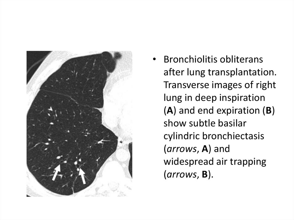
• Bronchiolitis obliteransafter lung transplantation.
Transverse images of right
lung in deep inspiration
(A) and end expiration (B)
show subtle basilar
cylindric bronchiectasis
(arrows, A) and
widespread air trapping
(arrows, B).
43. Other endotypes
ABPA• Blood eosinophilia
• thick sputum with black
• Bronchial obstruction with
wheeze,
• Asthma in case history
• recurrent exacerbations
Post-infective
• ulilateral, localized
• Severe infection in case
history
Immune deficiency
• Start at early age
• Infections from
childhood/infancy if inborn
• Frequent exacerbations
• Pneumonias
• non-respiratory infections
44. Allergic bronchopulmonary aspergillosis
American Journal of Roentgenology > Volume 193, Issue 3 > BronchiectasisSeptember 2009, Volume 193,Number 3 Bronchiectasis Luce Cantin1, Alexander A. Bankier1 and Ronald L. Eisenberg1 American Journal
of Roentgenology. 2009;193: W158-W171. 10.2214/AJR.09.3053
• Asthma
• Presence of transient pulmonary
infiltrates (fleeting shadows)
• Elevated total serum IgE
• Peripheral blood eosinophilia
• Elevated serum IgE and IgG to Af
Immediate cutaneous reactivity
to Af
• Precipitating antibodies against Af
• Central/proximal bronchiectasis
with normal tapering of distal
bronchi
• mucoid impaction (large arrow)
• distal bronchiolitis (small arrow)
45. Same
• central bronchiectasisand mucoid impaction,
so-called finger-in-glove
appearance
46. Other investigations: endotypes assessment
• Co-morbidities and past medical history toidentify relevant and possibly causative disease
• serum total IgE and specific IgE or skin prick test
to Aspergillus fumigatus in all patients with
bronchiectasis
• Serum IgG, IgA, IgM in all patients with BE
Adam T Hill,1 Anita L Sullivan,2 James D Chalmers British Thoracic Society Guideline for bronchiectasis in
adults Thorax 2019;74(Suppl 1):1–69. doi:10.1136/thoraxjnl-2018-212463
47.
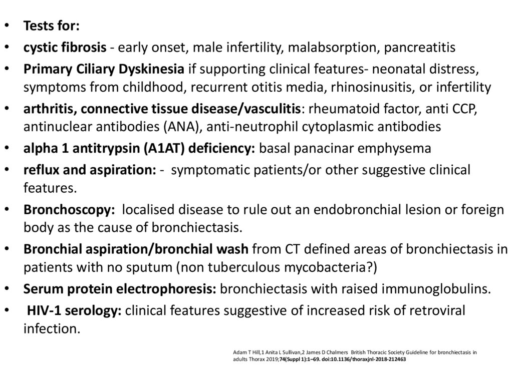
• Tests for:• cystic fibrosis - early onset, male infertility, malabsorption, pancreatitis
• Primary Ciliary Dyskinesia if supporting clinical features- neonatal distress,
symptoms from childhood, recurrent otitis media, rhinosinusitis, or infertility
• arthritis, connective tissue disease/vasculitis: rheumatoid factor, anti CCP,
antinuclear antibodies (ANA), anti-neutrophil cytoplasmic antibodies
• alpha 1 antitrypsin (A1AT) deficiency: basal panacinar emphysema
• reflux and aspiration: - symptomatic patients/or other suggestive clinical
features.
• Bronchoscopy: localised disease to rule out an endobronchial lesion or foreign
body as the cause of bronchiectasis.
• Bronchial aspiration/bronchial wash from CT defined areas of bronchiectasis in
patients with no sputum (non tuberculous mycobacteria?)
• Serum protein electrophoresis: bronchiectasis with raised immunoglobulins.
• HIV-1 serology: clinical features suggestive of increased risk of retroviral
infection.
Adam T Hill,1 Anita L Sullivan,2 James D Chalmers British Thoracic Society Guideline for bronchiectasis in
adults Thorax 2019;74(Suppl 1):1–69. doi:10.1136/thoraxjnl-2018-212463
48. Other investigations
• Spirogram/functional investigation of lungs,oxygen saturation, blood gases
• Daily protein loss, GFR, urine analysis – for
early diagnosis of inflammatory (SAA)
amyloidosis
• Other investigations if necessary
49. Focal idiopathic in left lower lobe
• non-TB mycobacteria• Perimenopausal
females
• Usually dry
bronchoectases
• Focal bronchiectasis
frequently
American Journal of Roentgenology > Volume 193, Issue 3 > BronchiectasisSeptember 2009, Volume 193, Number 3
Bronchiectasis Luce Cantin1, Alexander A. Bankier1 and Ronald L. Eisenberg1 American Journal of Roentgenology.
2009;193: W158-W171. 10.2214/AJR.09.3053
50.
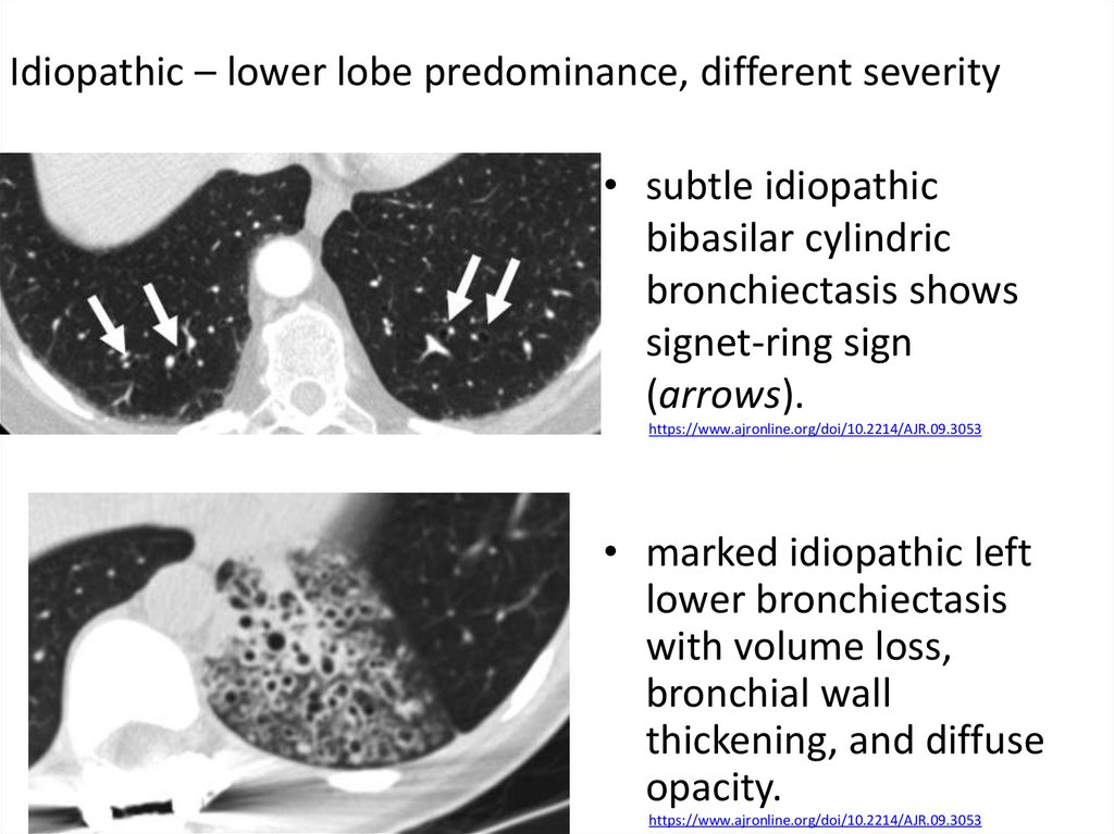
Idiopathic – lower lobe predominance, different severity• subtle idiopathic
bibasilar cylindric
bronchiectasis shows
signet-ring sign
(arrows).
https://www.ajronline.org/doi/10.2214/AJR.09.3053
• marked idiopathic left
lower bronchiectasis
with volume loss,
bronchial wall
thickening, and diffuse
opacity.
https://www.ajronline.org/doi/10.2214/AJR.09.3053
51. Mycobacterium avium- intracellulare infection
Mycobacterium avium- intracellulare infection• Bronchiectasis
(arrows)
predominantly
involves right middle
lobe and lingula.
American Journal of Roentgenology >
Volume 193, Issue 3 >
BronchiectasisSeptember 2009, Volume 193, Number 3 Bronchiectasis
Luce Cantin1, Alexander A. Bankier1 and Ronald L. Eisenberg1 American Journal of Roentgenology.
2009;193: W158-W171. 10.2214/AJR.09.3053
52. Obstruction
Tumor• More gradual onset (1-3
mo)
• Dyspnea progression from
expiratory to inspiratory
• Dry cough, hemopthisis
• More see “lung cancer”
Foreign body
• Usually in children
• May be acute suffocation
episode in case history with
stridor
• Relapsing pneumonias
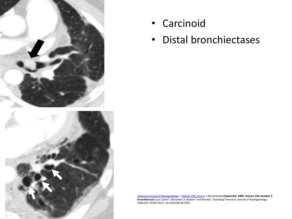
53. Carcinoid.
• Endobronchial growth• May arise berofe
bifurcation of lobar
bronchi
• Serotonin secretion
symptoms as following:
• Flushes up to 10-20 times
daily
• Bronchospasm
• Restritive CMP of
endomyocardial nature
American Journal of Roentgenology > Volume 193, Issue 3 > BronchiectasisSeptember 2009, Volume 193,
Number 3 Bronchiectasis Luce Cantin1, Alexander A. Bankier1 and Ronald L. Eisenberg1 American Journal
of Roentgenology. 2009;193: W158-W171. 10.2214/AJR.09.3053
54.
• Carcinoid• Distal bronchiectases
American Journal of Roentgenology > Volume 193, Issue 3 > BronchiectasisSeptember 2009, Volume 193, Number 3
Bronchiectasis Luce Cantin1, Alexander A. Bankier1 and Ronald L. Eisenberg1 American Journal of Roentgenology.
2009;193: W158-W171. 10.2214/AJR.09.3053
55. Tumors: postradiation fibrosis
• Right paramediastinalfibrotic changes,
developed after
treatment of lung
cancer, are associated
with traction
bronchiectasis (arrows).
American Journal of Roentgenology > Volume 193, Issue 3 > BronchiectasisSeptember 2009, Volume 193,
Number 3 Bronchiectasis Luce Cantin1, Alexander A. Bankier1 and Ronald L. Eisenberg1 American Journal
of Roentgenology. 2009;193: W158-W171. 10.2214/AJR.09.3053
56. Broncholithiasis: post TB
• Calcified left upper lobeendobronchial
broncholithiasis
• Results of lymph node
calcification (compression
and erosion of calcified
perigronchial lymph nodes
• Cause – TB, histoplasmosis
or other granulomatous
disease, more rare foreign
body
• 0.1% - 0.2% of all lung
diseases.
American Journal of Roentgenology > Volume 193, Issue 3 > BronchiectasisSeptember 2009, Volume 193,
Number 3 Bronchiectasis Luce Cantin1, Alexander A. Bankier1 and Ronald L. Eisenberg1 American Journal
of Roentgenology. 2009;193: W158-W171. 10.2214/AJR.09.3053
Broncholithiasis: An Uncommon Cause of Chronic Cough
Ungprasert, Patompong MD; Srivali, Narat MD; Bauer, Michael A. MD;
Edmonds, Lee C. MD
Author InformationJournal of Bronchology & Interventional
Pulmonology: January 2014 - Volume 21 - Issue 1 - p 102-103
57. Congenital abnormalities
• Congenital stenosis• (left mainstem
bronchus
American Journal of Roentgenology > Volume 193, Issue 3 > BronchiectasisSeptember 2009, Volume 193, Number 3
Bronchiectasis Luce Cantin1, Alexander A. Bankier1 and Ronald
• Bronchial atresia
focal bronchiectasis (arrow) distal to bronchial
atresia associated with hyperlucency and
hyperexpansion of left lung.
58. Other causes
• Mounier-Kuhn'ssyndrome. Enlarged
trachea (arrow).
• Enlarged mainstem
bronchi (black arrows)
and distal
bronchiectasis (white
arrows).
American Journal of Roentgenology > Volume 193, Issue 3 > BronchiectasisSeptember 2009, Volume 193,
Number 3 Bronchiectasis Luce Cantin1, Alexander A. Bankier1 and Ronald L. Eisenberg1 American Journal
of Roentgenology. 2009;193: W158-W171. 10.2214/AJR.09.3053
59. Williams-Campbell
• mostly varicose andcystic central
bronchiectasis (arrows).
American Journal of Roentgenology >
Volume 193, Issue 3 >
BronchiectasisSeptember 2009, Volume 193, Number 3 Bronchiectasis
Luce Cantin1, Alexander A. Bankier1 and Ronald L. Eisenberg1 American Journal of Roentgenology.
2009;193: W158-W171. 10.2214/AJR.09.3053
60.
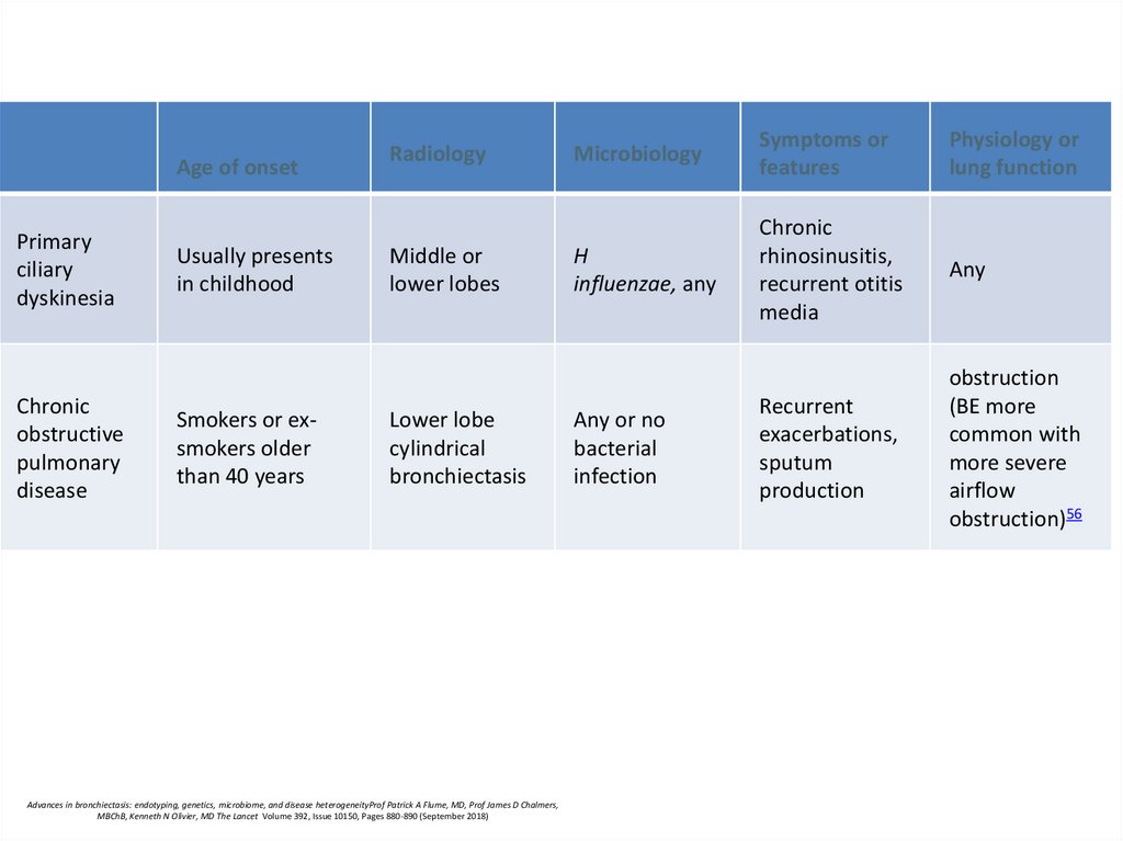
Age of onsetPrimary
ciliary
dyskinesia
Chronic
obstructive
pulmonary
disease
Usually presents
in childhood
Smokers or exsmokers older
than 40 years
Radiology
Middle or
lower lobes
Lower lobe
cylindrical
bronchiectasis
Advances in bronchiectasis: endotyping, genetics, microbiome, and disease heterogeneityProf Patrick A Flume, MD, Prof James D Chalmers,
MBChB, Kenneth N Olivier, MD The Lancet Volume 392, Issue 10150, Pages 880-890 (September 2018)
Microbiology
Symptoms or
features
Physiology or
lung function
H
influenzae, any
Chronic
rhinosinusitis,
recurrent otitis
media
Any
Recurrent
exacerbations,
sputum
production
obstruction
(BE more
common with
more severe
airflow
obstruction)56
Any or no
bacterial
infection
61.
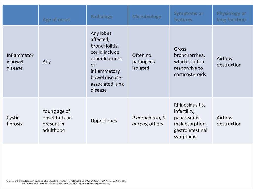
Age of onsetInflammator
y bowel
disease
Cystic
fibrosis
Any
Young age of
onset but can
present in
adulthood
Radiology
Any lobes
affected,
bronchiolitis,
could include
other features
of
inflammatory
bowel diseaseassociated lung
disease
Upper lobes
Advances in bronchiectasis: endotyping, genetics, microbiome, and disease heterogeneityProf Patrick A Flume, MD, Prof James D Chalmers,
MBChB, Kenneth N Olivier, MD The Lancet Volume 392, Issue 10150, Pages 880-890 (September 2018)
Microbiology
Symptoms or
features
Physiology or
lung function
Often no
pathogens
isolated
Gross
bronchorrhea,
which is often
responsive to
corticosteroids
Airflow
obstruction
P aeruginosa, S
aureus, others
Rhinosinusitis,
infertility,
pancreatitis,
malabsorption,
gastrointestinal
symptoms
Airflow
obstruction
62. Cystic fibrosis
• Cystic fibrosis (CF) is an autosomal recessive disease• Loss of function of the cystic fibrosis transmembrane
conductance regulator (CFTR) at the apical
membrane of airway epithelial cells
American Journal of Respiratory and Critical Care Medicine
Vol. 187, No. 7 | Apr 01, 2013
Cystic Fibrosis Pulmonary Guidelines Chronic Medications for Maintenance of Lung Health
Peter J. Mogayzel Jr.1, Edward T. Naureckas 2,
Advances in bronchiectasis: endotyping, genetics, microbiome, and disease
heterogeneity The lancet Volume 392, Issue 10150, 8–14 September 2018, Pages
880-890
63. Pathogenesis: hypothesis
• chemical shield’ hypothesis: in normalcondition airway epithelium produces low salt
(<50 mM NaCl) airway surface liquid, so
defensin-like antimicrobial activities are
performed
• importance of isotonic (i.e. ∼150 mM NaCl)
airway surface liquid volume normally
performs efficient mucus clearance
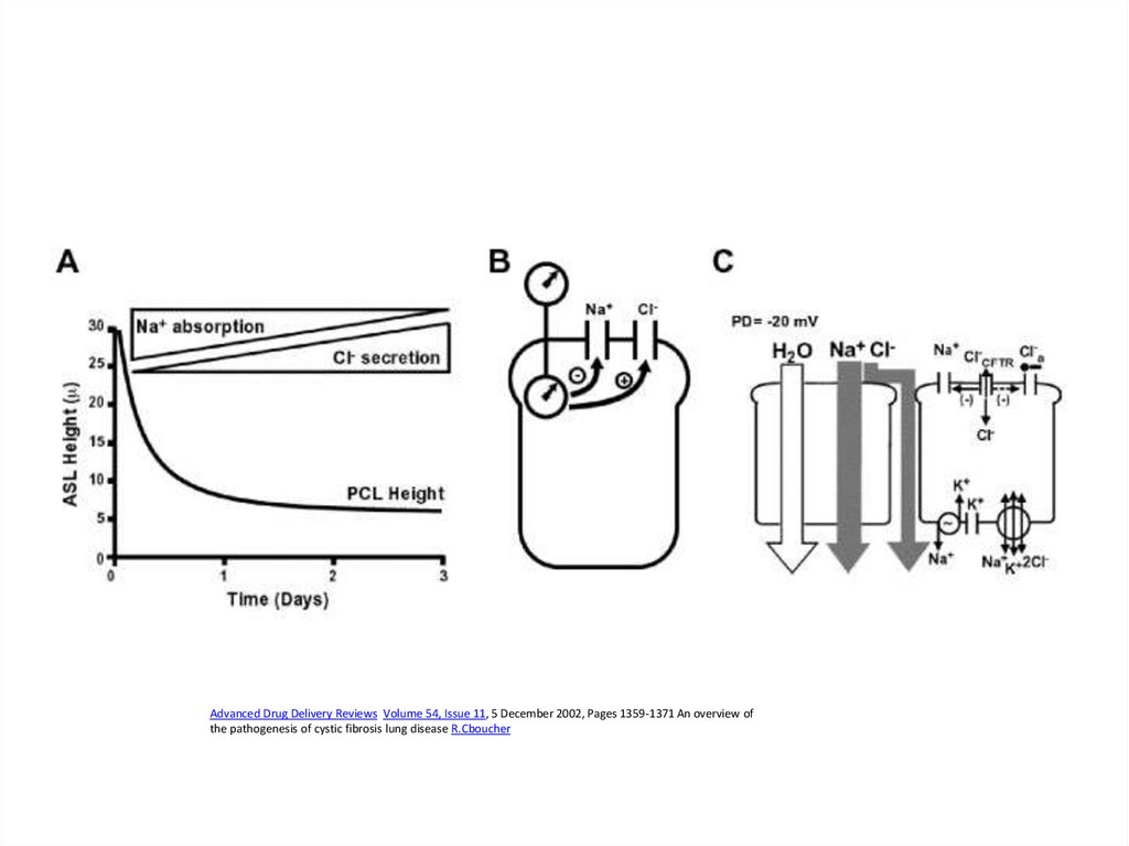
64. Periciliary liquid layer
The mucus layer as a liquid reservoir.Upper panel depicts normal geometry of mucus and periciliary liquid (PCL) layers
Lower left: additional liquid expands the mucus layer
Lower right: removal of liquid can remove ∼50% of the mucus layer without perturbing PCL
volume
65.
Advanced Drug Delivery Reviews Volume 54, Issue 11, 5 December 2002, Pages 1359-1371 An overview ofthe pathogenesis of cystic fibrosis lung disease R.Cboucher
66. CFTR mutations classifications
Advances in bronchiectasis: endotyping, genetics, microbiome, and disease heterogeneity The lancetVolume 392, Issue 10150, 8–14 September 2018, Pages 880-890
67.
• Median age at diagnosis- 6-8 months; twothirds of patients are diagnosed by 1 year of
age
68. Primary ciliary dyskinesia
• multiple genes69. Idiopathic bronchiectasis associated with non-tuberculous mycobacteria (NTM)
Idiopathic bronchiectasis associated with nontuberculous mycobacteria (NTM)post-menopausal non-smoker females
chronic cough
No predisposing factors
share characteristics with other endotypes, notably a
high prevalence of CFTR mutations and ciliary
dysfunction, but do not meet diagnostic criteria for
cystic fibrosis or primary ciliary dyskinesia.
• tall, asthenic type, with scoliosis, pectus
excavatum, mitral valve prolapse, dural ectasia, minor
features overlapping with Marfan and EhlersDanlos syndromes
Advances in bronchiectasis: endotyping, genetics, microbiome, and disease heterogeneity The lancet
Volume 392, Issue 10150, 8–14 September 2018, Pages 880-890
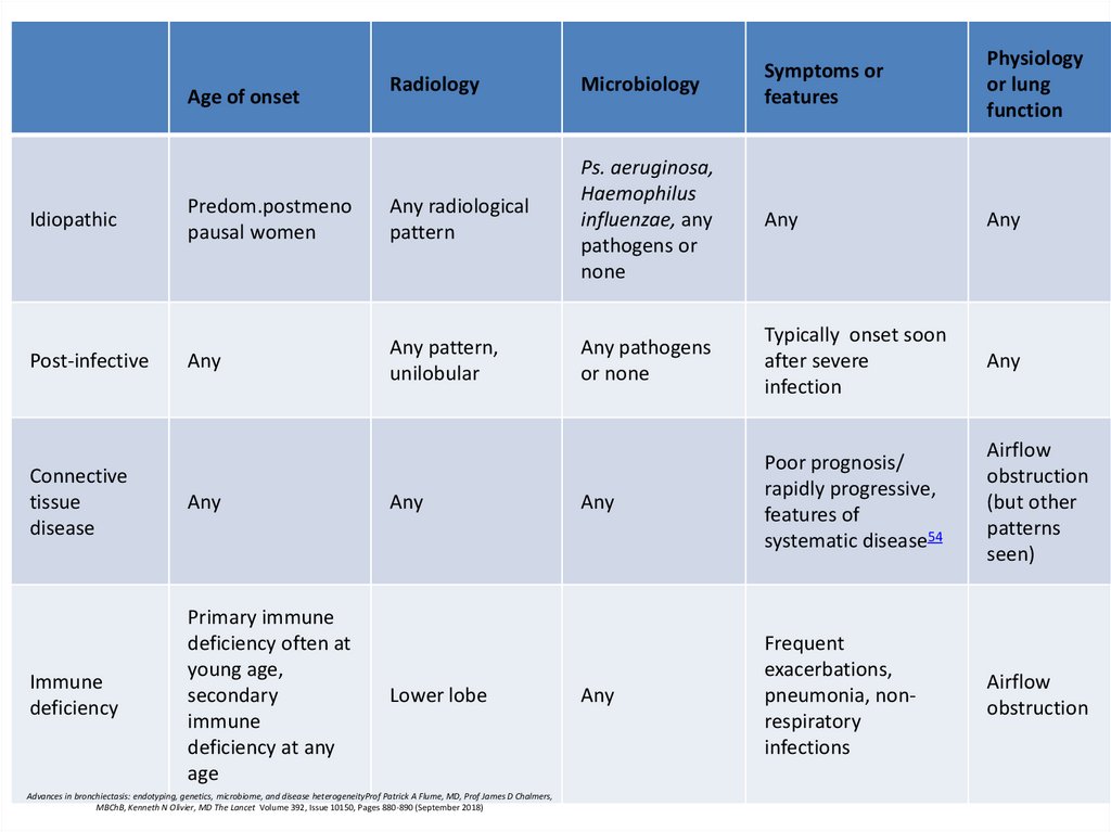
70.
RadiologyMicrobiology
Predom.postmeno
pausal women
Any radiological
pattern
Ps. aeruginosa,
Haemophilus
influenzae, any
pathogens or
none
Any
Any
Any
Any pattern,
unilobular
Any pathogens
or none
Typically onset soon
after severe
infection
Any
Any
Poor prognosis/
rapidly progressive,
features of
systematic disease54
Airflow
obstruction
(but other
patterns
seen)
Any
Frequent
exacerbations,
pneumonia, nonrespiratory
infections
Airflow
obstruction
Age of onset
Idiopathic
Post-infective
Physiology
or lung
function
Symptoms or
features
Connective
tissue
disease
Any
Immune
deficiency
Primary immune
deficiency often at
young age,
secondary
immune
deficiency at any
age
Any
Lower lobe
Advances in bronchiectasis: endotyping, genetics, microbiome, and disease heterogeneityProf Patrick A Flume, MD, Prof James D Chalmers,
MBChB, Kenneth N Olivier, MD The Lancet Volume 392, Issue 10150, Pages 880-890 (September 2018)
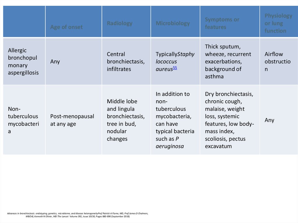
71.
Age of onsetAllergic
bronchopul
monary
aspergillosis
Nontuberculous
mycobacteri
a
Any
Post-menopausal
at any age
Microbiology
Symptoms or
features
Physiology
or lung
function
Central
bronchiectasis,
infiltrates
TypicallyStaphy
lococcus
aureus55
Thick sputum,
wheeze, recurrent
exacerbations,
background of
asthma
Airflow
obstructio
n
Middle lobe
and lingula
bronchiectasis,
tree in bud,
nodular
changes
In addition to
nontuberculous
mycobacteria,
can have
typical bacteria
such as P
aeruginosa
Dry bronchiectasis,
chronic cough,
malaise, weight
loss, systemic
features, low bodymass index,
scoliosis, pectus
excavatum
Any
Radiology
Advances in bronchiectasis: endotyping, genetics, microbiome, and disease heterogeneityProf Patrick A Flume, MD, Prof James D Chalmers,
MBChB, Kenneth N Olivier, MD The Lancet Volume 392, Issue 10150, Pages 880-890 (September 2018)
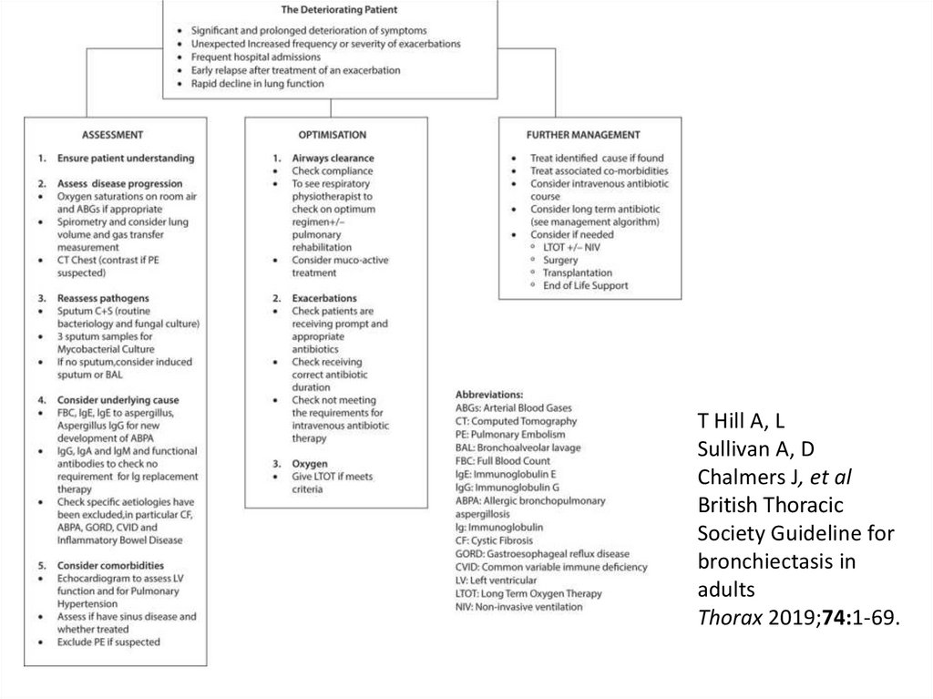
72.
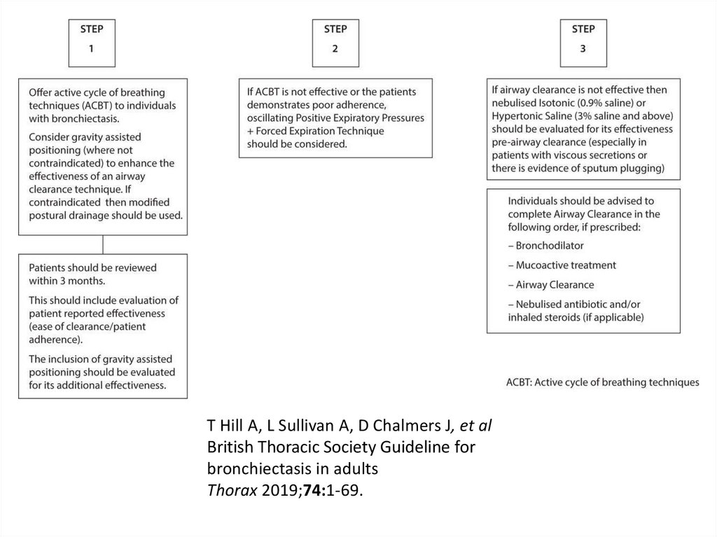
T Hill A, LSullivan A, D
Chalmers J, et al
British Thoracic
Society Guideline for
bronchiectasis in
adults
Thorax 2019;74:1-69.
73. Initial treatment
European Respiratory Society guidelines for the management of adult bronchiectasisEva Polverino, Pieter C. Goeminne, Melissa J. McDonnell, Stefano Aliberti, Sara E. Marshall, Michael
R. Loebinger, European Respiratory Journal 2017 50: 1700629;
74.
• Offer annual influenza immunisation to allpatients with bronchiectasis. (D)
• Offer polysaccharide pneumococcal
vaccination to all patients with bronchiectasis.
T Hill A, L Sullivan A, D Chalmers J, et al
British Thoracic Society Guideline for
bronchiectasis in adults
Thorax 2019;74:1-69.
75. Physiotherapy – drainage promotion
AD: autogenic drainage; ELTGOL: total slow expiration withopen glottis and infralateral position; ACBT: active cycle of
breathing techniques; PEP: positive expiratory pressure; T-PEP:
temporary positive expiratory pressure; HFCWO: high
frequency chest wall oscillation.
European Respiratory Society guidelines for the management of adult bronchiectasis
Eva Polverino, Pieter C. Goeminne, Melissa J. McDonnell, Stefano Aliberti, Sara E. Marshall, Michael
R. Loebinger, European Respiratory Journal 2017 50: 1700629;
76. Airway clearance techniques
• should be taught by a respiratory physiotherapist.• Patients admitted with an exacerbation of
bronchiectasis should be seen daily by a
respiratory physiotherapist until their airway
clearance is optimised.
• CT imaging should be reviewed to complement
the physiotherapy assessment. Where indicated,
this information could be used in order to teach
the patient the appropriate postural drainage
position(s) for their affected bronchopulmonary
segment(s).
T Hill A, L Sullivan A, D Chalmers J, et al
British Thoracic Society Guideline for bronchiectasis in adults
Thorax 2019;74:1-69.
77.
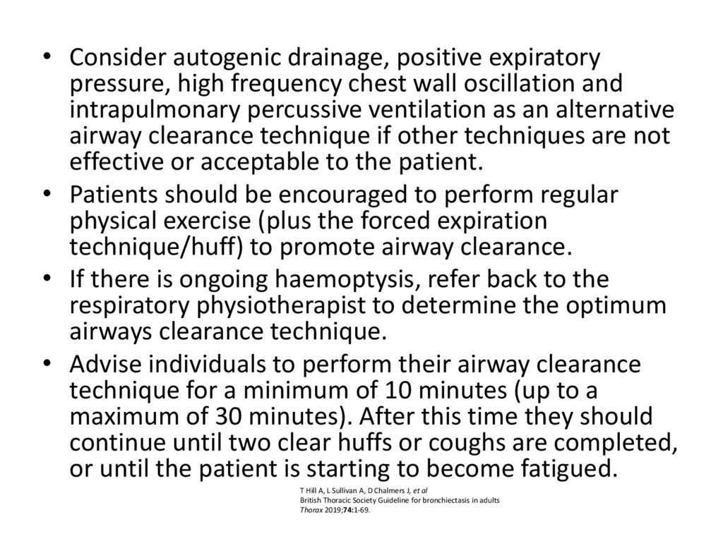
• Consider autogenic drainage, positive expiratorypressure, high frequency chest wall oscillation and
intrapulmonary percussive ventilation as an alternative
airway clearance technique if other techniques are not
effective or acceptable to the patient.
• Patients should be encouraged to perform regular
physical exercise (plus the forced expiration
technique/huff) to promote airway clearance.
• If there is ongoing haemoptysis, refer back to the
respiratory physiotherapist to determine the optimum
airways clearance technique.
• Advise individuals to perform their airway clearance
technique for a minimum of 10 minutes (up to a
maximum of 30 minutes). After this time they should
continue until two clear huffs or coughs are completed,
or until the patient is starting to become fatigued.
T Hill A, L Sullivan A, D Chalmers J, et al
British Thoracic Society Guideline for bronchiectasis in adults
Thorax 2019;74:1-69.
78. Airway clearance techniques during an acute exacerbation
• Manual techniques may be offered to enhancesputum clearance when the patient is fatigued or
undergoing an exacerbation.
• Consider intermittent positive pressure breathing
or non-invasive ventilation during an acute
exacerbation to offload the work of breathing so
fatigued and/or breathless patients can tolerate a
longer treatment session and can adopt postural
drainage positions. T Hill A, L Sullivan A, D Chalmers J, et al
British Thoracic Society Guideline for
bronchiectasis in adults
Thorax 2019;74:1-69.
79. Mucoactives in bronchiectasis
Do not routinely use recombinant human DNase in adults with bronchiectasis.
Consider the use of humidification with sterile water or normal saline to facilitate
airway clearance.
Consider a trial of mucoactive treatment in patients with bronchiectasis who have
difficulty in sputum expectoration.
Perform an airway reactivity challenge test when inhaled mucoactive treatment is
first administered.
Consider pre-treatment with a bronchodilator prior to inhaled or nebulised
mucoactive treatments especially in individuals where bronchoconstriction is likely
(patients with asthma or bronchial hyper-reactivity and those with severe airflow
obstruction FEV1<1 litre).
If carbocysteine is prescribed, a 6 month trial should be given and continued if
there is ongoing clinical benefit.
T Hill A, L Sullivan A, D Chalmers J, et al
British Thoracic Society Guideline for bronchiectasis in adults
Thorax 2019;74:1-69.
80.
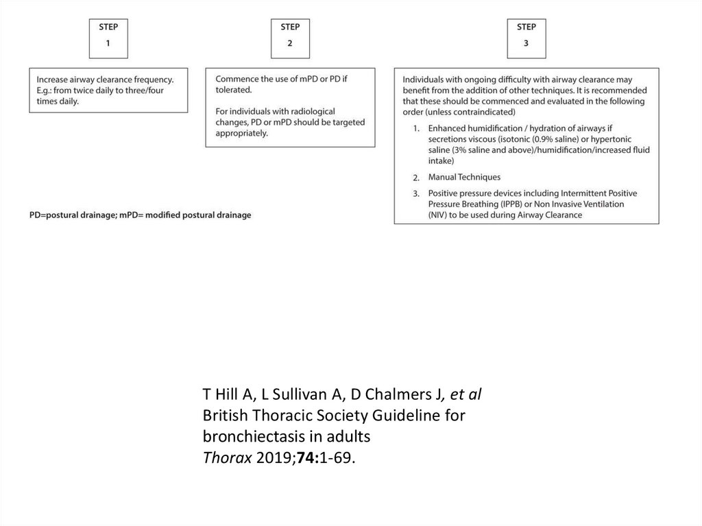
T Hill A, L Sullivan A, D Chalmers J, et alBritish Thoracic Society Guideline for
bronchiectasis in adults
Thorax 2019;74:1-69.
81.
T Hill A, L Sullivan A, D Chalmers J, et alBritish Thoracic Society Guideline for
bronchiectasis in adults
Thorax 2019;74:1-69.
82. Inhaled GCS:
• Do not offer long-term oral corticosteroids forpatients with bronchiectasis without other
indications (such as ABPA, chronic asthma, COPD,
inflammatory bowel disease). (D)
• Inhaled corticosteroids have an established role
in the management of asthma and in a
proportion of patients with COPD which are
common co-morbid conditions in bronchiectasis.
T Hill A, L Sullivan A, D Chalmers J, et al
British Thoracic Society Guideline for bronchiectasis in adults
Thorax 2019;74:1-69.
83. PDE inhibitors, CXCR2 antagonists, statins etc
• Do not routinely offer phosphodiesterase type4 (PDE4) inhibitors, methylxanthines or
leukotriene receptor antagonists for
bronchiectasis treatment. (D)
• Do not routinely offer CXCR2 antagonists,
neutrophil elastase inhibitors or statins for
bronchiectasis treatment. (B)
T Hill A, L Sullivan A, D Chalmers J, et al
British Thoracic Society Guideline for
bronchiectasis in adults
Thorax 2019;74:1-69.
84. Antibiotics
European Respiratory Society guidelines for the management of adult bronchiectasisEva Polverino, Pieter C. Goeminne, Melissa J. McDonnell, Stefano Aliberti, Sara E. Marshall, Michael
R. Loebinger, European Respiratory Journal 2017 50: 1700629;
85.
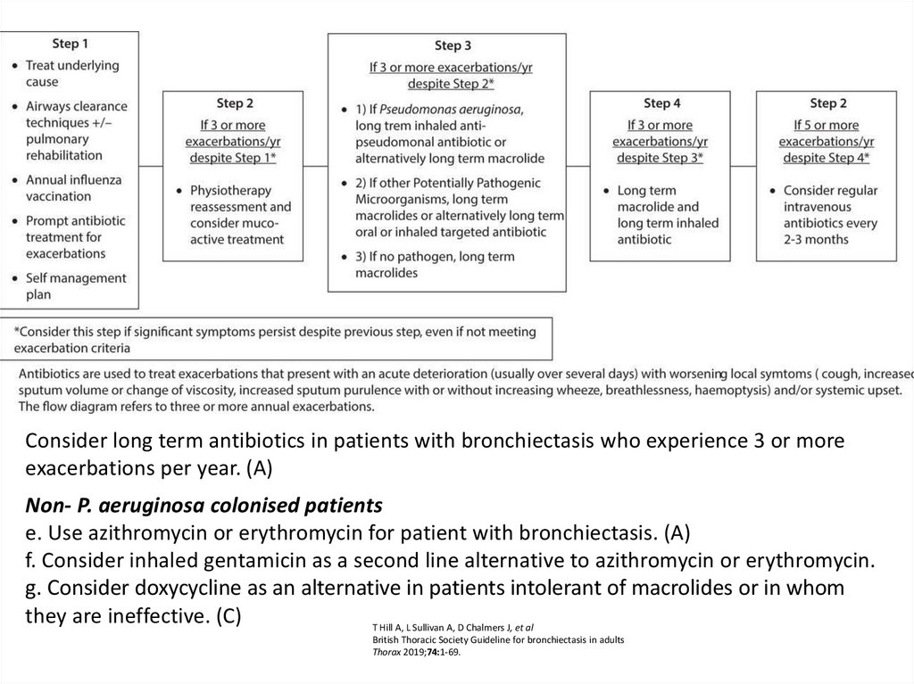
Consider long term antibiotics in patients with bronchiectasis who experience 3 or moreexacerbations per year. (A)
Non- P. aeruginosa colonised patients
e. Use azithromycin or erythromycin for patient with bronchiectasis. (A)
f. Consider inhaled gentamicin as a second line alternative to azithromycin or erythromycin.
g. Consider doxycycline as an alternative in patients intolerant of macrolides or in whom
they are ineffective. (C)
T Hill A, L Sullivan A, D Chalmers J, et al
British Thoracic Society Guideline for bronchiectasis in adults
Thorax 2019;74:1-69.
86. Safety
• Prior to starting long term macrolides, for safety reasons:• (1) ensure no active NTM infection with at least one
negative respiratory NTM culture;
• (2) use with caution if the patient has significant hearing
loss needing hearing aids or significant balance issues.
• Prior to starting long term inhaled aminoglycosides, for
safety reasons:
• (1) avoid using if creatinine clearance <30ml/min;
• (2) use with caution if the patient has significant hearing
loss needing hearing aids or significant balance issues;
• (3) avoid concomitant nephrotoxic medications.
T Hill A, L Sullivan A, D Chalmers J, et al
British Thoracic Society Guideline for
bronchiectasis in adults
Thorax 2019;74:1-69.
87. Ps.aeruginosa
European Respiratory Society guidelines for the management of adult bronchiectasisEva Polverino, Pieter C. Goeminne, Melissa J. McDonnell, Stefano Aliberti, Sara E. Marshall, Michael
R. Loebinger, European Respiratory Journal 2017 50: 1700629;
88.
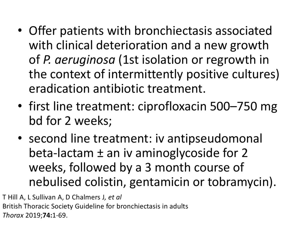
• Offer patients with bronchiectasis associatedwith clinical deterioration and a new growth
of P. aeruginosa (1st isolation or regrowth in
the context of intermittently positive cultures)
eradication antibiotic treatment.
• first line treatment: ciprofloxacin 500–750 mg
bd for 2 weeks;
• second line treatment: iv antipseudomonal
beta-lactam ± an iv aminoglycoside for 2
weeks, followed by a 3 month course of
nebulised colistin, gentamicin or tobramycin).
T Hill A, L Sullivan A, D Chalmers J, et al
British Thoracic Society Guideline for bronchiectasis in adults
Thorax 2019;74:1-69.
89.
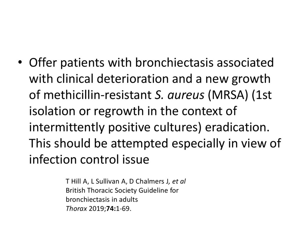
• Offer patients with bronchiectasis associatedwith clinical deterioration and a new growth
of methicillin-resistant S. aureus (MRSA) (1st
isolation or regrowth in the context of
intermittently positive cultures) eradication.
This should be attempted especially in view of
infection control issue
T Hill A, L Sullivan A, D Chalmers J, et al
British Thoracic Society Guideline for
bronchiectasis in adults
Thorax 2019;74:1-69.
90.
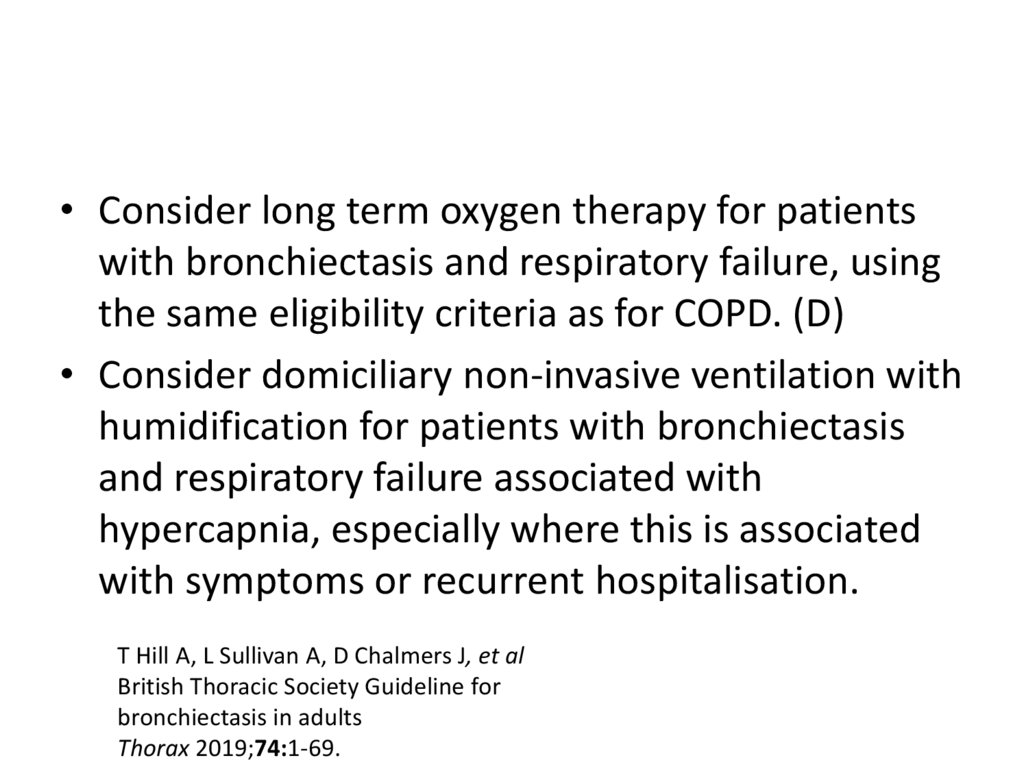
• Consider long term oxygen therapy for patientswith bronchiectasis and respiratory failure, using
the same eligibility criteria as for COPD. (D)
• Consider domiciliary non-invasive ventilation with
humidification for patients with bronchiectasis
and respiratory failure associated with
hypercapnia, especially where this is associated
with symptoms or recurrent hospitalisation.
T Hill A, L Sullivan A, D Chalmers J, et al
British Thoracic Society Guideline for
bronchiectasis in adults
Thorax 2019;74:1-69.
91.
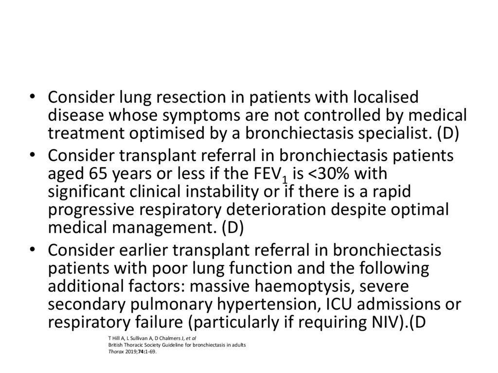
• Consider lung resection in patients with localiseddisease whose symptoms are not controlled by medical
treatment optimised by a bronchiectasis specialist. (D)
• Consider transplant referral in bronchiectasis patients
aged 65 years or less if the FEV1 is <30% with
significant clinical instability or if there is a rapid
progressive respiratory deterioration despite optimal
medical management. (D)
• Consider earlier transplant referral in bronchiectasis
patients with poor lung function and the following
additional factors: massive haemoptysis, severe
secondary pulmonary hypertension, ICU admissions or
respiratory failure (particularly if requiring NIV).(D
T Hill A, L Sullivan A, D Chalmers J, et al
British Thoracic Society Guideline for bronchiectasis in adults
Thorax 2019;74:1-69.
92. allergic broncho-pulmonary aspergillosis
allergic broncho-pulmonaryaspergillosis
• Offer oral corticosteroid to patients with active
ABPA. An initial dose of 0.5 mg/kg/d, for 2 weeks
is recommended. Wean steroids according to
clinical response and serum IgE levels. (D)
• Consider itraconazole as a steroid sparing agent
for patients dependent on oral corticosteroids
where difficulty in weaning is experienced. (B)
• Monitor patients with active ABPA with total IgE
level to assess treatment response
T Hill A, L Sullivan A, D Chalmers J, et al
British Thoracic Society Guideline for bronchiectasis in adults
Thorax 2019;74:1-69.