


 medicine
Similar presentations:


MeDiMa 3D, digital X-ray tomosynthesis mammography system
1.
digital X-raytomosynthesis
mammography
system
2.
Breast cancer is made up to 32% of all malignancies in women and is the cause of 18% ofall cancer deaths. Today one in eight women Russia may have breast cancer and very
often it is due to the incorrect or late diagnosis.
In the great majority of Russian medical institutions mammography is represented by a
film or digital mammography systems allowing to obtain an overview plain image of the
breast. A plain film may lead to loss of important information, and consequently to a
wrong diagnosis.
Digital 3D mammography system “MEDIMA 3D” allows to obtain true three dimensional
mammography exams, enabling to have an accurate view of the spatial structure of
mammary gland. 3D mammogram provides maximum diagnostic value, reveals the
smallest initial lesions, malignancy potential and possible spread of disease.
3.
X-ray digital mammograph “MEDIMA 3D” developed by “Mosrentgenprom” provides 3types of x-ray imagine modes:
• 2D mammogram;
• 3D mammogram;
• reconstruction of 2D mammograms from 3D mammograms adjusted for correlation
analysis.
Mammograph performs stationary imaging with the patient in lying position that gives
sharp images by excluding unintentional patient moves and using circumferential view at
360°.
Revolutionary possibilities of the “MEDIMA 3D” provide full comprehensive range of
mammographic breast examinations of superior quality, the best diagnostic performance
and the highest among available competitors spatial resolution of 20 pairs of lines
per mm.
4.
Features1.
Working field – 220 х300 mm.
2.
Spatial resolution of 2D mammogram (vertical and horizontal)
screening mode – 10 pairs of lines per mm;
diagnostic mode – 20 pairs of lines per mm.
3.
4.
Pixel size in 3D mammogram (tomosynthesis)
screening mode – 54х54х1000 mm;
diagnostic mode – 27х27х500 mm.
The mean absorbed dose to mammary gland meets the requirements of European
guidelines for quality assurance in breast cancer screening and diagnosis (Fourth
Edition) as for minimal allowed dose.
5.
Test patternvertical
horizontal
6.
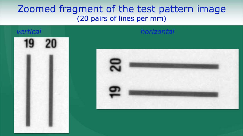
Zoomed fragment of the test pattern image(20 pairs of lines per mm)
vertical
horizontal
7.
8.
9.
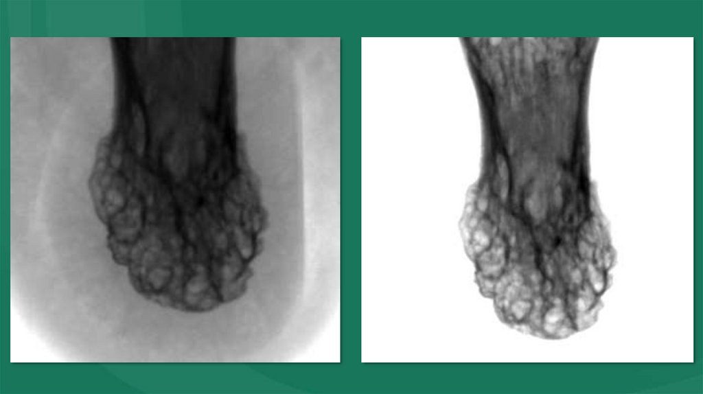
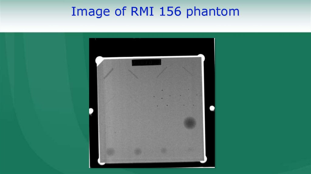
Image of RMI 156 phantom10.
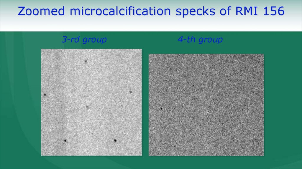
Zoomed microcalcification specks of RMI 1563-rd group
4-th group
11.
Breast image12.
Breast image(ROI)
13.
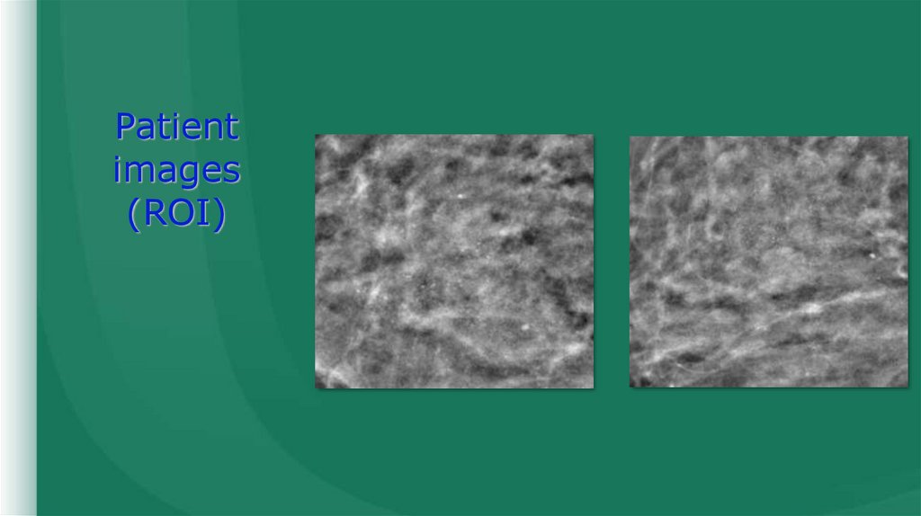
Patient images14.
Patientimages
(ROI)
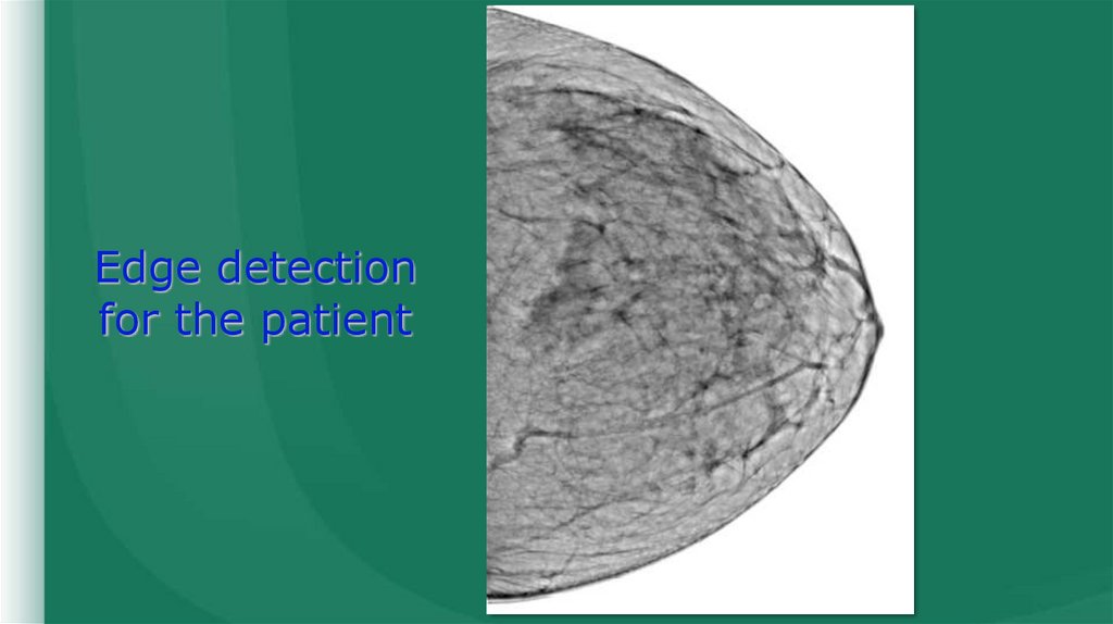
15.
Edge detectionfor the patient
16.
Patient image(ROI)
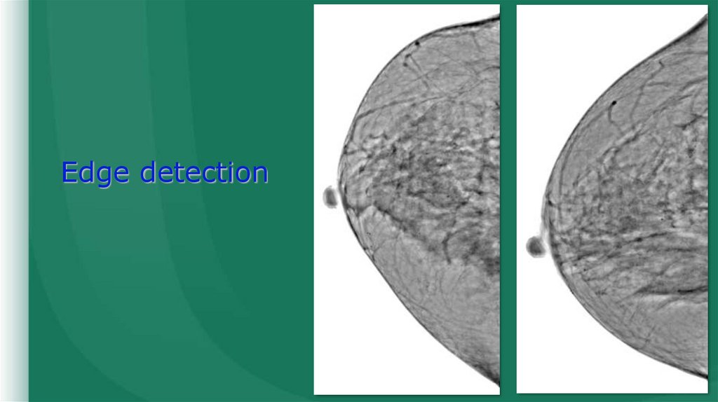
17.
Edge detection18.
Patientimage
(ROI)
19.
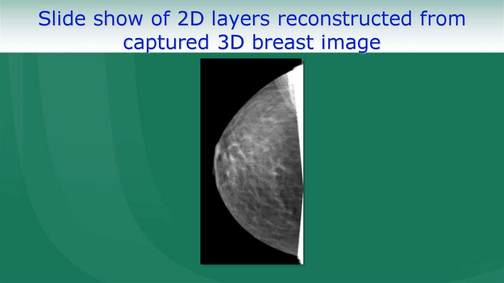
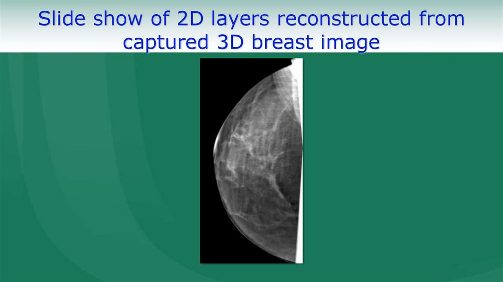
Slide show of 2D layers reconstructed fromcaptured 3D breast image
20.
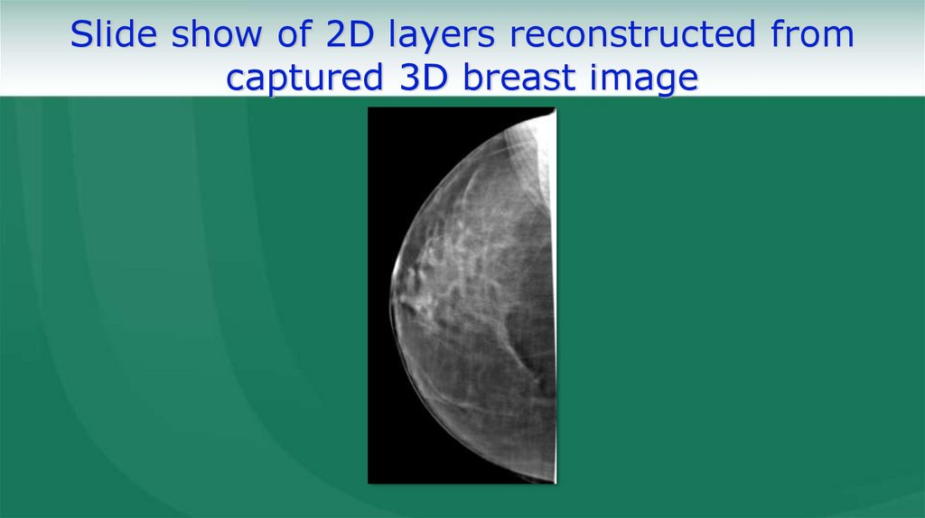
Slide show of 2D layers reconstructed fromcaptured 3D breast image
21.
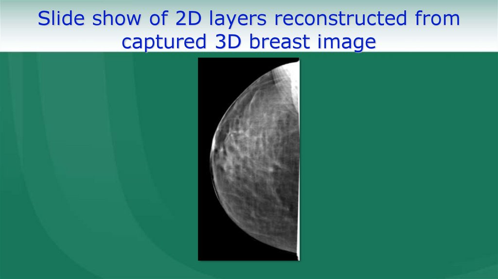
Slide show of 2D layers reconstructed fromcaptured 3D breast image
22.
Slide show of 2D layers reconstructed fromcaptured 3D breast image
23.
Slide show of 2D layers reconstructed fromcaptured 3D breast image
24.
Slide show of 2D layers reconstructed fromcaptured 3D breast image
25.
Russia,125252, Moscow, 21 Novopestchanaya St., bld. 2,
tel. (499) 157-06-92, 157-24-18,
fax (495) 157-62-56
248001, Kaluga, 85A Saltykova-Schedrina St.,
tel./fax (4842) 57-09-48
www.mosrentgenprom.ru,
e-mail: mosrentgenprom@mail.ru, m.prom@rambler.ru