


 medicine
Similar presentations:


 of thebreast")


Anatomy of the Breast
1.
Breast Lump And NippleDischarge
2.
Anatomy of theBreast
3.
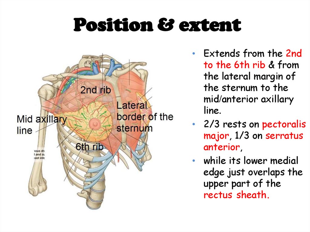
Position & extent• Extends from the 2nd
to the 6th rib & from
the lateral margin of
the sternum to the
mid/anterior axillary
line.
• 2/3 rests on pectoralis
major, 1/3 on serratus
anterior,
• while its lower medial
edge just overlaps the
upper part of the
rectus sheath.
4.
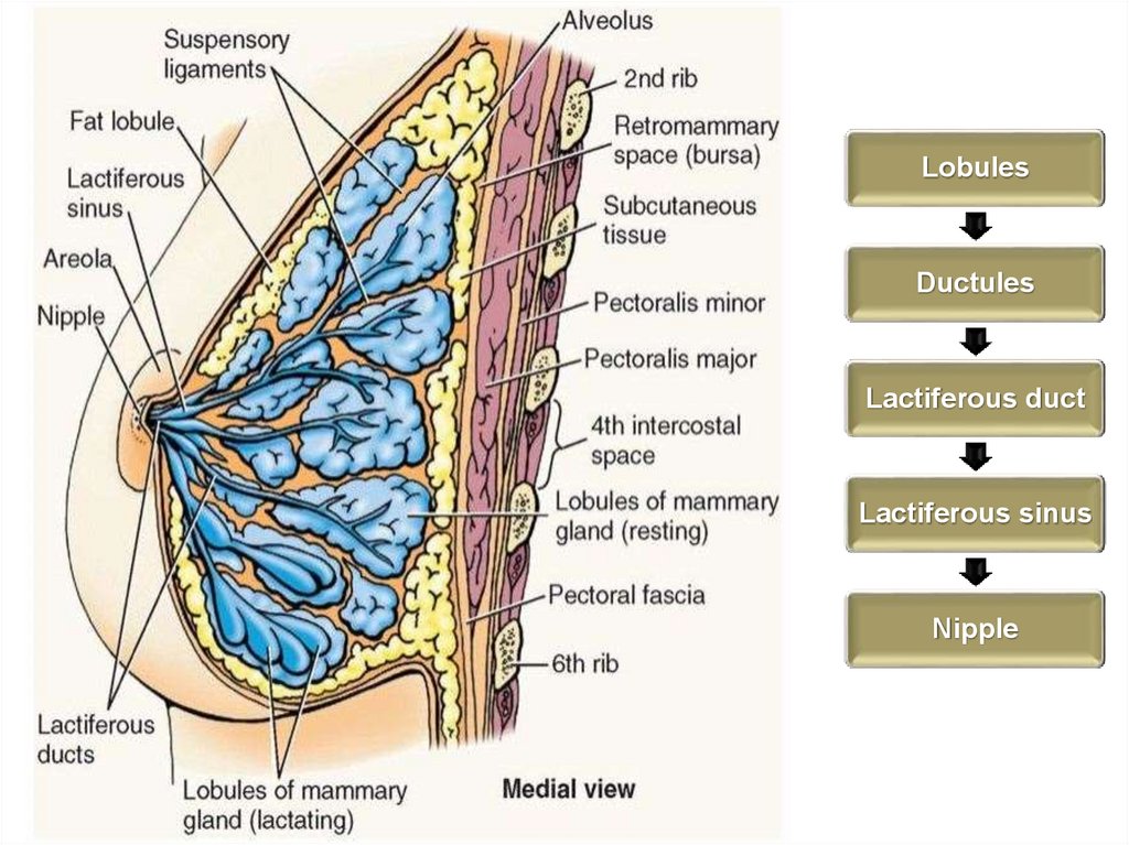
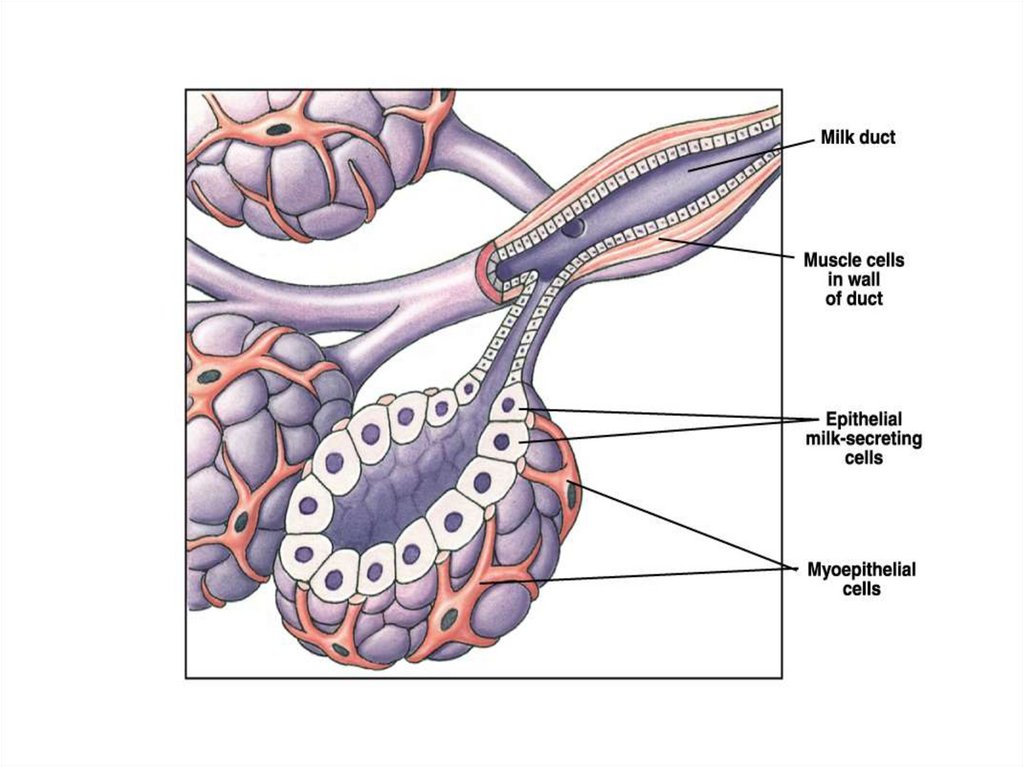
Structure of the Breast• The lobule is the basic structural unit of the
mammary gland.The number and size of the lobules
vary enormously: they are most numerous in young
women. From 10 to over 100 lobules empty via
ductules into a lactiferous duct, of which there are
15–20. Each lactiferous duct is lined with a spiral
arrangement of contractile myoepithelial cells and is
provided with a terminal ampulla, a reservoir for
milk or abnormal discharges.
• The nipple is covered by thick skin with
corrugations. Near its apex lie the orifices of the
lactiferous ducts. The nipple contains smooth
muscle fibres arranged concentrically and
longitudinally; thus, it is an erectile structure, which
points outwards.
5.
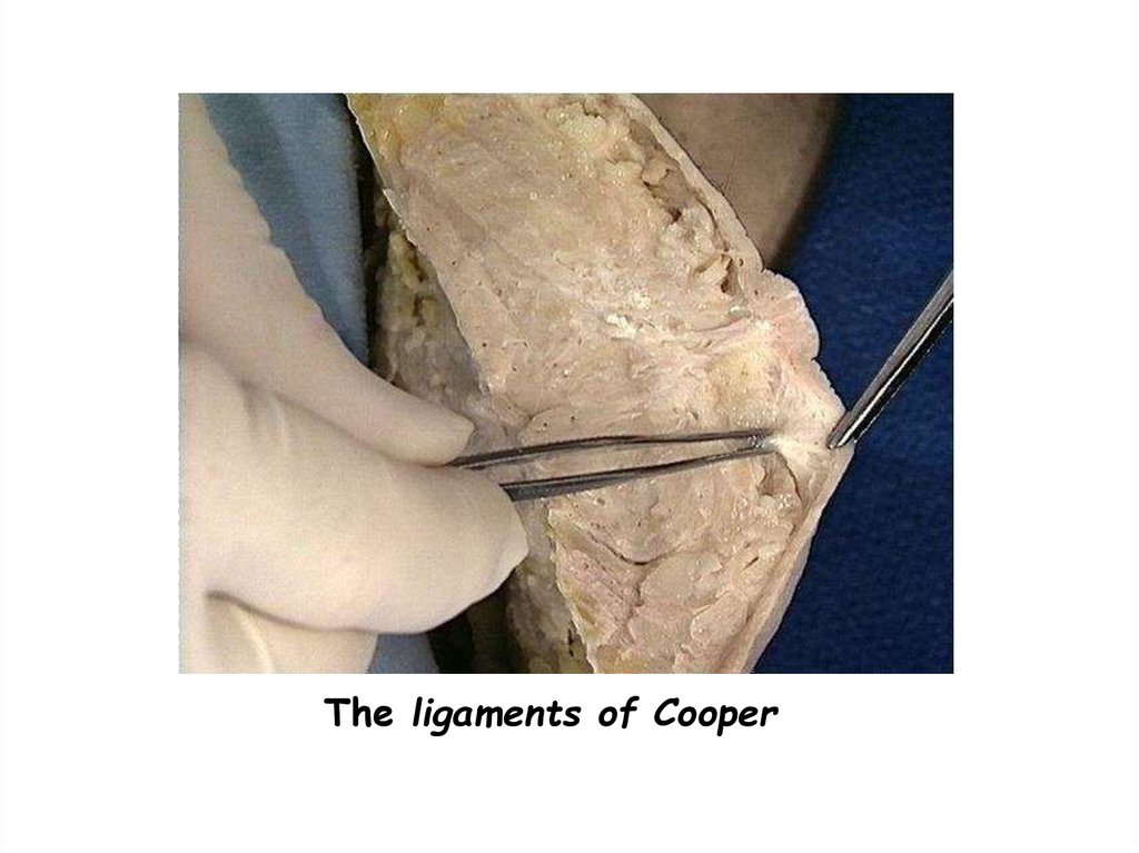
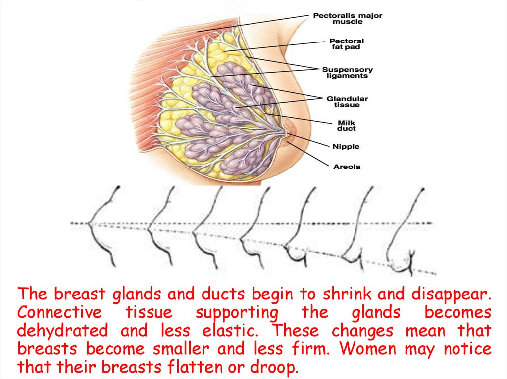
• The ligaments of Cooper are hollow conicalprojections of fibrous tissue filled with
breast tissue; the apices of the cones are
attached firmly to the superficial fascia and
thereby to the skin overlying the breast.
The shape of the breasts is naturally
determined by the support of the suspensory
Cooper's ligaments
6.
LobulesDuctules
Lactiferous duct
Lactiferous sinus
Nipple
7.
The ligaments of Cooper8.
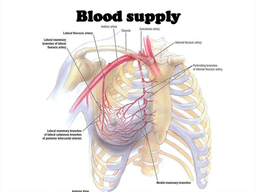
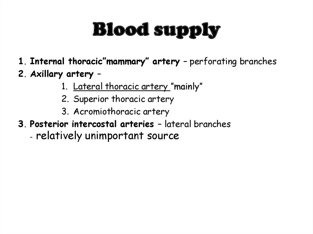
Blood supply9.
Blood supply1. Internal thoracic”mammary” artery – perforating branches
2. Axillary artery –
1. Lateral thoracic artery ”mainly”
2. Superior thoracic artery
3. Acromiothoracic artery
3. Posterior intercostal arteries – lateral branches
-
relatively unimportant source
10.
Venous drainage• Sub areolar venous plexus
• Posterior intercostal veins
communicate with internal vertebral
venous plexus veins - therefore
cancers can spread to vertebra- may
cause back pain
11.
Nerves of the Breast• Cutaneous innervation
• Medial pectoral nerve
• Lateral pectoral nerve
• Long thoracic nerve
12.
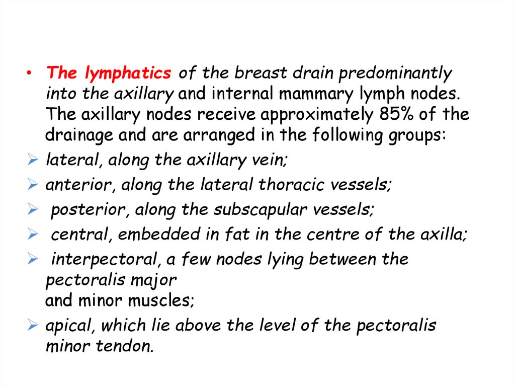
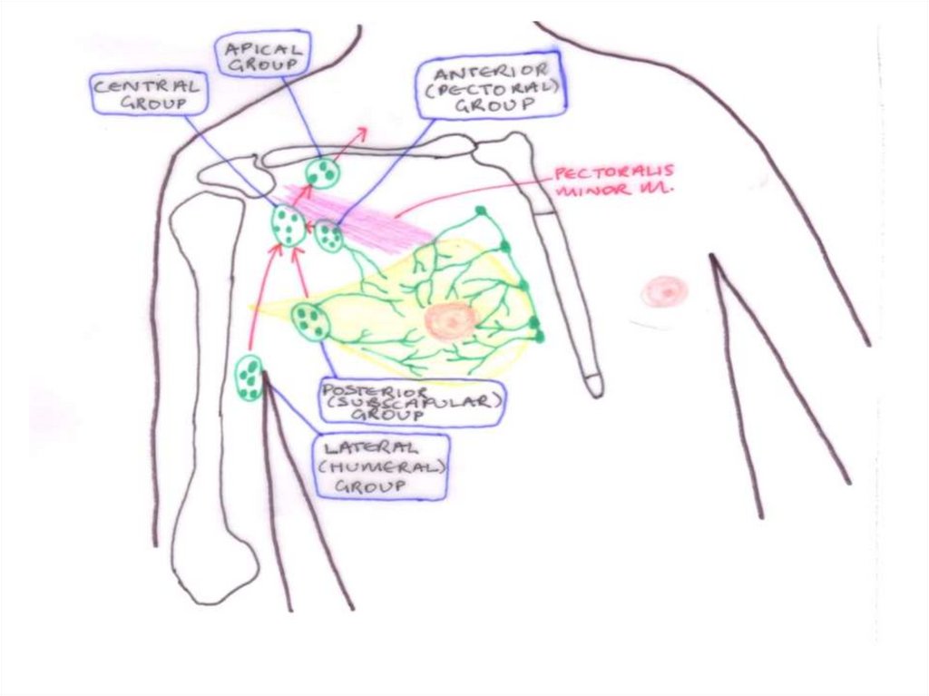
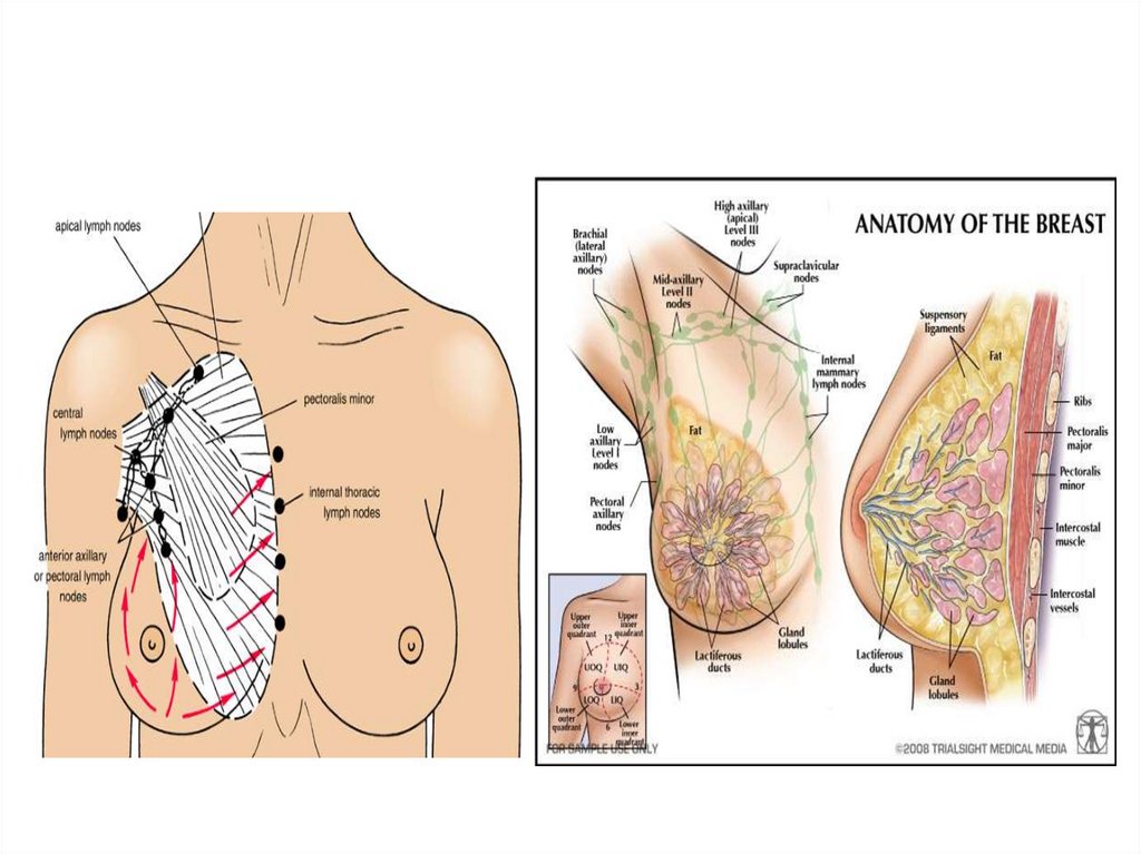
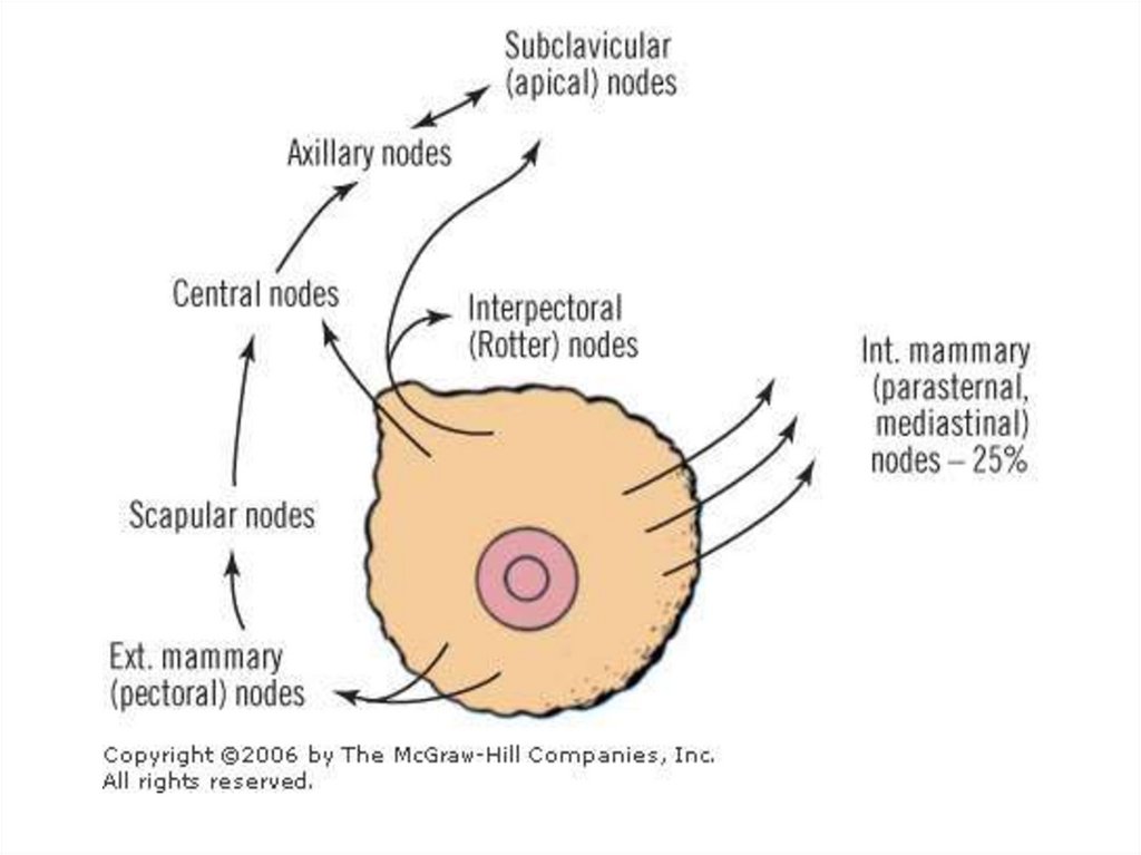
• The lymphatics of the breast drain predominantlyinto the axillary and internal mammary lymph nodes.
The axillary nodes receive approximately 85% of the
drainage and are arranged in the following groups:
lateral, along the axillary vein;
anterior, along the lateral thoracic vessels;
posterior, along the subscapular vessels;
central, embedded in fat in the centre of the axilla;
interpectoral, a few nodes lying between the
pectoralis major
and minor muscles;
apical, which lie above the level of the pectoralis
minor tendon.
13.
14.
15.
16.
17.
Physiology of theBreast
18.
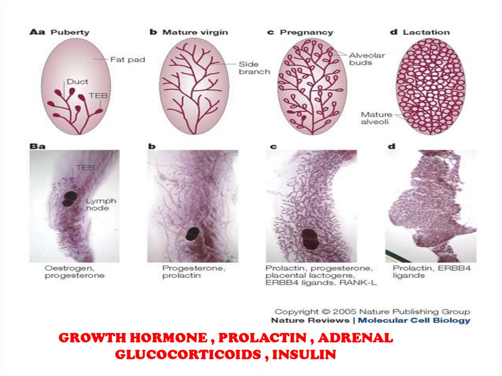
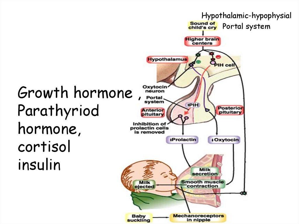
GROWTH HORMONE , PROLACTIN , ADRENALGLUCOCORTICOIDS , INSULIN
19.
20.
Hypothalamic-hypophysialPortal system
Growth hormone ,
Parathyriod
hormone,
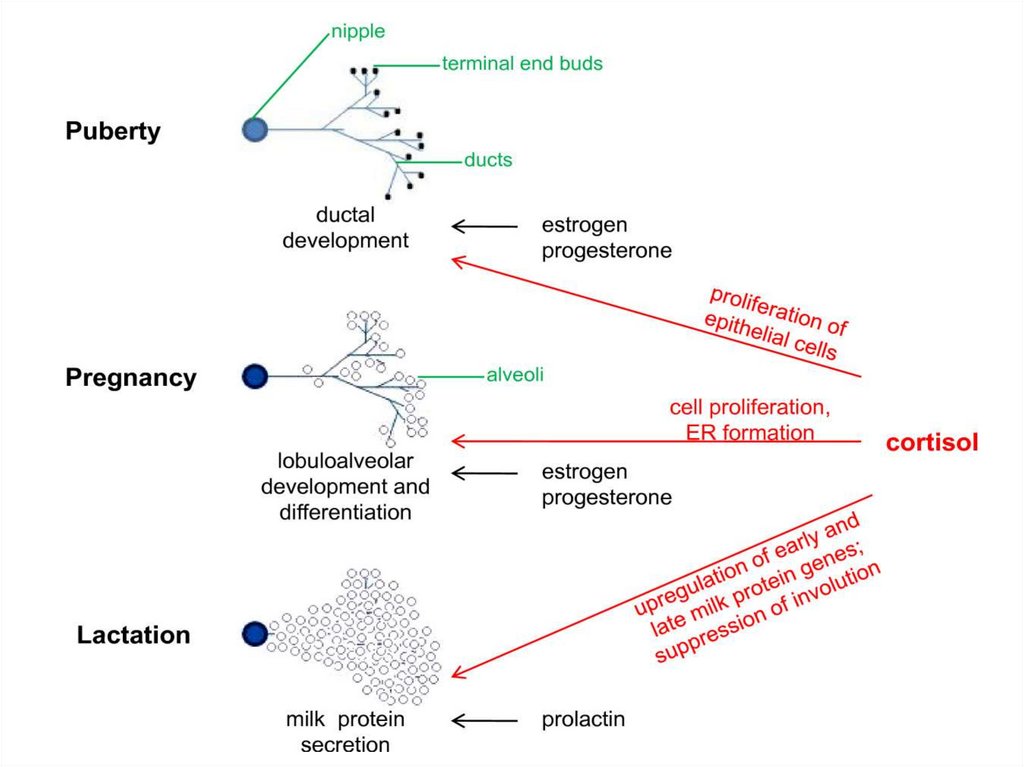
cortisol
insulin
21.
22.
The breast glands and ducts begin to shrink and disappear.Connective tissue supporting the glands becomes
dehydrated and less elastic. These changes mean that
breasts become smaller and less firm. Women may notice
that their breasts flatten or droop.
23.
Benign Breast Disease24.
Benign Breast Disease• The most common cause of breast
problems.
•30% of women will suffer from a benign
disorder requiring treatment some time
in their lives.
•Most common symptoms are Pain,
Lumpiness or a Lump.
25.
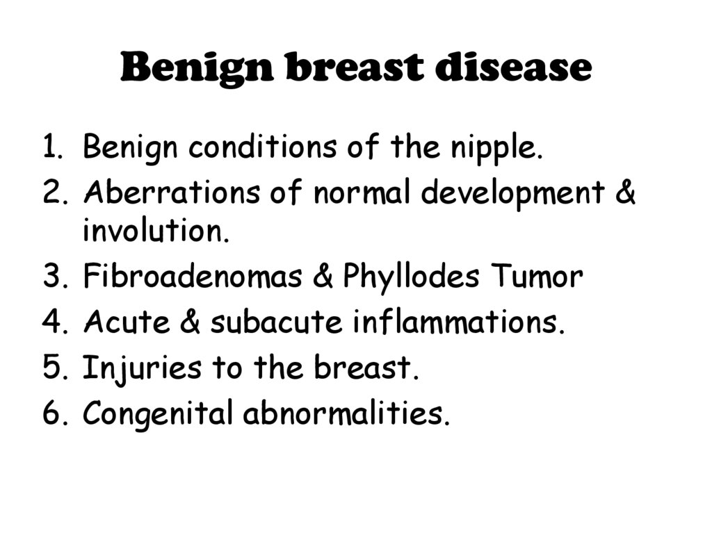
Benign breast disease1. Benign conditions of the nipple.
2. Aberrations of normal development &
involution.
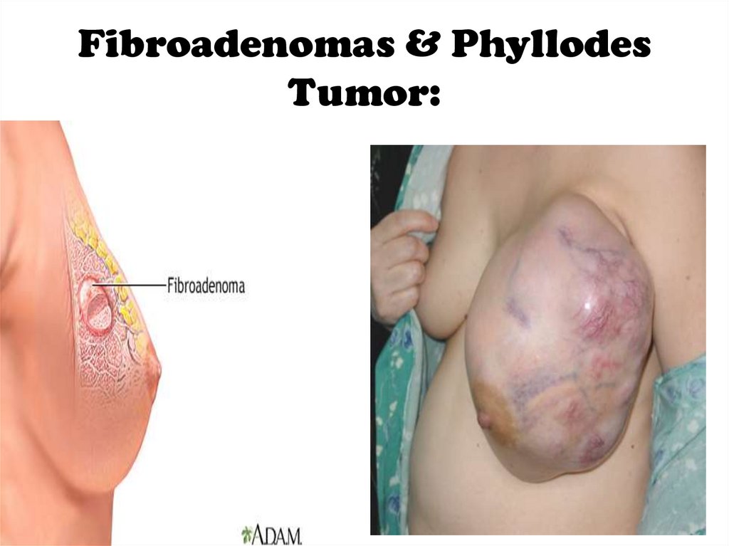
3. Fibroadenomas & Phyllodes Tumor
4. Acute & subacute inflammations.
5. Injuries to the breast.
6. Congenital abnormalities.
26.
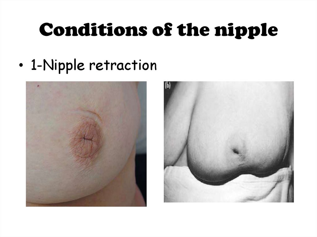
Conditions of the nipple• 1-Nipple retraction
27.
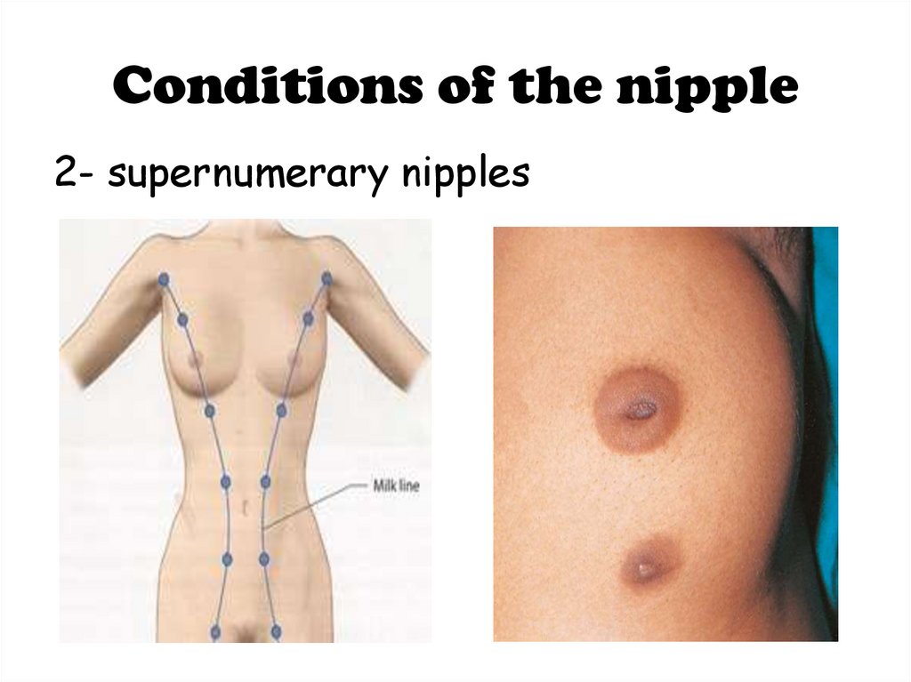
Conditions of the nipple2- supernumerary nipples
28.
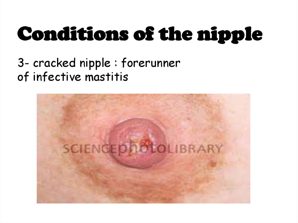
Conditions of the nipple3- cracked nipple : forerunner
of infective mastitis
29.
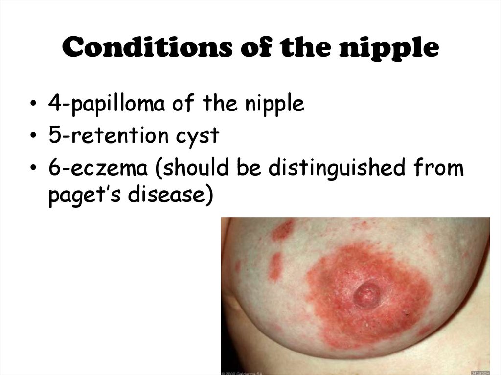
Conditions of the nipple• 4-papilloma of the nipple
• 5-retention cyst
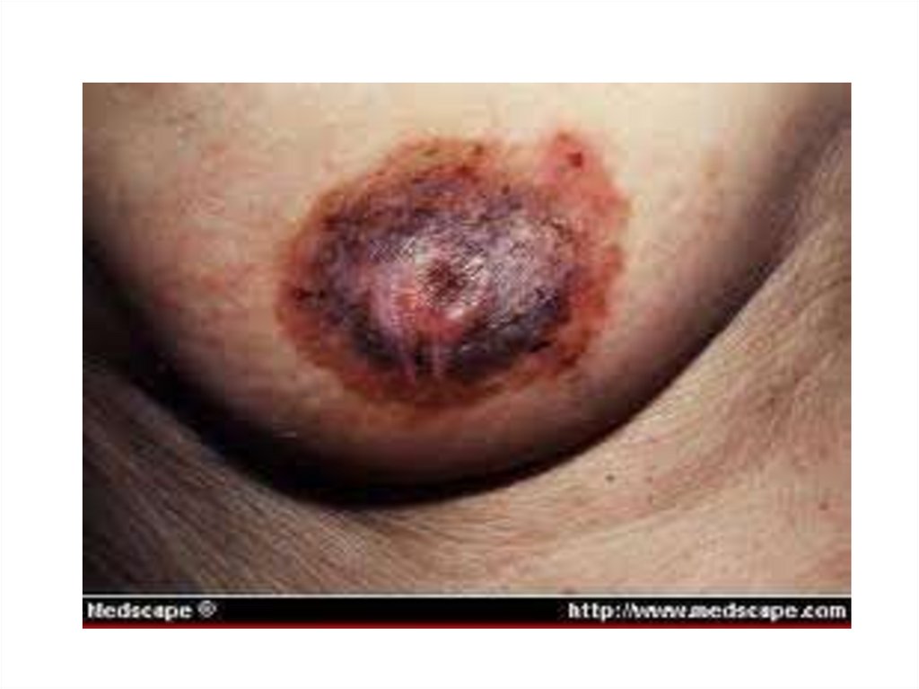
• 6-eczema (should be distinguished from
paget’s disease)
30.
31.
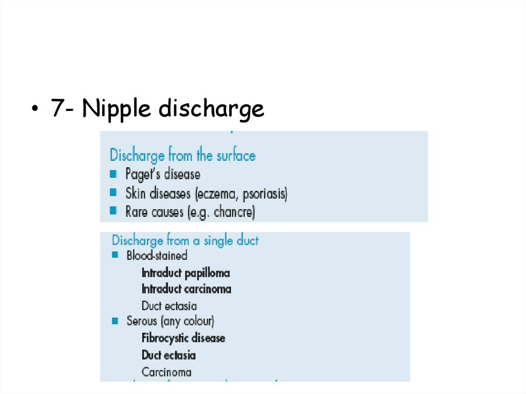
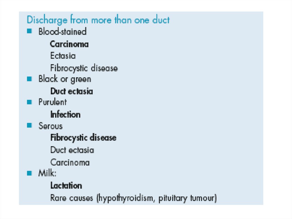
• 7- Nipple discharge32.
33.
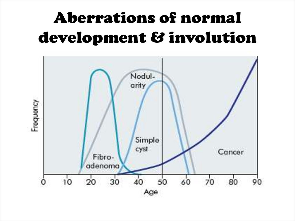
Aberrations of normaldevelopment & involution
34.
Aberrations of normaldevelopment & involution
1.
2.
3.
4.
Cyst formation
Fibrosis
Hyperplasia
Papillomatosis
35.
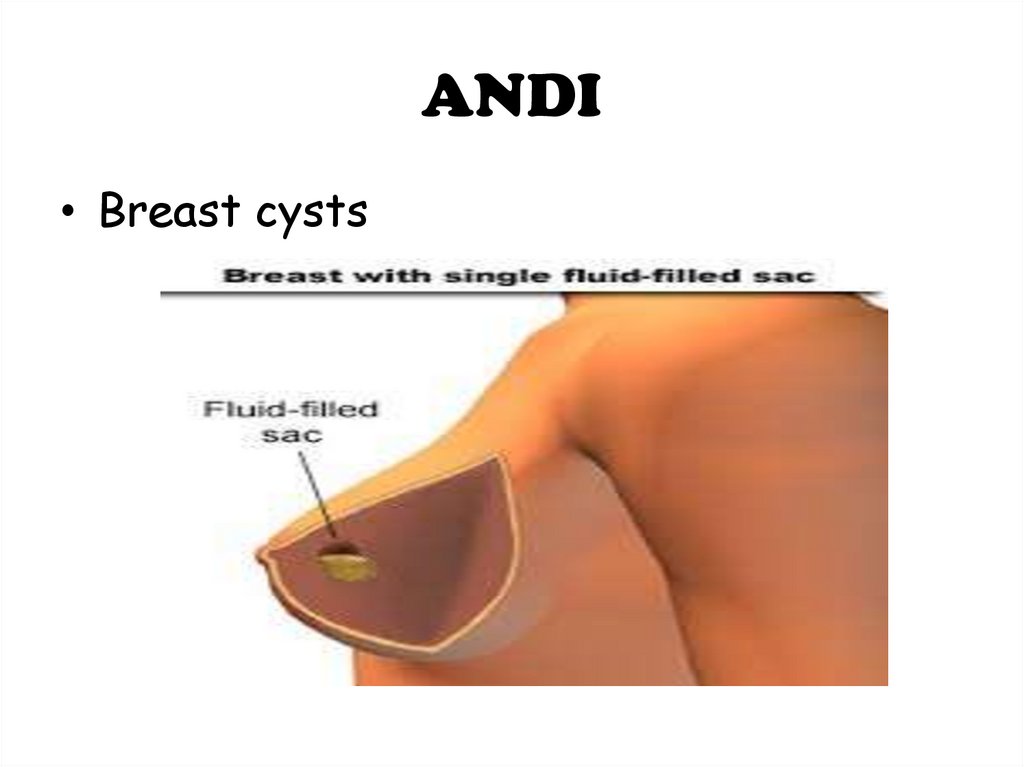
ANDI• Breast cysts
36.
Fibroadenomas & PhyllodesTumor:
37.
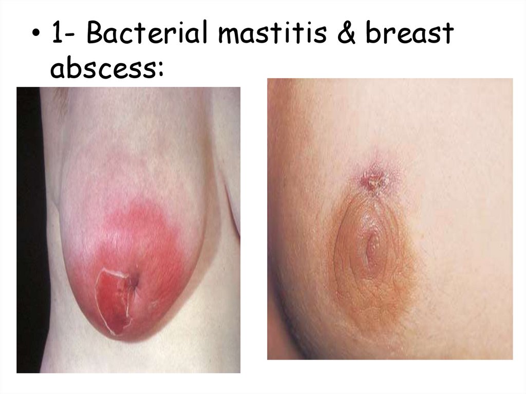
• Acute and SubacuteInflammations of the Breast:
38.
• 1- Bacterial mastitis & breastabscess:
39.
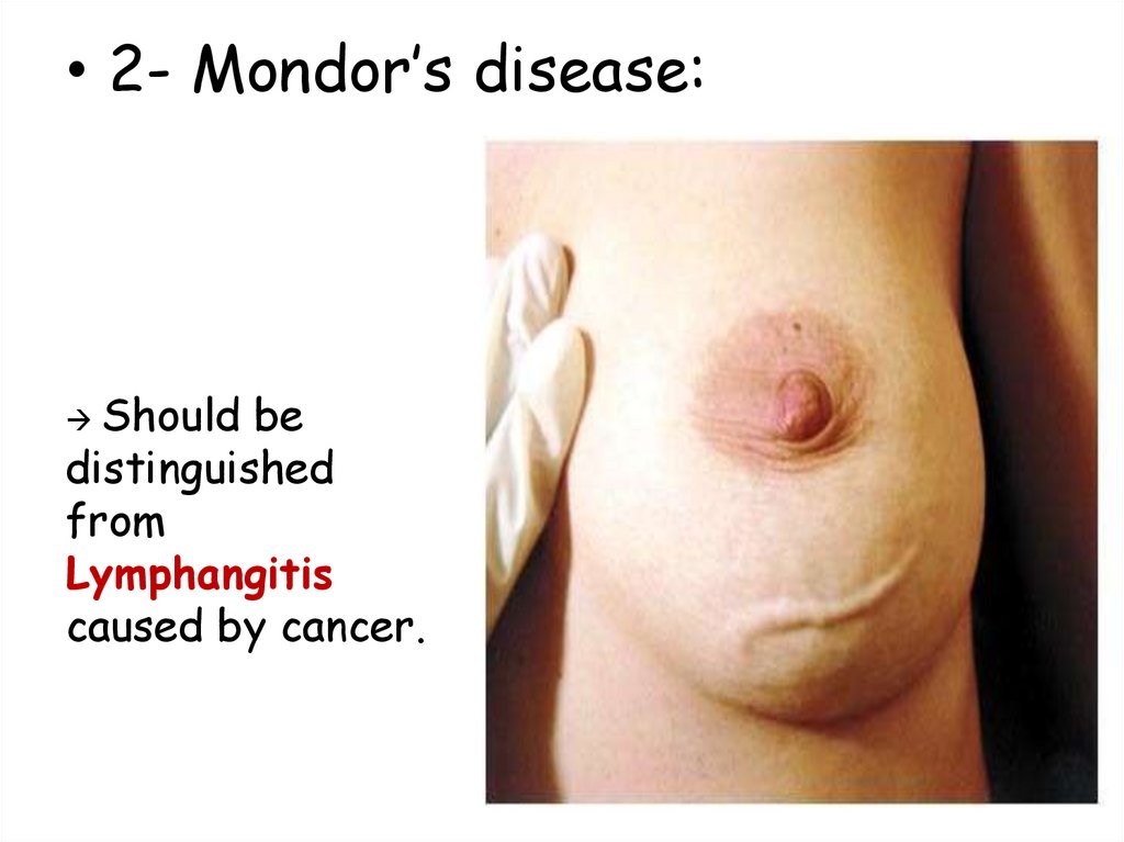
• 2- Mondor’s disease:Should be
distinguished
from
Lymphangitis
caused by cancer.
40.
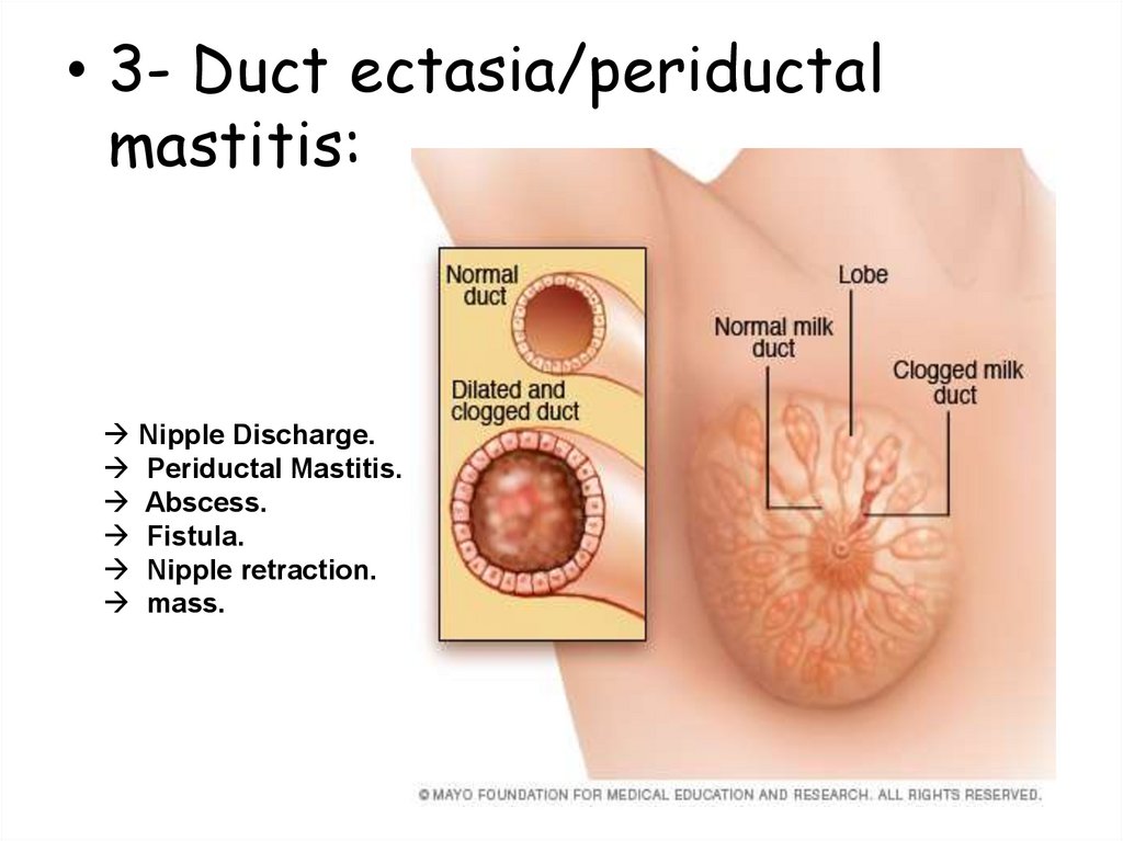
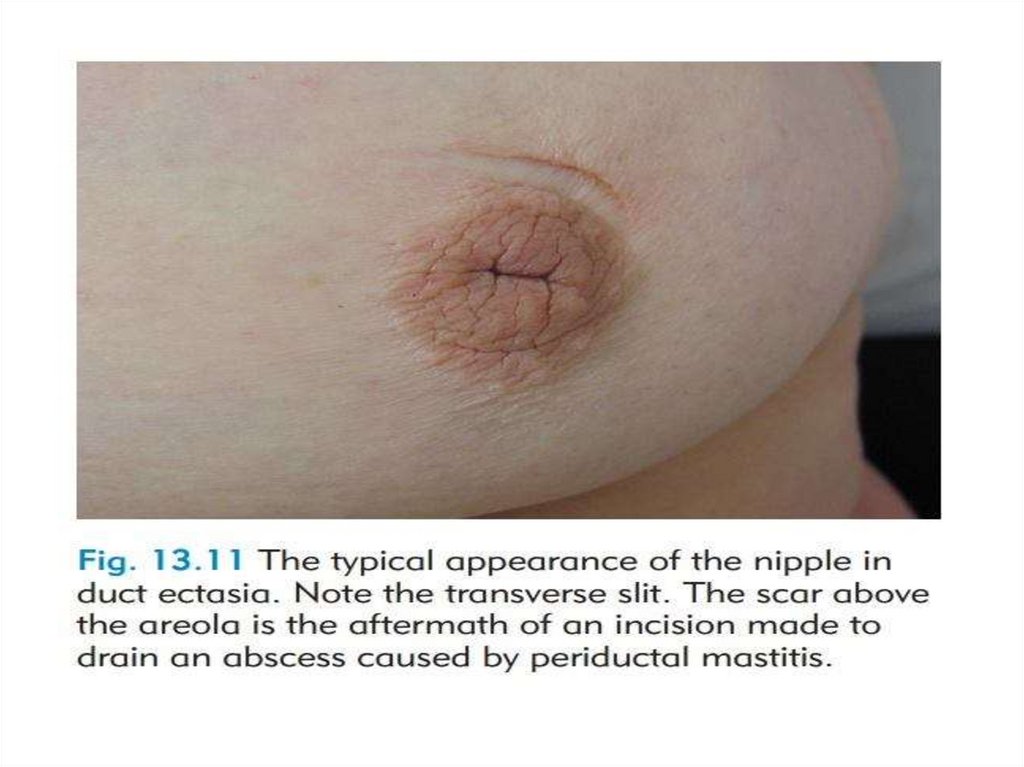
• 3- Duct ectasia/periductalmastitis:
Nipple Discharge.
Periductal Mastitis.
Abscess.
Fistula.
Nipple retraction.
mass.
41.
42.
• Injuries to the Breast:1- Haematoma.
2-Traumatic fat necrosis.
43.
• Congenital Abnormalities:44.
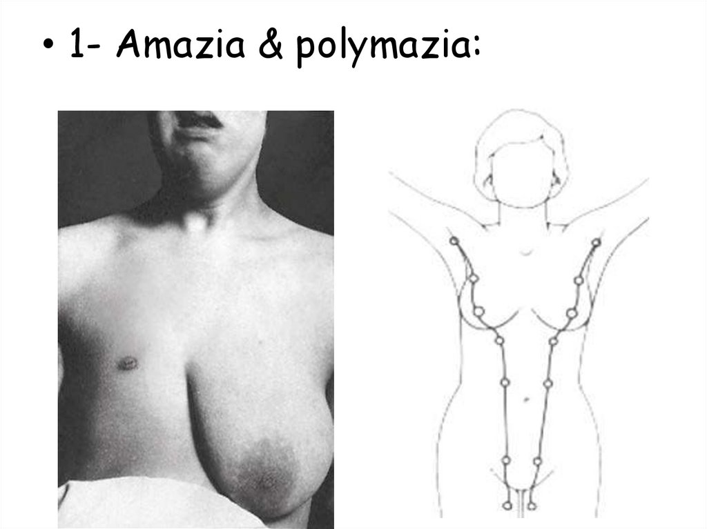
• 1- Amazia & polymazia:45.
• 2- Mastitis of infants:46.
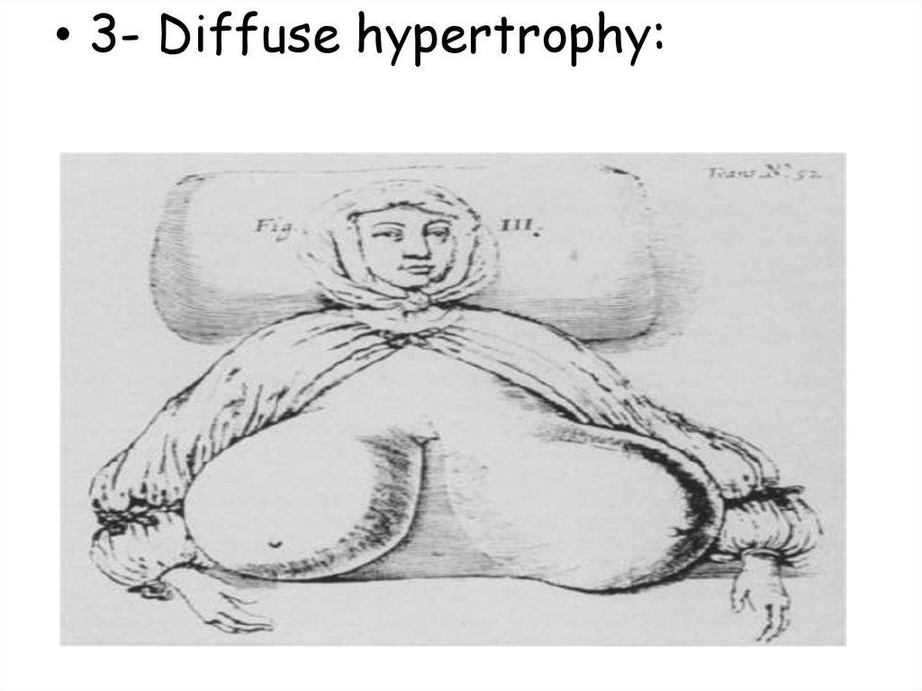
• 3- Diffuse hypertrophy:47.
Malignant diseasesCARCINOMA OF THE BREAST
48.
• Breast cancer is the second mostcommon cancer with nearly 1.7 million
new cases in 2012.
• Most common cancer in women.
• Most common cause of death in middleaged women.
49.
Aetiological factorsGeographical…
Age…
Gender…
Genetic…
Diet…
Endocrine…
Previous radiation…
50.
Pathogenesis• Genetic factor…
• Hormonal factor…
• Enviromental factor…
51.
Histopathologic classificationDuctal ------ Lobular
Invasive ------ In situ
52.
Breast carcinoma in situ53.
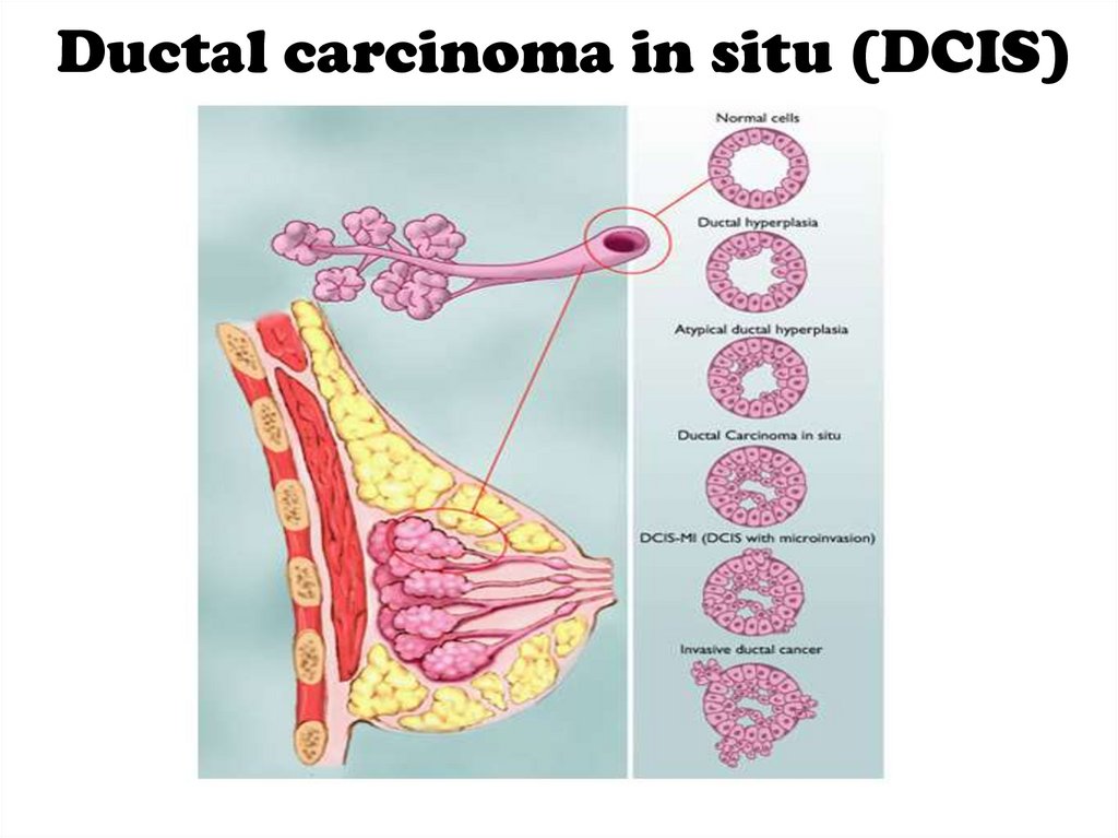
Ductal carcinoma in situ (DCIS)54.
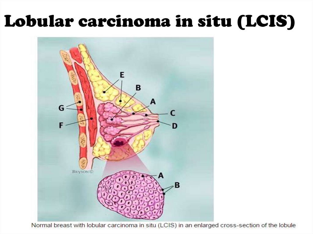
Lobular carcinoma in situ (LCIS)55.
• In situ carcinoma is pre-invasive cancer.• Becoming increasingly common.
• At least 20% of patients will develop
invasive cancer.
56.
Treatment• Surgical excision
Mastectomy?
Partial mastectomy with safety margins > 1cm
• Radiotherapy?
57.
Invasive breast carcinoma58.
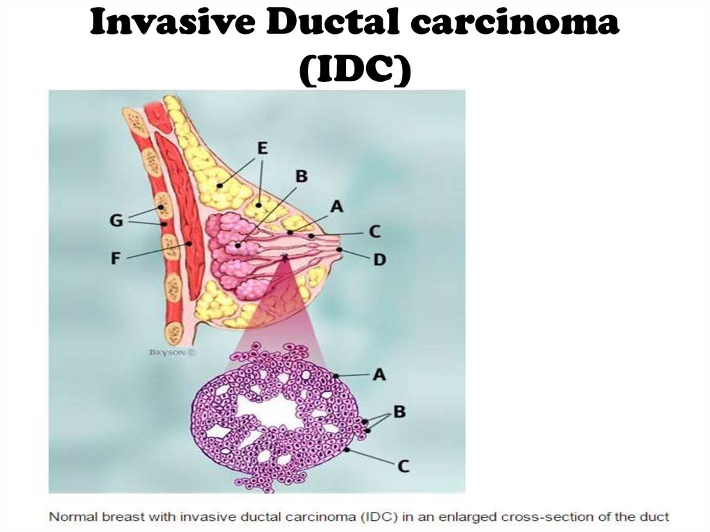
Invasive Ductal carcinoma(IDC)
59.
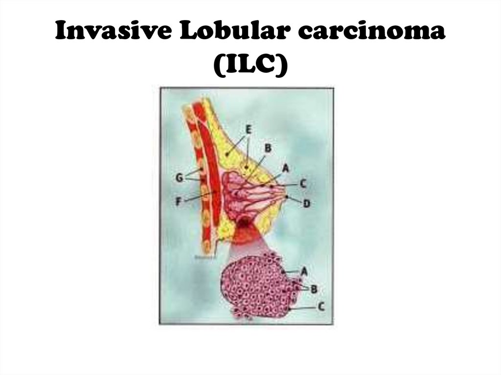
Invasive Lobular carcinoma(ILC)
60.
Other rarer variants• Colloid (mucinous) carcinoma: produce
abundant mucin.
• Medullary carcinoma: solid sheets of
large cells often associated with a
marked lymphocytic reaction.
• Tubular carcinoma.
• Papillary carcinoma.
61.
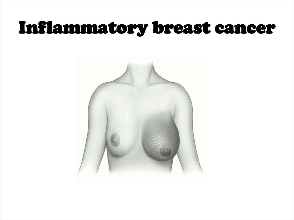
Inflammatory breast cancer62.
• Rare, highly aggressive cancer thatpresents as a painful, swollen breast,
which is warm with cutaneous oedema.
• Biopsy…
• Aggressive chemotherapy, radiotherapy
and salvage surgery.
63.
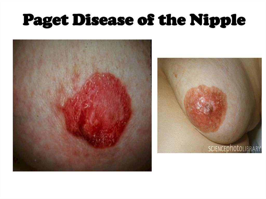
Paget Disease of the Nipple64.
• It is a superficial manifestation of anunderlying breast carcinoma (IDC or
DCIS).
• Presents as an eczema-like condition of
the nipple and areola, which persists
despite local treatment.
65.
The spread of breast cancer• Local spread…
• Lymphatic metastasis…
• Hematogenous spread…
66.
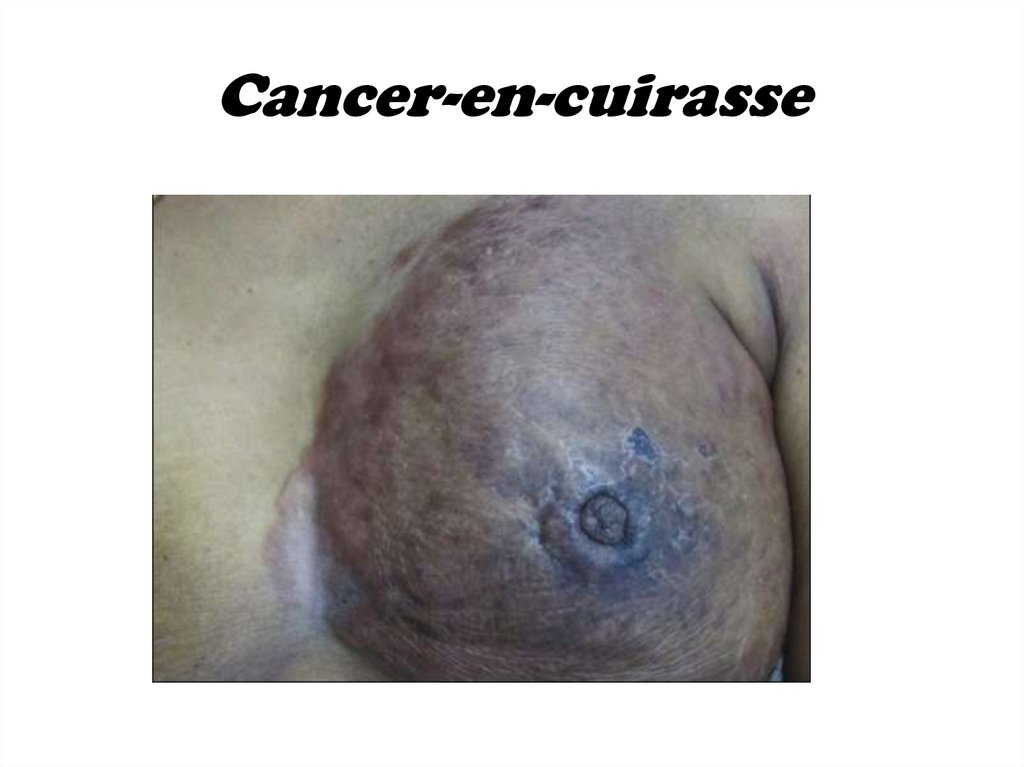
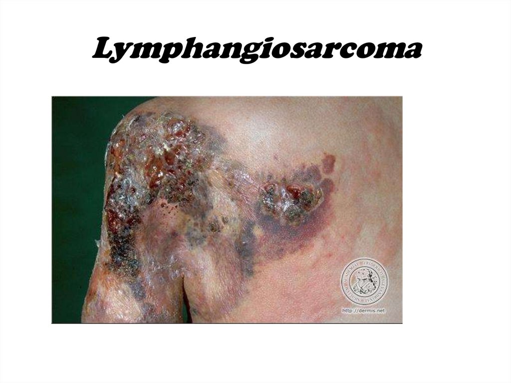
Phenomena resulting fromlymphatic obstruction in
advanced breast cancer:
67.
Peau d’orange68.
Cancer-en-cuirasse69.
Lymphangiosarcoma70.
71.
Breast Carcinoma GradingThe degree of differentiation:
• Well differentiated.
• Moderately differentiated.
• Poorly differentiated.
72.
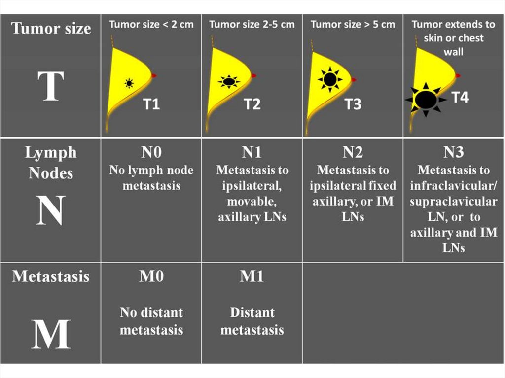
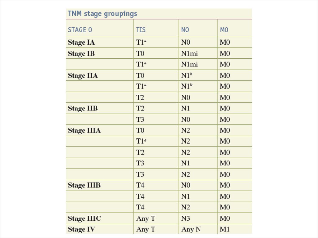
Breast cancer stagingTNM staging takes into account:
1. The size of the tumour (T).
2. Whether the cancer has spread to the
lymph glands (lymph nodes) (N).
3. Whether the tumour has spread
anywhere else in the body (M – for
metastases).
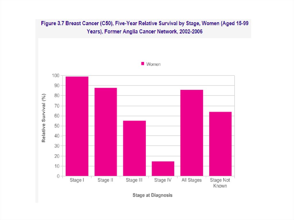
73.
74.
75.
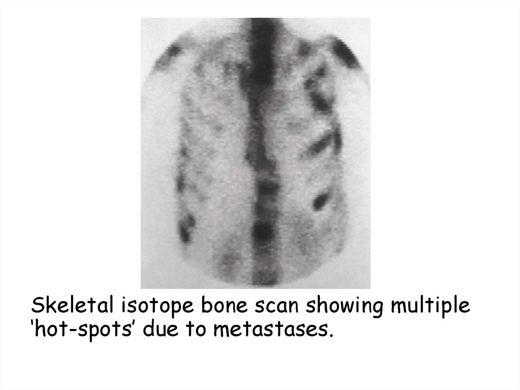
Skeletal isotope bone scan showing multiple‘hot-spots’ due to metastases.
76.
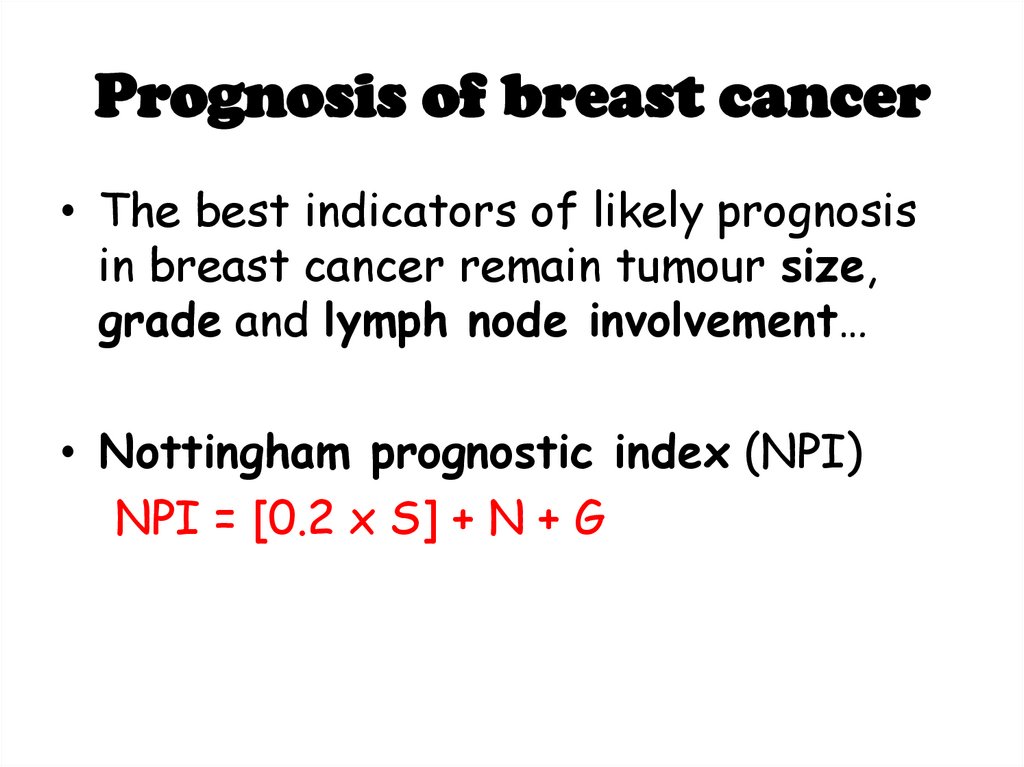
Prognosis of breast cancer• The best indicators of likely prognosis
in breast cancer remain tumour size,
grade and lymph node involvement…
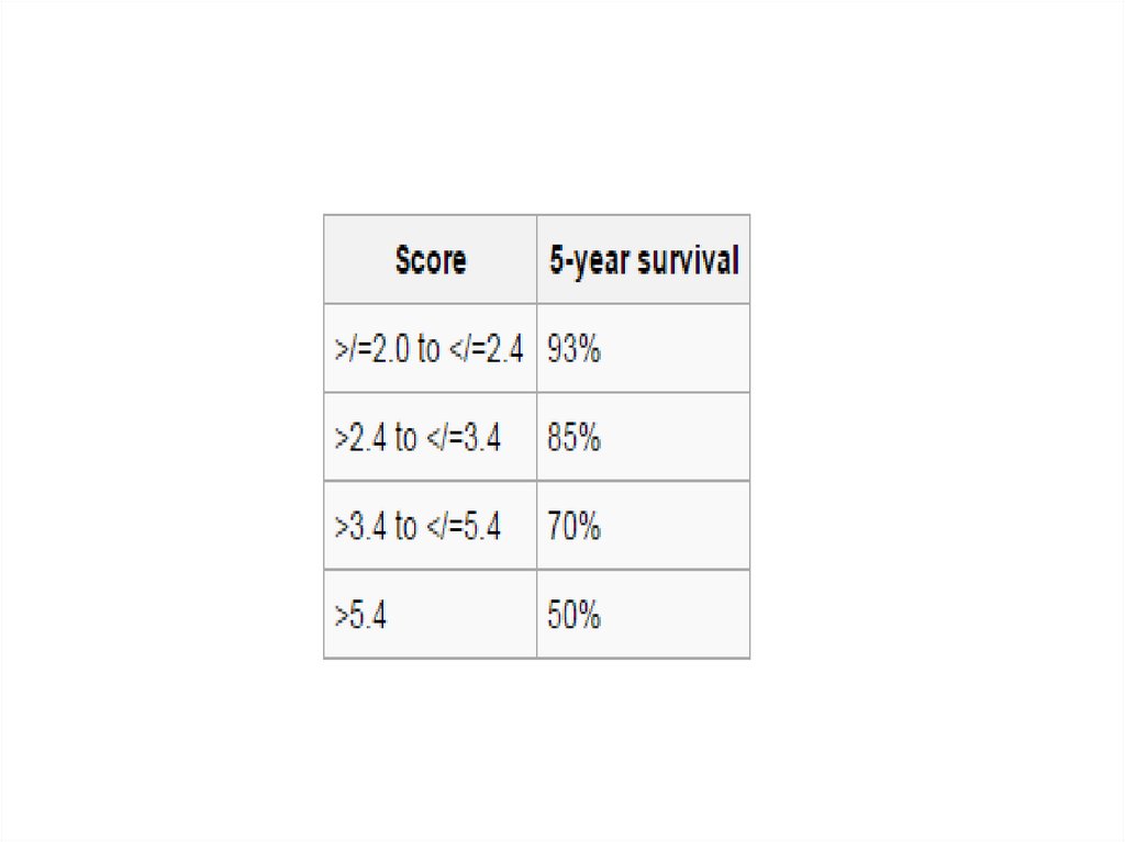
• Nottingham prognostic index (NPI)
NPI = [0.2 x S] + N + G
77.
78.
79.
Breast Cancer in MenBreast Cancer in Men accounts for less than 1% of male
cancers and less than 1% of all breast cancers. BRCA2
mutations are associated with approximately 5% of these
cancers.
Patients generally present with a nontender hard mass.
Mammography distinguishes gynecomastia from
malignancy. Malignant lesions are more likely to be
eccentric, with irregular margins, and are often
associated with nipple retraction and microcalcifications.
Biopsy of suspicious lesions is essential.
85% of malignancies are infiltrating ductal carcinoma and
are +ve for ER.
Adjuvant hormonal, chemotherapy, and radiation
treatment criteria are the same as in women.
80.
Screening & Imaging• Breast screening aims to find breast
cancers early. It uses an X-ray test called
a mammogram that can spot cancers when
they are too small to see or feel.
• Most common screening tests are:
1. Mammogram.
2. Clinical Breast Exam.
3. Self Breast Exam.
81.
82.
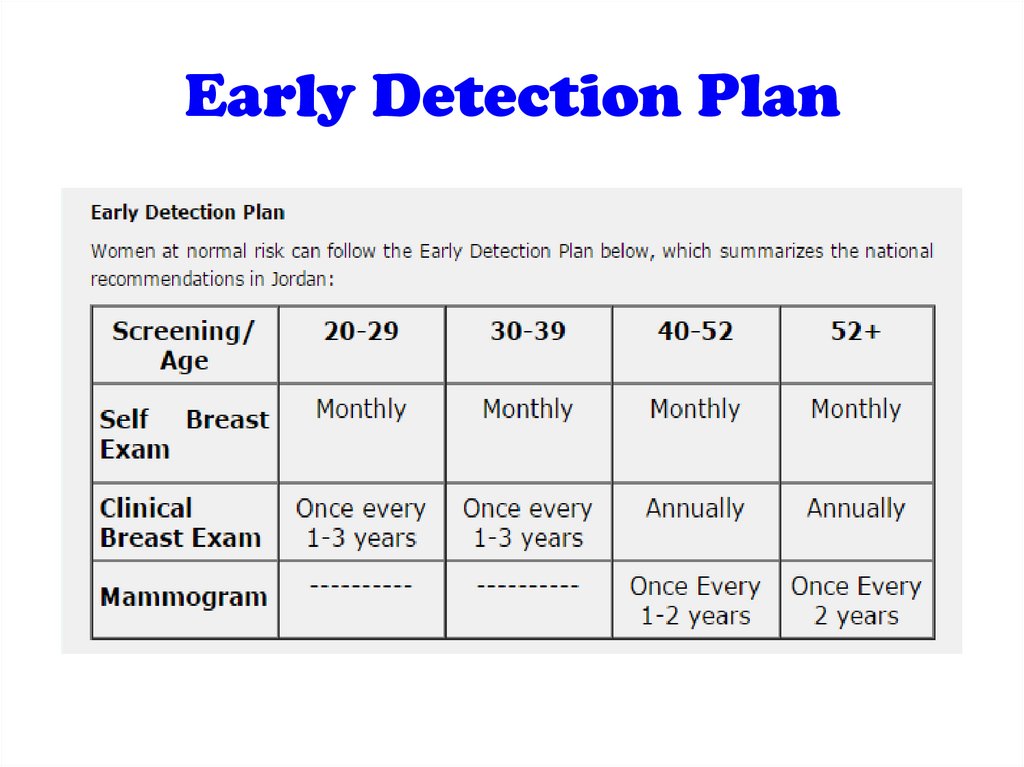
Early Detection Plan83.
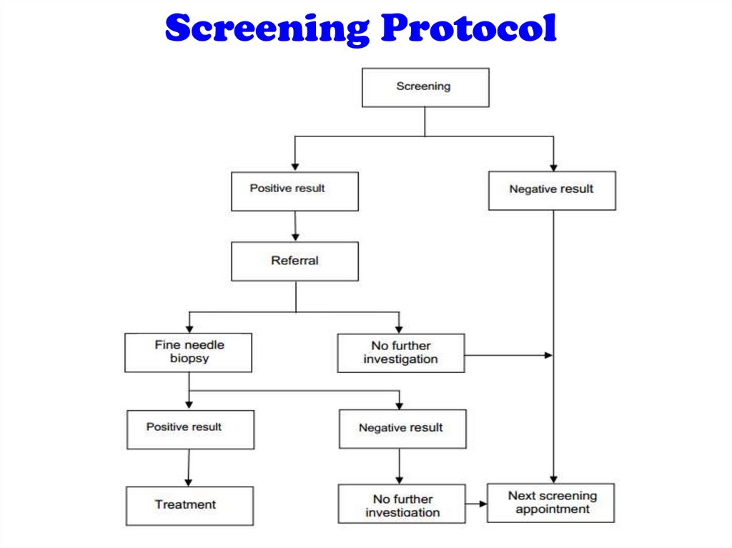
Screening Protocol84.
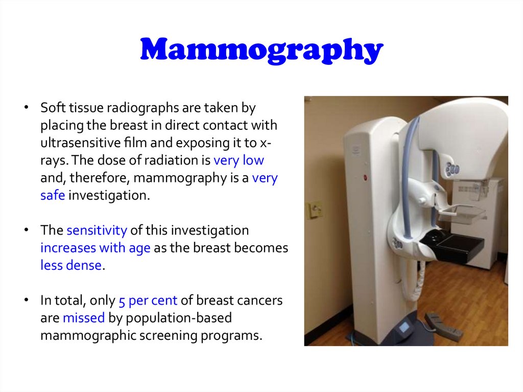
Mammography• Soft tissue radiographs are taken by
placing the breast in direct contact with
ultrasensitive lm and exposing it to xrays. The dose of radiation is very low
and, therefore, mammography is a very
safe investigation.
• The sensitivity of this investigation
increases with age as the breast becomes
less dense.
• In total, only 5 per cent of breast cancers
are missed by population-based
mammographic screening programs.
85.
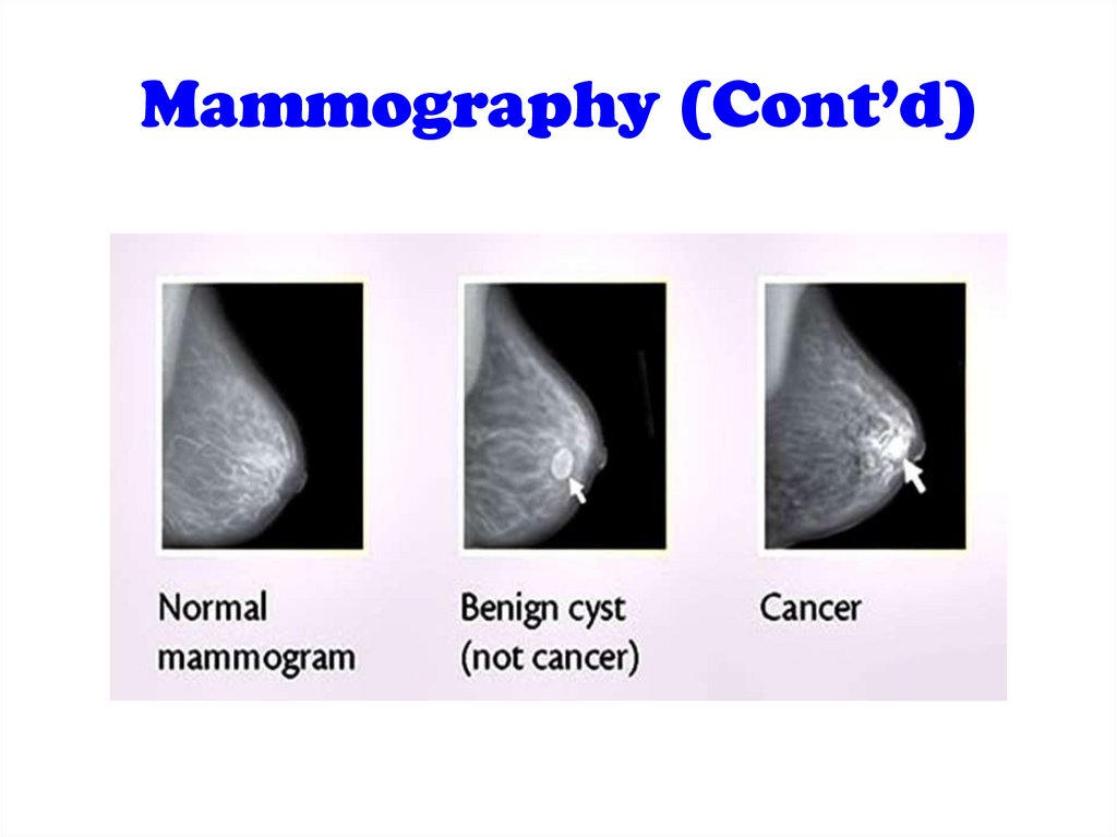
Mammography (Cont’d)86.
Ultrasonography• Ultrasound is particularly useful in young
women with dense breasts in whom
mammograms are dif cult to interpret,
and in distinguishing cysts from solid
lesions.
• It can also be used to localize impalpable
areas of breast pathology.
• It is not useful as a screening tool and
remains operator dependent.
87.
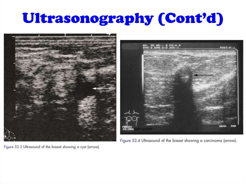
Ultrasonography (Cont’d)88.
Magnetic Resonance Imaging• Magnetic resonance imaging (MRI) is of
increasing interest to breast surgeons in a
number of settings:
1. It can be useful to distinguish scar from recurrence in
women who have had previous breast conservation
therapy for cancer.
2. It is becoming the standard of care when a lobular cancer
is diagnosed to assess for multifocality and
multicentricity.
3. It has proven to be useful as a screening tool in high-risk
women (because of family history).
89.
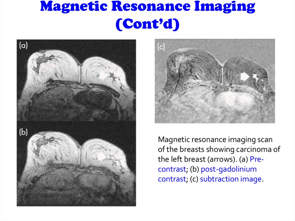
Magnetic Resonance Imaging(Cont’d)
Magnetic resonance imaging scan
of the breasts showing carcinoma of
the left breast (arrows). (a) Precontrast; (b) post-gadolinium
contrast; (c) subtraction image.
90.
History:91.
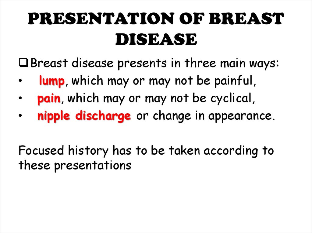
PRESENTATION OF BREASTDISEASE
Breast disease presents in three main ways:
• lump, which may or may not be painful,
• pain, which may or may not be cyclical,
• nipple discharge or change in appearance.
Focused history has to be taken according to
these presentations
92.
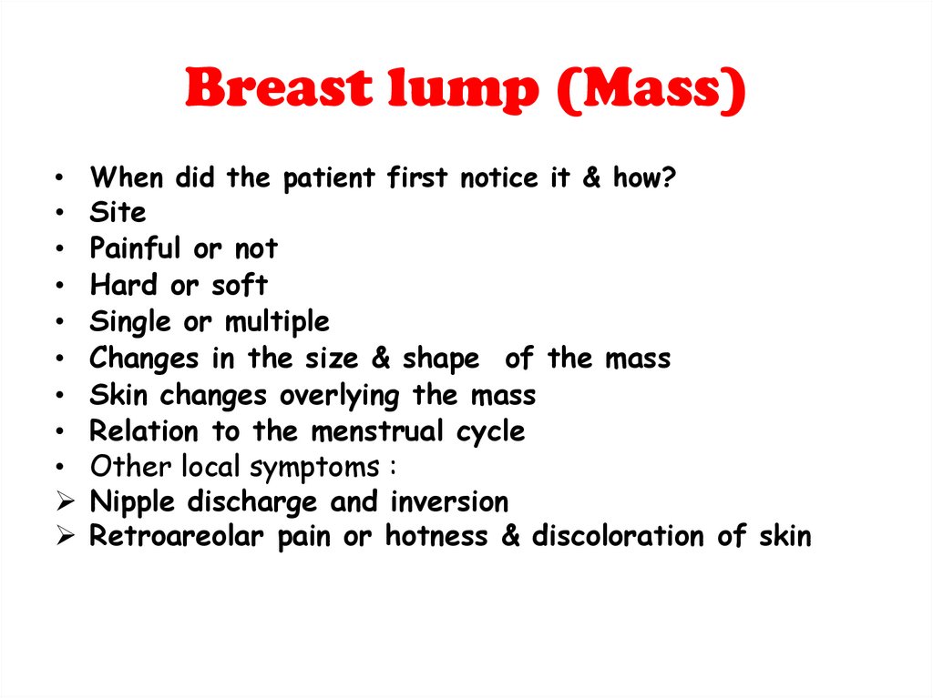
Breast lump (Mass)When did the patient first notice it & how?
Site
Painful or not
Hard or soft
Single or multiple
Changes in the size & shape of the mass
Skin changes overlying the mass
Relation to the menstrual cycle
Other local symptoms :
Nipple discharge and inversion
Retroareolar pain or hotness & discoloration of skin
93.
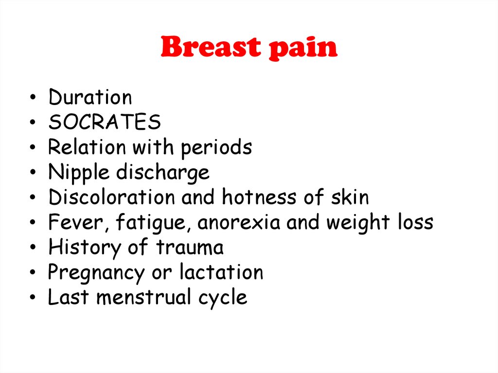
Breast painDuration
SOCRATES
Relation with periods
Nipple discharge
Discoloration and hotness of skin
Fever, fatigue, anorexia and weight loss
History of trauma
Pregnancy or lactation
Last menstrual cycle
94.
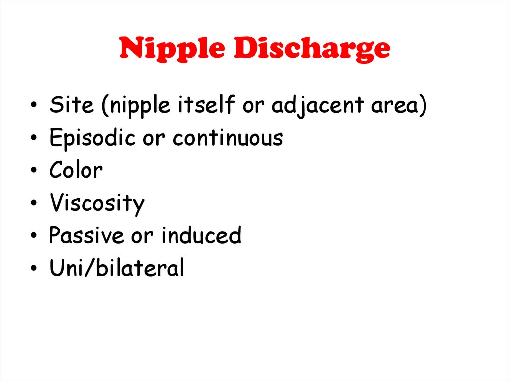
Nipple DischargeSite (nipple itself or adjacent area)
Episodic or continuous
Color
Viscosity
Passive or induced
Uni/bilateral
95.
Skin changesSkin dimple
Eczema
Indrawing of the skin
Ulceration
Discoloration
Redness and hotness
Overall swelling of the breast
96.
Nipple changes• Is it retracted or destroyed
• Uni/bilateral
• Can it be everted easily
97.
• Gynecological symptoms :Last menstrual cycle
duration
menarche
menopause
Any changes: Increased blood, clots or
irregularity
previous pregnancies and lactation:
o How many children has the patient had?
o Age of the pt when she had her 1st child
o Were the children breast-fed, and if so, for
how long?
98.
• Past History (e.g breast cyst)• Drug History (e.g oral contraceptives,
hormone replacement therapy)
• Family History ( breast or ovarian Ca)
• Previous Irradiation ( Hodgkins
lymphoma )
99.
PhysicalExamination:
100.
PositionInspection
Palpation
101.
position• The patient must be fully undressed to
the waist.
• sitting 45 degrees
• Patients sometimes say that their lump
can only be felt when they adopt a
certain posture and they should
therefore be examined in this position
as well.
102.
Inspection• Stand or sit directly in front of the patient,
inspect both breasts and look for the
following features
A) With the patient’s hands resting on thighs
:
1. Size
2. Symmetry
3. Skin :
-ulceration -puckering
-nodules -peau d’orange -discoloration
103.
4. Nipples & Areolae:Depression
Destruction
Discoloration
Displacement
Deviation
Discharge
104.
• To check for accessory nipple: check thenipple line ( axilla-->groin), if the nipple is
inverted ask the patient to evert it.
• Normal direction downward and outward
(if not deviated).
• To check if there is discharge or not:
ask her if there was discharge on her
underwear.
ask her to squeeze the nipple.
105.
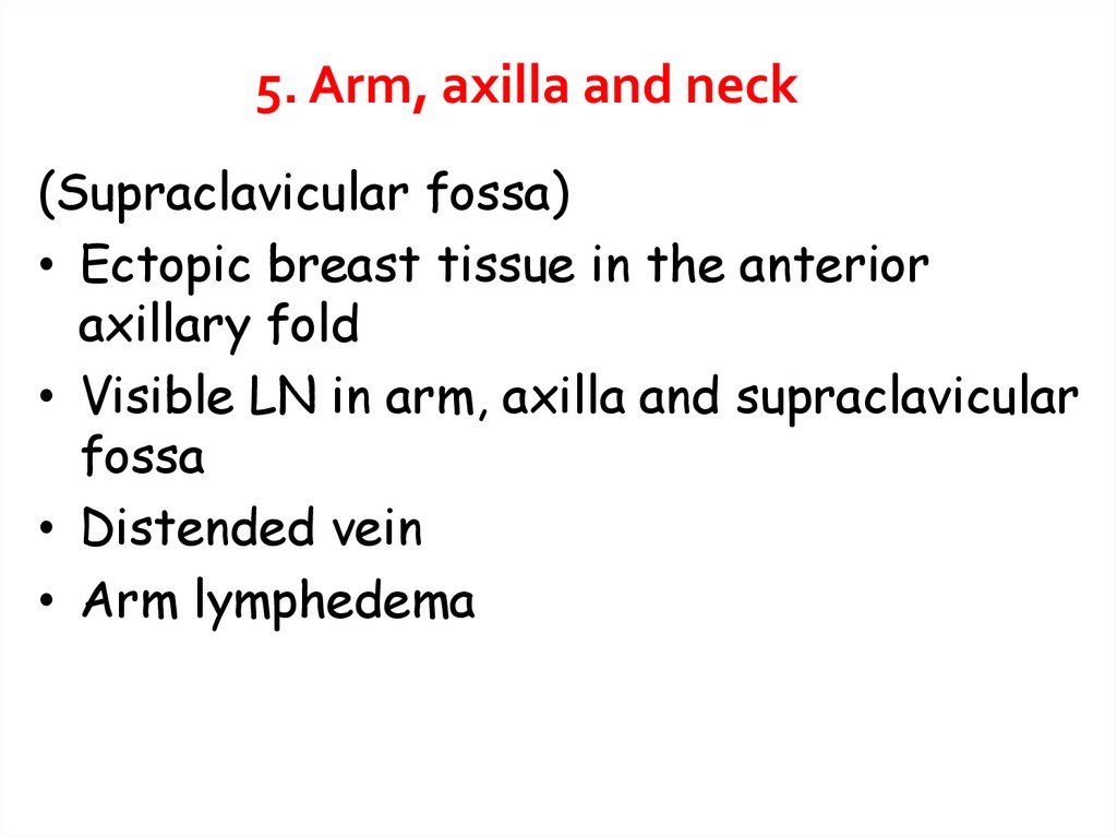
5. Arm, axilla and neck(Supraclavicular fossa)
• Ectopic breast tissue in the anterior
axillary fold
• Visible LN in arm, axilla and supraclavicular
fossa
• Distended vein
• Arm lymphedema
106.
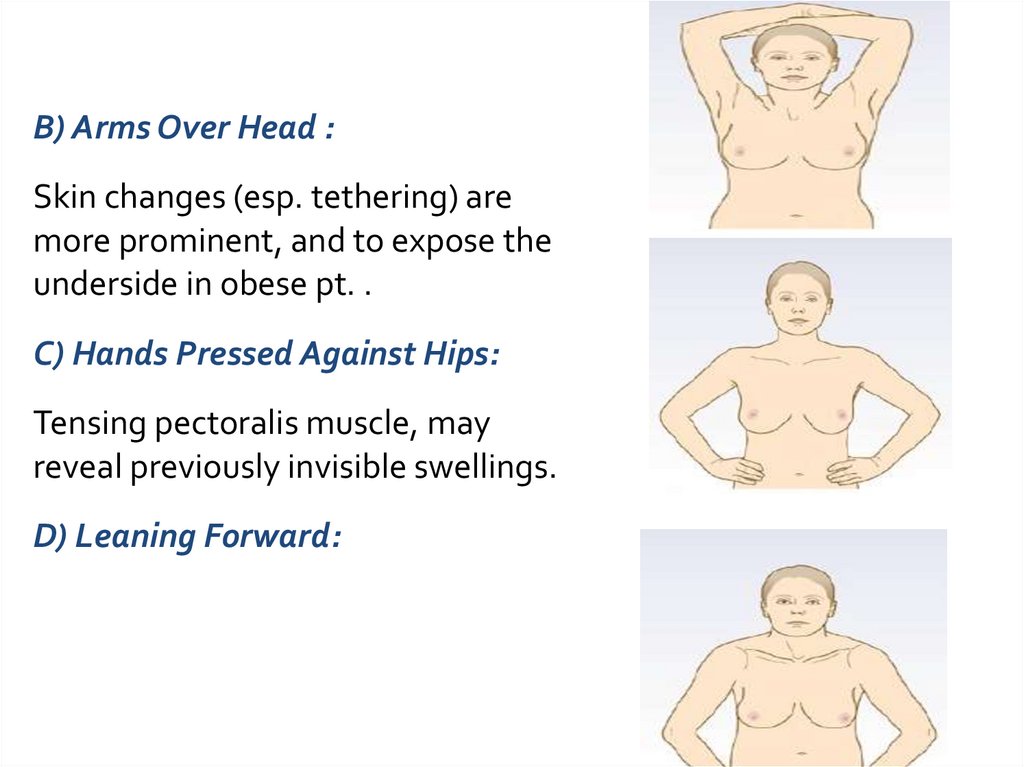
B) Arms Over Head :Skin changes (esp. tethering) are
more prominent, and to expose the
underside in obese pt. .
C) Hands Pressed Against Hips:
Tensing pectoralis muscle, may
reveal previously invisible swellings.
D) Leaning Forward:
107.
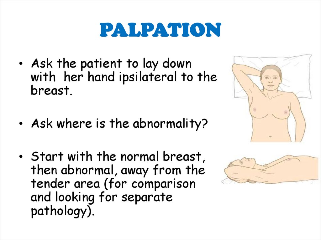
PALPATION• Ask the patient to lay down
with her hand ipsilateral to the
breast.
• Ask where is the abnormality?
• Start with the normal breast,
then abnormal, away from the
tender area (for comparison
and looking for separate
pathology).
108.
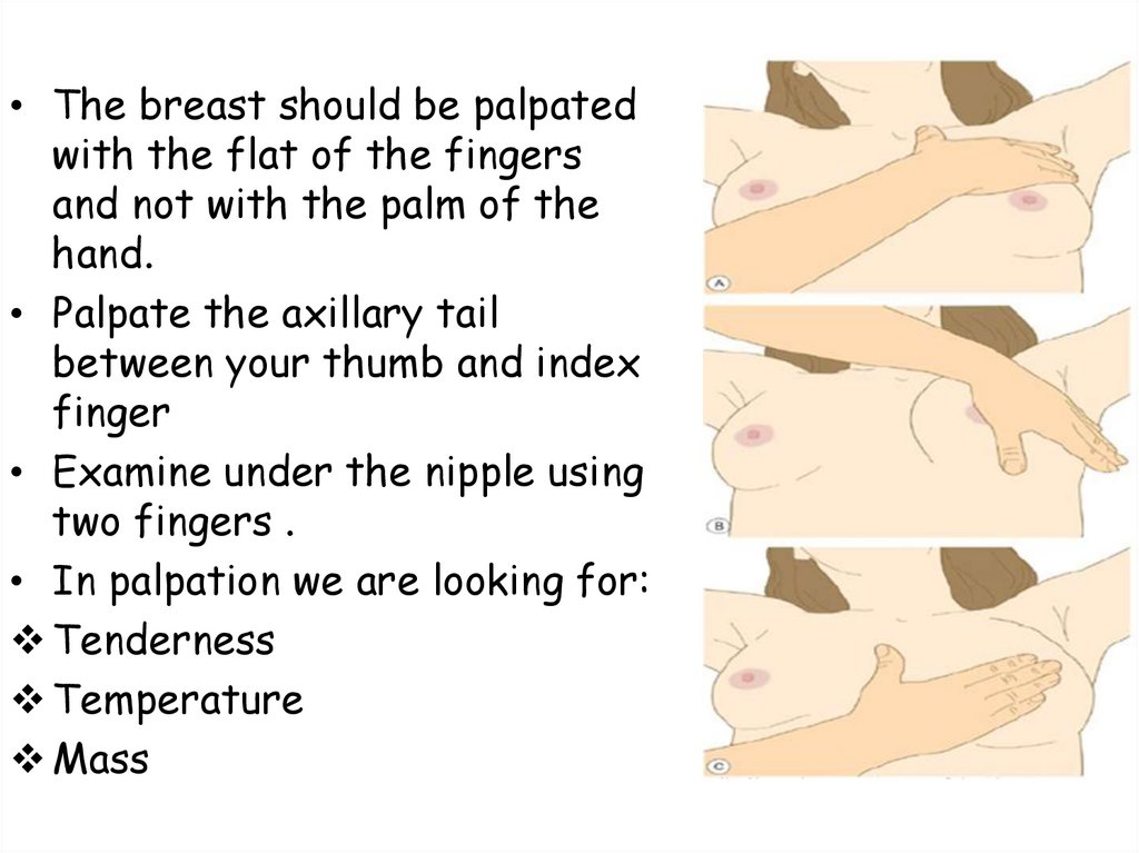
• The breast should be palpatedwith the flat of the fingers
and not with the palm of the
hand.
• Palpate the axillary tail
between your thumb and index
finger
• Examine under the nipple using
two fingers .
• In palpation we are looking for:
Tenderness
Temperature
Mass
109.
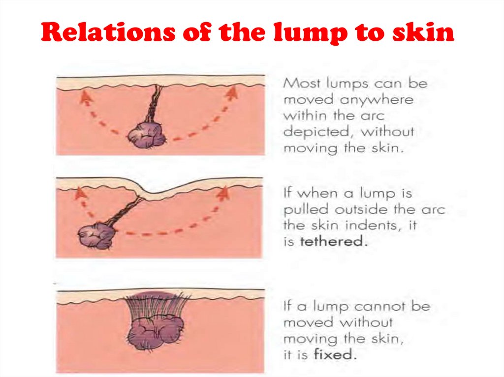
* If there is a lump we should analyze it :Lump
Site
Size
Shape Edge Surface Consistency
Fixed or
Tethered