


 and T2 -weighted (B) i mages in a patient with bilateral single lumen silicon implants. Note extracapsular rupture of the right breast implant, with a collection of silicon lying in the lateral aspect of the breast. There is intraca")


.")


 The position of a spiculate mass in the upper part of the left breast is marked with a localising wire. (B) Peroperative specimen radiography confirms that the mass has been excised.")


 lesions")


. On the lateral view the calcifications show a straight upper border, the 'tea cup' sign (B).")


 Irregular linear branching microcalcification.")


 medicine
Similar presentations:


Radiology and imaging of the mammаry gland
1. Radiology and imaging of the mammаry gland.
2. Normal anatomy
Normal StructuresNormal breast is composed:
mainly of parenchyma (lobules and ducts)
connective tissue
fat
Lobules are drained by ducts. There are
about 15 to 20 lobes in the breast. The lobar
ducts converge upon the nipple.
3. Parenchyma
The lobules are glandular units and are seenas ill-defined, splotchy opacities of medium
density. Their size varies from 1 to several
millimeters, and larger opacities result from
conglomerates of lobules with little
interspersed fat.
4.
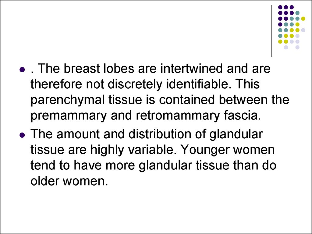
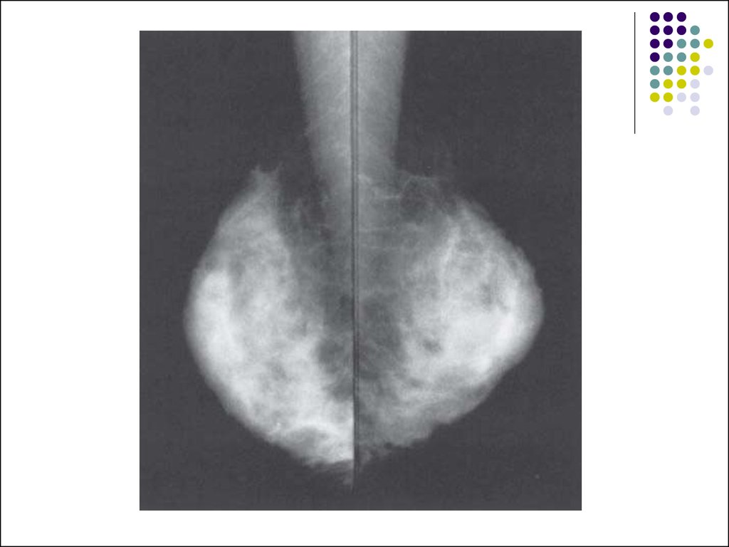
. The breast lobes are intertwined and aretherefore not discretely identifiable. This
parenchymal tissue is contained between the
premammary and retromammary fascia.
The amount and distribution of glandular
tissue are highly variable. Younger women
tend to have more glandular tissue than do
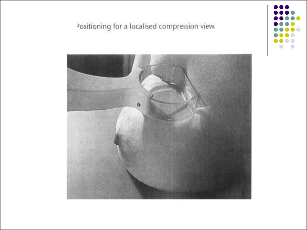
older women.
5.
6.
7. Connective tissue
Trabecular structures, which arecondensations of connective tissue, appear
as thin (1 mm) linear opacities of medium to
high density. Cooper’s ligaments are the
supporting trabeculae over the breast that
give the organ its characteristic shape, and
are thus seen as curved lines around fat
lobules along the skin-parenchyma interface
within any one breast.
8. Fat
The breast is composed of a large amount offat, which is lucent, or almost black, on
mammograms. Fat is distributed in the
subcutaneous layer, in among the
parenchymal elements centrally, and in the
retromammary layer anterior to the pectoral
muscle.
9. Lymph Nodes
Lymph nodes are seen in the axillae andoccasionally in the breast itself.
10. Veins
Veins are seen traversing the breast asuniform, linear opacities, about 1 to 5 mm in
diameter
11. Arteries
Arteries appear as slightly thinner, uniform,linear densities and are best seen when
calcified, as in patients with atherosclerosis,
diabetes, or renal disease.
12. Skin
Skin lines are normally thin and are not easilyseen without the aid of a bright light for filmscreen mammograms. Various processing
algorithms with digital mammography allow
better visualization of the skin.
13. Normal variants
The normal anatomical variants of the brest resultfrom the embryological development of the brest
from the band of ectoderm on the ventral surface of
the embryo extending from clavicle to groin, the
`nipple line'. An area of accessory breast tissue is
commonly seen in the axillary tail, or occasionally in
the inframammary fold. An accessory nipple may
occur at any site along the nipple line. Congenital
absence or hyperplasia of the pectoralis muscle may
occur and is seen in Poland's syndrome.
14.
15. The dense breast
Diffuse increase in the density of the breast tissue iscaused
by oedema (see the `Oedematous breast' below)
by an increase in the glandular tissue
or fibrous tissue
This is commonly seen in benign breast change,
and may be accompanied by evidence of cysts, and
in women who are taking hormone replacement
therapy (HRT) for menopausal symptoms.
16.
The increased density of the parenchymaseen as a result of HRT has been shown to
be associated with a decrease in the
sensitivity of screening mammography for
cancer detection. Diffuse increase in
parenchymal density is also occasionally
seen due to loss of fat due to severe weight
loss or cachexia, or lack of fat due to
lipodystrophy.
17. Mammography
The film-screen mammogram is created withx-rays, radiographic film, and intensifying
screens adjacent to the film within the
cassette; hence the term film-screen
mammography.
The digital mammogram is created using a
similar system, but replacing the film and
screen with a digital detector.
18.
The routine examination consists of twoviews of each breast:
the craniocaudal (C-C) view
the mediolateraloblique (MLO) view, with a
total of four films.
19.
The C-C view can be considered the “topdown” view, and the MLO an angled viewfrom the side.
The patient undresses from the waist up and
stands for the examination, leaning slightly
against the mammography unit.
The technologist must mobilize, elevate, and
pull the breast to place as much breast tissue
as possible on the surface of the film cassette
holder.
20.
A flat, plastic compression paddle is thengently but firmly lowered onto the breast
surface to compress the breast into as thin a
layer as possible.
This compression achieves both
immobilizations during exposure and
dispersion of breast tissue shadows over a
larger area, thereby permitting better visual
separation of imaged structures
21.
22.
23.
Compression may be uncomfortable, and may evenbe painful in a small proportion of patients.
However, most patients accept this level of
discomfort for the few seconds required for each
exposure, particularly if they understand the need
for compression and know what to expect during the
examination.
Mammography has proved to be more costeffective, while maintaining resolution high enough
to demonstrate early malignant lesions, than any
other breast imaging technique.
24. Compression
Firm compression is essential for high-qualitymammograms and is applied using a
powered system operated by a foot control. It
is important that there is even compression of
the entire breast.
25.
The effects of compression are:(i) reduced dose;
(ii) reduced scatter-improved contrast;
(iii) reduced geometric unsharpness;
(iv) reduced movement unsharpness;
(v) reduced range of breast thickness;
(vi) reduced tissue overlap improved
resolution.
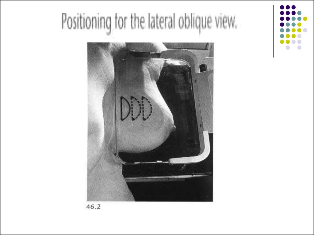
26. Mammography projections and normal appearances
The standard examination for womenundergoing either symptomatic
mammography or their first screening
examination consists of a lateral oblique and
a cranio-caudal view of each breast.
The lateral oblique view is usually obtained
with the tube angled at 45° to the horizontal,
but tube angulation from 30° to 60° may be
needed depending on the build of the
woman.
27.
More breast tissue is demonstrated on thelateral oblique projection than on any other
projection.
Careful positioning is essential for
satisfactory demonstration of the breast.
28.
The standard craniocaudal film is obtainedwith a vertical X-ray beam and the nipple
should be in profile. The craniocaudal
projection demonstrates the subareolar,
medial, and lateral portions of the breast.
However, tissue in the posterolateral aspect
of the breast may be incompletely
demonstrated.
29. Supplementary views
For demonstration of tissue in the mostposterolateral part of the breast, an extended
craniocaudal view is used with the patient
rotated medially to bring the lateral aspect of
the breast and axillary tail over the film. When
the posteromedial portion of the breast is not
satisfactorily demonstrated, an extended
craniocaudal view with lateral rotation of the
patient is obtained.
30. Magnification views
Magnification views are obtained byincreasing the object-film distance, producing
an `air gap', and using a fine focal spot to
increase resolution. A magnification factor of
1.5 is usual and the increased resolution
obtained is particularly helpful for detailed
analysis of microcalcifications and the
margins of small mass lesions.
31. Localized compression views
Localized compression views are obtained byusing a small paddle compression device and may
be used together with magnification. By
compressing one area of the breast, tissue overlying
a small lesion is displaced, allowing better
demonstration of its features. The technique is also
very helpful in analysing asymmetrical soft-tissue
shadows, either by confirming that the shadow has
the appearance of normal glandular tissue or by
demonstrating that an underlying lesion is present.
32.
33. Screening Mammography
The standard mammogram (along with appropriatehistory taking) makes up the entire screening
mammogram. The indication for this examination is
the search for occult carcinoma in an asymptomatic
patient. Physical examination by the patient’s
physician, known as the clinical breast examination
(CBE), is an indispensable element in complete
breast screening.
Such patients should be referred for diagnostic
mammography.
34. Diagnostic Mammography
The diagnostic mammogram begins with thetwo-view standard mammogram. Additional
maneuvers are then used as appropriate in
each case, dictated by history, physical
examination, and findings on initial
mammography.
35. Indications for diagnostic mammography are:
(1) a palpable mass or other symptom orsign (e.g., skin dimpling, nipple retraction,
or nipple discharge that is clear or bloody)
(2) a radiographic abnormality on a
screening mammogram.
Additionally, patients with a personal history
of breast cancer may be considered in the
diagnostic category.
36. Indications of mammography
• Screening asymptomatic women aged 50 yearsand over
• Screening asymptomatic women aged 35 years
and over who have a high risk of developing breast
cancer:
-women who have one or more first degree relatives
who have been diagnosed with premenopausal
breast cancer
-women with histologic risk factors found at previous
surgery, e.g. atypical ductal hyperplasia
37.
• Investigation of symptomatic women aged35 years and over with a breast lump or other
clinical evidence of breast cancer
• Surveillance of the breast following local
excision of breast carcinoma
• Evaluation of a breast lump in women
following augmentation mammoplasty
• Investigation of a suspicious breast lump in
a man
38. Patient Preparation
For the mammogram, two-piece clothing ismost convenient as the patient will need to
undress from the waist up. Patients should
not apply antiperspirant to the breast or axilla
because it may cause artifacts.
39.
Mammography is generally limited toambulatory, cooperative patients because of
the difficulties in proper positioning and
because mammography units are not
portable. If a debilitated patient has a
palpable mass, then ultrasound would be a
reasonable first step, followed by bedside
needle aspiration or biopsy if the mass is
solid. Screening mammography in markedly
debilitated patients rarely has clinical utility.
40. Computer-Aided Detection
Computer-aided detection (CAD) utilizescomplex algorithms to analyze the data from
a mammogram for suspicious:
calcifications
masses
architecture distortion
41. _ Ultrasonography
TechniqueHigh-quality images of the normal and abnormal
breast can be obtained with modern ultrasound
equipment. At the minimum, a 7.5 MHz linear array
probe should be used, though digital broadbandwidth transducers using higher frequency (midrange exceeding 7.5 MHz) are now widely available
and allow higher resolution imaging. The patient is
examined in the supine oblique position.
42.
The side being examined is raised and thearm placed above the head to ensure that the
breast tissue is evenly distributed over the
chest wall. In addition to conventional
orthogonal scanning directions, canning in
the radial and antiradial planes are of value in
demonstrating ductal abnormalities.
43. The indications for ultrasonography are:
(1) a mammographically detected mass, thenature of which is indeterminate
(2) a palpable mass that is not seen on
mammography
(3) a palpable mass in a patient below the
age recommended for routine mammography
(4) guidance for intervention.
44.
Ultrasonography is a highly reliable techniquefor differentiating cystic from solid masses.
Although certain features have been
described as indicative of benign or
malignant solid masses, this determination is
more difficult to make and less accurate than
the determination of the cystic nature of a
mass.
45. A limitation
A limitation of ultrasonography is that it isvery operatordependent.
Also, it images only a small part of the breast
at any one moment. Therefore, an overall
inclusive survey is not possible in one image,
and lesions may easily be missed.
46. Normal breast ultrasound: 1 = skin; 2 = subcutaneous fat; 3 = glandular tissue; 4 = retromammary fat; 5 = pectoralis muscle; 6 = rib.
47. Magnetic Resonance Imaging
The role of MRI in mammography continues toexpand, with common applications including:
(1) staging of and surgical planning for breast
tumors
(2) searching for a primary tumor in patients who
present with cancerous axillary lymph nodes
(3) evaluating tumor response to neoadjuvant
chemotherapy
(4) differentiating tumor recurrence from
posttreatment changes in patients with previous
breast-conserving surgery and radiation
48.
(5) screening of high-risk patients(6) evaluating implants
(7) evaluating difficult (dense or fibrous) breasts
In addition, the technology for MR-guided breast
biopsies is increasingly available.
MRI can show whether a lesion is solid or contains
fat or fluid. Dynamic scanning after administration of
intravenous contrast shows whether structures
enhance and at what rate.
49.
50. Axial T1 -weighted (A) and T2 -weighted (B) i mages in a patient with bilateral single lumen silicon implants. Note extracapsular rupture of the right breast implant, with a collection of silicon lying in the lateral aspect of the breast. There is intraca
Axial T1 -weighted (A) and T2 -weighted (B) i mages in a patient with bilateralsingle lumen silicon implants. Note extracapsular rupture of the
right breast implant, with a collection of silicon lying in the lateral aspect of the
breast. There is intracapsular rupture of the left breast implant, with a
classical linguine sign.
51. _ Ductography
Ductography, or galactography, usesmammographic imaging with contrast
injection into the breast ducts.
52. The indication
The indication for use is a profuse,spontaneous, nonmilky nipple discharge from
a single duct orifice.
If these conditions are not present, the
ductogram is likely to be of little help. The
purpose is to reveal the location of the ductal
system involved.
The cause of the discharge is frequently not
identified.
53.
The patient lies in supine position while thedischarging duct is cannulated with a blunttipped needle or catheter under visual
inspection and with the aid of a magnifying
glass. A small amount of contrast material
(usually not more than 1 mL) is injected
gently by hand into the duct. Several
mammographic images are then made. The
procedure requires about 30 minutes and is
not normally painful.
54. A ductogram showing small filling defects due to an intraductal carcinoma (arrows).
55. Image-Guided Needle Aspiration and Biopsy
The first indication is aspiration of cysticlesions to confirm diagnosis, to relieve pain,
or both. Nonpalpable cysts require either
ultrasound or mammography to be seen. A
fine needle (20- to 25-gauge) usually suffices
to extract the fluid. The cystic fluid is not
routinely sent for cytology unless it is bloody.
The second indication concerns solid
lesions.
56. Needle biopsy is used in this case
(1) to confirm benignity of a lesion carrying a lowsuspicion of malignancy mammographically
(2) to confirm malignancy in a highly suspicious
lesion prior to initiating further surgical planning and
treatment
(3) to evaluate any other relevant mammographic
lesion for which either follow-up imaging or surgical
excision is a less desirable option for further
evaluation
Guidance for needle biopsy can be accomplished with
stereotactic mammography, ultrasound, and MR.
57. 1 4G needle and automated biopsy device used for ultrasound and stereotactic core breast biopsy.
58. Stereotactic-guided fine needle aspiration. The check pair of films shows the tip of the needle positioned within the small cluster of microcalcification on both views.
59. Stereotactic core biopsy. Stereo film pair showing 'post fire' position of needle during biopsy of microcalcification
Stereotactic core biopsy. Stereo film pair showing 'post fire'position of needle during biopsy of microcalcification
60. Image-Guided Needle Localization
When a nonpalpable breast lesion must beexcised, imaging is used to guide placement
of a needle into the breast, with the needle tip
traversing or flanking the lesion. Either
ultrasonographic or mammographic guidance
can be used, and the choice again depends
on lesion characteristics and personal
preference.
61.
Once the needle is in the appropriateposition, a hook wire is inserted through the
needle to anchor the device in place. This
prevents migration during patient transport
and surgery. After needle placement, the
patient is taken to the operating theater for
excision of the lesion by the surgeon.
62. Wire localisation and surgical excision of a nonpalpable carcinoma. (A) The position of a spiculate mass in the upper part of the left breast is marked with a localising wire. (B) Peroperative specimen radiography confirms that the mass has been excised.
63. Patient Preparation
Patients for whom stereotactic biopsy isbeing considered should be able to lie in
prone position without moving for about 1
hour.
64. Approach to the Palpable Lump
When a breast lump is found, several questionsmust be answered before proceeding with breast
imaging.
First, given that lumpy breasts are a normal variant,
when is a lump significant?
Experts in CBE advise palpation with the flat surface
of two to three fingers, and not with the fingertips.
With this technique, nonsignificant lumps will
disperse into background breast density, but a
significant lump will stand out as a dominant mass.
65.
Second, is the lump new or enlarged? A newlump is more suspicious than a lump that has
not changed over a few years.
66.
Third, how big is the lump? Tiny pea-sized orsmaller lumps, particularly in young women,
are often observed closely with repeated
CBE, because small breast nodules are
extremely common, frequently resolve
spontaneously, and are usually benign.
Repeating CBE in 6 weeks allows for interval
menses, which frequently causes waning or
resolution of the lump. If the lump persists,
diagnostic mammography is indicated.
67.
Fourth, how old is the patient? If the patient is lessthan 35 years of age, then radiation is avoided
unless specifically indicated, because the younger
breast is more sensitive to radiation.
For patients over the age of 35 years, breast
imaging begins with a diagnostic mammogram at
the time a lump is deemed to be significant. The
mammogram provides a view of the lump, as well as
of the remainder of the involved breast and the
opposite breast, where associated findings may aid
in diagnosis and treatment planning.
68.
If the patient is below 35 years of age, a significantlump is usually first examined with ultrasonography
to determine whether a simple cyst is present. If
there is no cyst, and the patient is below 30 years of
age, the radiologist may choose to obtain a
mammogram, but the density of the breast in such a
young patient may limit the usefulness of
radiomammography, so the mammogram may be
limited to one breast or to a single view.
69.
For women between the ages of 30 and 40years, judgment is needed as to whether
other imaging is indicated. Several factors
should be weighed, including age, family
history of breast carcinoma, reproductive
history, and findings at CBE.
If the primary care physician is uncertain of
the significance of the findings of CBE,
evaluation by a breast specialist may be
helpful prior to requesting radiologic tests.
70. Bi - rads assessment categories
category 0 - need additional imaging evaluationcategory 1 - negative
category 2 - benign finding, noncancerous
category 3 - probably benign finding, short interval
follow-up suggested
category 4 - suspicious abnormality, biopsy
considered
category 5 - highly suggestive of malignancy,
appropriate action needed
71. Circumscribed mass
A circumscribed mass is analysed according to the following features:I. Density:
(i) radiolucent
(ii) mixed density
(iii) radiopaque (soft-tissue density)
2. Contour:
(i) sharply outlined capsule - `halo' sign
(ii) ill-defined outline
3. Interval change
4. Number:
(i) single
(ii) multiple.
72. Radiolucent lesions
LipomaOil cyst
Galactocele.
73. Mixed density lesions
adenolipoma hamartomagalactocele
hematoma
lymph node
74. Radiopaque (soft-tissue density) lesions
Benign lesions* Cyst
* Fibroadenoma
* Papilloma
*Phyllodes tumour
* Abscess
* Lymph node
• rheumatoid arthritis
• sarcoidosis
* Sebaceous cyst
75. Malignant lesions
* Mucinous carcinoma* Medullary carcinoma
* Papillary carcinoma
* Invasive ductal carcinoma
* Intracystic carcinoma
* Metastasis
• melanoma
• lung
• ovary
* Lymphoma
* Sarcoma
* Pathological lymph node
• breast cancer
• Phyllodes tumour
• lymphoma
• metastasis
Recurrent breast cancer
76. Calcifications
Arterial: curvilinear, parallel line calcificationsalong the course of a blood vessel.
Skin calcification: multiple small ringshaped calcifications.
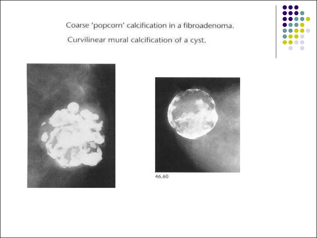
Fibroadenoma: coarse `popcorn' type
calcification associated with a soft-tissue
mass. Less commonly the calcifications may
he fine, irregular or curvilinear `eggshell' type
related to the periphery of the lesion.
77.
Cyst: curvilinear calcification may occur inthe wall of a cyst.
Carcinoma: the calcification particles of
ductal carcinoma in situ are typically variable
in density and shape: linear, casting,
branching, and irregular shapes may be
present, with variation of the density from
particle to particle.
78. Ductal carcinoma in situ. Irregular pleomorphic microcalcification
79. Milk of calcium in benign cystic change. On the craniocaudal view the calcifications appear as round 'smudge' shadows (A). On the lateral view the calcifications show a straight upper border, the 'tea cup' sign (B).
Milk of calcium in benign cystic change. On the craniocaudalview the calcifications appear as round 'smudge' shadows (A). On the
lateral view the calcifications show a straight upper border, the 'tea cup'
sign (B).
80. Skin calcification. Multiple small ring-shaped calcifications
81. Course calcification due to fat necrosis from previous surgery
82. Renal failure. Extensive stromal and vascular calcification.
83.
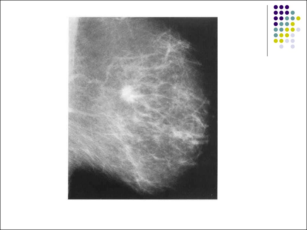
84. Spiculate mass
A spiculate mass is the commonestmammographic appearance of invasive
breast carcinoma.
1. It consists of a central soft-tissue tumor
mass from the surface of which spicules
extend into the surrounding breast tissue.
There is often associated distortion of the
surrounding breast tissue with straightening
of the trabeculae due to retraction.
85.
2. Large or superficially positioned tumors may beassociated with localized skin thickening and
retraction.
3. Deeply positioned tumors may be associated with
tethering of the pectoralis muscle.
4. Irregular microcalcifications due to associated
ductal carcinoma in situ may be found within the
tumour or in the surrounding breast tissue,
sometimes extending to the nipple.
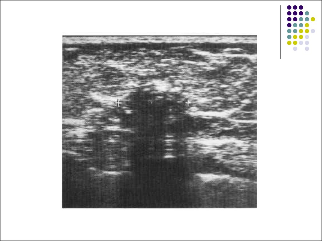
86. The typical ultrasound features are
Most spiculate carcinomas of I cm diameteror more can be demonstrated by ultrasound.
of an echo-poor mass, with poorly defined
margins and posterior acoustic shadowing
distortion of the surrounding breast tissue
may be visible and a rim of increased
reflectivity around the tumour mass may be
seen
87.
the presence of these signs, however, isvariable: acoustic shadowing may be absent;
an echo-poor mass may not be visible with
very small tumors.
similar suspicious ultrasound appearances
may be caused by a sclerosing fibroadenoma
or benign complex sclerosing lesion
88.
89.
90.
91.
92.
93. Non-invasive intracystic carcinoma.
94. Ductal carcinoma in situ-high-grade comedo type. (A-C) Irregular linear branching microcalcification.
95. Interval cancers are classified radiologically as follows:
I. True interval: there is no evidence of the canceron the screening films but the cancer is
demonstrated on clinical mammograms at
presentation.
2. Occult: there is no evidence of the cancer either
on the screening mammograms or on the clinical
mammograms.
3. False negative: there is evidence of the cancer
on the original screening films which corresponds
with the abnormal signs shown on clinical
mammograms at the time of diagnosis.
96.
4. Minimal sign: there are subtle features on thescreening mammograms which correspond to the
position of the carcinoma shown on the clinical films
but are only recognisable on retrospective review or
for which recall would not have been indicated.
5. Unclassified: mammography was not performed
at the time of diagnosis and therefore the presence
of mammographic signs of malignancy on the
previous screening films cannot he verified.
97. Fibroadenoma
Fibroadenoma are characteristicallysharply outlined
low soft tissue density lesions, sometimes with a
lobulated outline
they are usually solitary but may be multiple with
increasing age, they may undergo
fibroadenoma can, however, show very fine
calcifications with some pleomorphism which can
raise the suspicion of malignancy
fibroadenoma do not arise de novo in women aged
40 years or more but may grow in menopausal
women who are taking HRT
98. The typical ultrasound appearance of a fibroadenoma is
a well circumscribed round or oval massshowing posterior acoustic enhancement and
with a homogeneous internal echo pattern
the ultrasound findings alone therefore
cannot be used to confirm the diagnosis of a
circumscribed solid lesion found on
mammography
99.
100.
101. Cyst
Cyst are the most common cause of a discrete breastmass.
they are often multiple and bilateral
they are common between the ages of 20 and 50
years, with a peak incidence between 40 and 50
years
simple cysts are not associated with an increased
risk of malignancy and have no malignant potential
On mammography they are seen as well-defined,
round or oval masses. Sometimes a characteristic
halo is visible on mammography
102.
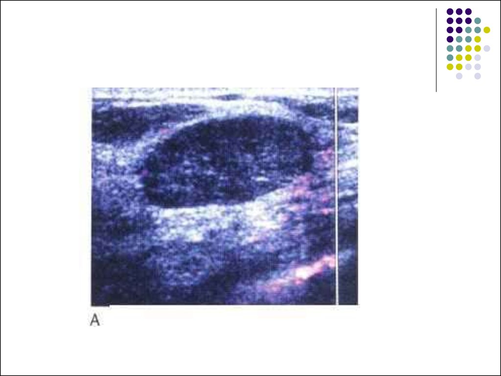
Cysts can be readily diagnosed with ultrasound.They have:
well-defined margins
are oval or round in shape
show an absence of internal echoes indicating the presence of
fluid
the area of breast tissue behind a cyst appears bright on
ultrasound (posterior enhancement) due to improved
transmission on the ultrasound beam through the cyst fluid When
these features are present, a cyst can be diagnosed with
certainty. Aspiration is easily performed under ultrasound
guidance to alleviate symptoms or when there is diagnostic
uncertainty. Cytology on cyst fluid is not routinely performed
unless there are atypical imaging features or the aspirate is
bloodstained
103.
When these features are present, a cyst canbe diagnosed with certainty. Aspiration is
easily performed under ultrasound guidance
to alleviate symptoms or when there is
diagnostic uncertainty.
Cytology on cyst fluid is not routinely
performed unless there are atypical imaging
features or the aspirate is bloodstained