


 medicine
Similar presentations:


Ultrasound Diagnosis
1.
ULTRASOUNDDIAGNOSIS
2.
ULTRASOUND METHOD OFDIAGNOSIS
The ultrasound diagnostic method (ultrasound,
sonography, ultrasonography) is a method of
obtaining a medical image based on the
registration and computer analysis of
ultrasonic waves reflected from biological
structures, i.e., based on the echo effect. The
method is often called echography.
3.
4.
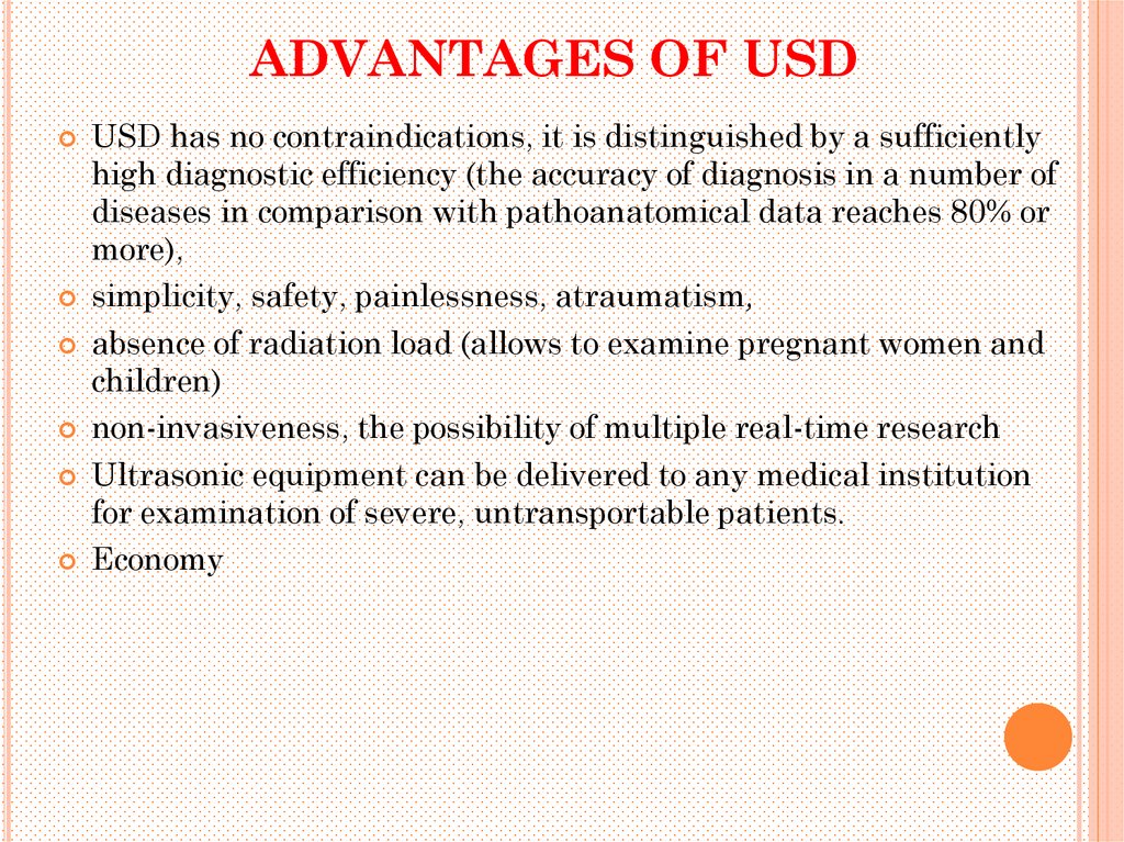
ADVANTAGES OF USDUSD has no contraindications, it is distinguished by a sufficiently
high diagnostic efficiency (the accuracy of diagnosis in a number of
diseases in comparison with pathoanatomical data reaches 80% or
more),
simplicity, safety, painlessness, atraumatism,
absence of radiation load (allows to examine pregnant women and
children)
non-invasiveness, the possibility of multiple real-time research
Ultrasonic equipment can be delivered to any medical institution
for examination of severe, untransportable patients.
Economy
5.
DISADVANTGES OF USDHowever, the ultrasound method has some
disadvantages:
o limitations in the study of a number of organs
and systems (lungs, internal bone structure,
adult brain, intestine, filled with gas)
o subjectivity in the interpretation of the
obtained images, i.e., the dependence of the
accuracy of diagnosis on the qualifications of
the physician;
6.
USE OF ULTRASOUNDUltrasound is widely used to diagnose diseases of
various organs and systems, especially ultrasound has
a high diagnostic efficiency in the study
digestive (liver, gall bladder, bile ducts, pancreas),
cardiovascular,
genitourinary (kidney, uterus, ovary, prostate) systems,
in obstetrics (prenatal diagnosis),
in the course of examination of superficial organs
(mammary glands, thyroid gland, lymph nodes), etc.
7.
The tasks, which are solved by the doctor of ultrasoundduring the research:
o assessment of the position of the body, its relation to
other bodies and systems;
o determination of the size, shape, contours of the
organ under study, comparing them with normal
indices;
o evaluation of organ structure (echogenicity), search
for foci, zones of pathological echogenicity (structural
disorders);
o the study of the function of the organ or system;
o assessment of blood flow of the organ, region;
o comparison of the received ultrasound picture with
clinical, laboratory and other data for drawing up of
an ultrasound conclusion
8.
History of development1880 - Pierre and Jacques Curie - opening of the direct piezoelectric effect
1881 - Lipman - description of the inverse piezoelectric effect phenomenon
1916 - France, England - installation of echolocators on submarines
1929 - Russia, Sokolov S.Ya. - laid the foundation of ultrasonic defectoscopy
1942 - Creation of the first instruments for ultrasonic research in medicine
1950 - ultrasound in ophthalmology
1958 - Baum and Greenwood used ultrasound in gastroenterology
1972 - Kossov applied a gray scale and a real time scale with ultrasound
9.
PHYSICAL AND BIOPHYSICAL BASISOF US
Ultrasound refers to sound vibrations lying above the
threshold of perception by the human hearing organ,
i.e., having a frequency of more than 20 kHz.
It is known that sound is a mechanical longitudinal
wave in which the oscillations of particles are in the
same plane as the direction of propagation of energy.
The physical basis of ultrasound is the piezoelectric
effect discovered in 1881 by the Curie brothers.
10.
The essence of the piezoelectric effect is that whencertain single chemical compounds (quartz, barium
titanate, cadmium sulphate, etc.) are deformed, in
particular, under the influence of ultrasonic waves,
electric charges of opposite sign appear on the surfaces of
these crystals. This is the so-called direct piezoelectric
effect
Conversely, when an electric charge is applied to these
single crystals, mechanical oscillations arise with the
emission of ultrasonic waves.
Thus, the same piezoelectric element can alternately be a
receiver, then a source of ultrasonic waves.
This part in ultrasonic devices is called an acoustic
transducer, transducer or sensor.
11.
Ultrasonic waves have certain properties usedfor diagnostics:
are distributed rectilinearly, therefore, it is
possible to obtain images of the organs under
study with practically no distortion, while
maintaining their linear dimensions and shape;
are able to focus;
penetrate into the organs;
differently reflected from the boundaries of
different densities as external contours of
biological tissues, and their internal structure are able to carry certain information about the
internal structure and function of organs
12.
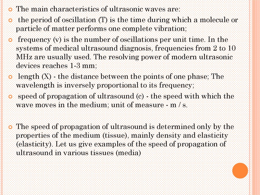
The main characteristics of ultrasonic waves are:the period of oscillation (T) is the time during which a molecule or
particle of matter performs one complete vibration;
frequency (v) is the number of oscillations per unit time. In the
systems of medical ultrasound diagnosis, frequencies from 2 to 10
MHz are usually used. The resolving power of modern ultrasonic
devices reaches 1-3 mm;
length (X) - the distance between the points of one phase; The
wavelength is inversely proportional to its frequency;
speed of propagation of ultrasound (c) - the speed with which the
wave moves in the medium; unit of measure - m / s.
The speed of propagation of ultrasound is determined only by the
properties of the medium (tissue), mainly density and elasticity
(elasticity). Let us give examples of the speed of propagation of
ultrasound in various tissues (media)
13.
PHYSICAL CHARACTERISTICS OFBIOLOGICAL ENVIRONMENT
Reflection
Refraction is a change in the direction of wave
propagation in the transition from one
environment in another
Dispersion - the occurrence of multiple changes in the
direction of propagation of ultrasound in inhomogeneities
in the biological environment
Absorption - the transfer of energy of ultrasonic waves
into other types of energy
The higher the frequency, the greater
the attenuation coefficient
fading
14.
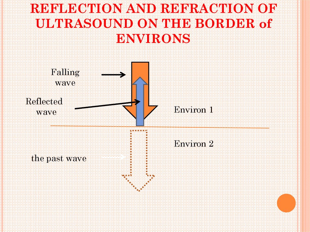
REFLECTION AND REFRACTION OFULTRASOUND ON THE BORDER of
ENVIRONS
Falling
wave
Reflected
wave
Environ 1
Environ 2
the past wave
15.
REFLECTION AND REFRACTION OFULTRASOUND ON THE BORDER of
ENVIRONS
Reflected
wave
Falling
wave
environ 1
α
αотр
environ 2
β
Refracted
wave
16.
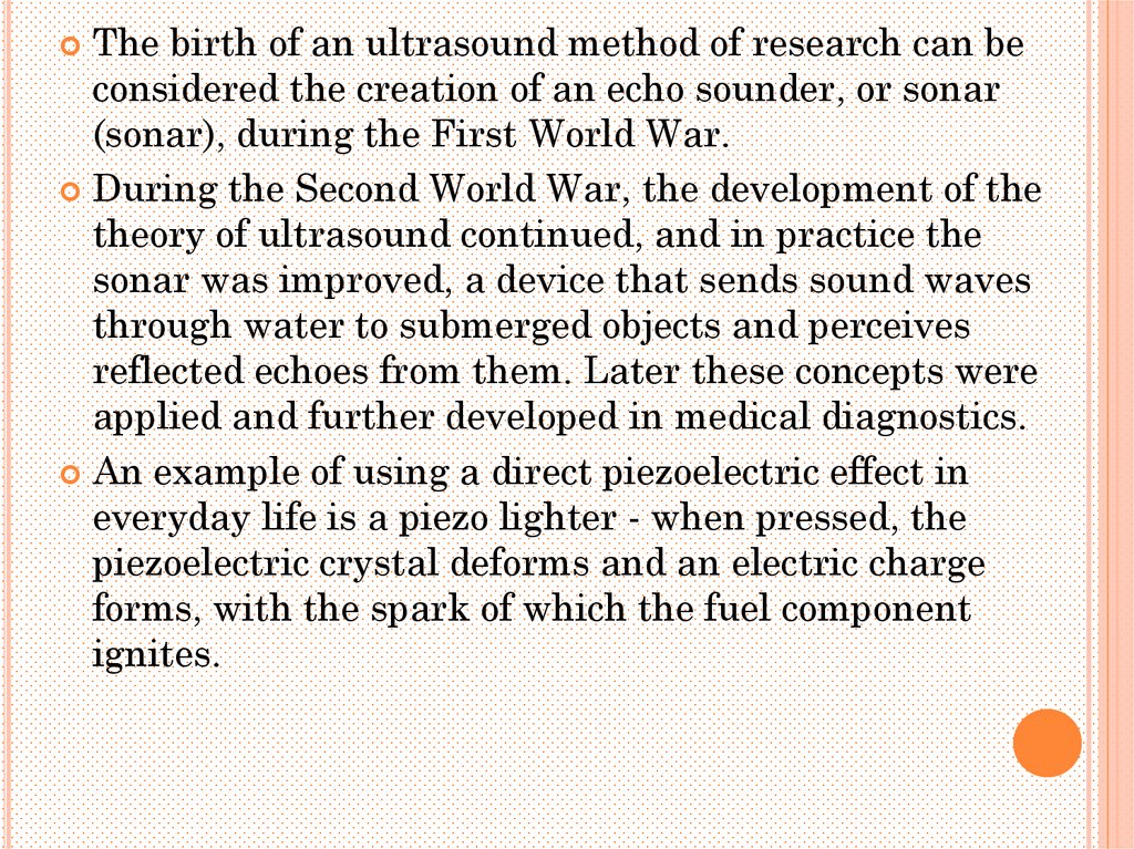
The birth of an ultrasound method of research can beconsidered the creation of an echo sounder, or sonar
(sonar), during the First World War.
During the Second World War, the development of the
theory of ultrasound continued, and in practice the
sonar was improved, a device that sends sound waves
through water to submerged objects and perceives
reflected echoes from them. Later these concepts were
applied and further developed in medical diagnostics.
An example of using a direct piezoelectric effect in
everyday life is a piezo lighter - when pressed, the
piezoelectric crystal deforms and an electric charge
forms, with the spark of which the fuel component
ignites.
17.
In modern ultrasonic devices, there are several basictypes of ultrasonic sensors that differ in operating
frequency (respectively, the depth of scanning and the
quality of the image obtained, or the resolution), as well
as the size and shape of the scanning surface.
The main types of ultrasonic sensors are
linear,
convective
sector.
18.
Linear sensor - high-frequency sensor with afrequency of 5-15 MHz, more often 7.5 MHz,
is used mainly for the investigation of
superficially located organs (thyroid, breast,
lymph nodes, surface vessels, etc.).
It has minimal distortion of the resulting
image, since the position of the transducer on
the surface of the body completely
corresponds to the size of the organ under
study.
Due to the higher frequency, linear sensors
allow the image of the investigated area to be
obtained at a high resolution, but are limited
to a small depth of scanning - no more than 810 cm.
19.
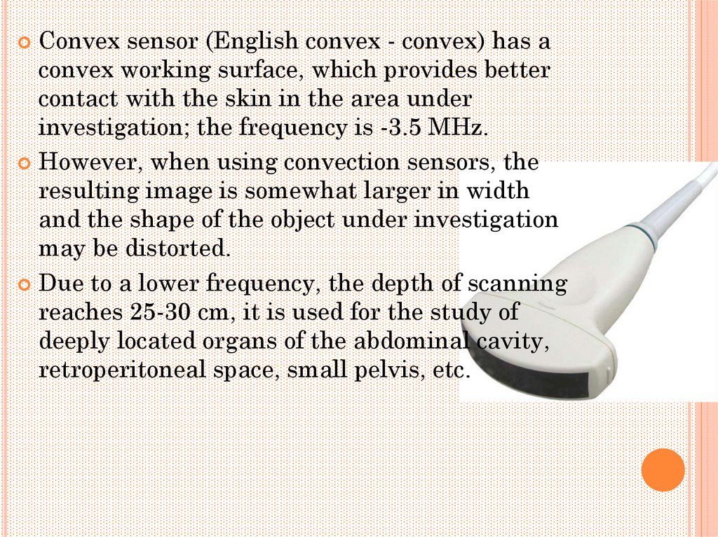
Convex sensor (English convex - convex) has aconvex working surface, which provides better
contact with the skin in the area under
investigation; the frequency is -3.5 MHz.
However, when using convection sensors, the
resulting image is somewhat larger in width
and the shape of the object under investigation
may be distorted.
Due to a lower frequency, the depth of scanning
reaches 25-30 cm, it is used for the study of
deeply located organs of the abdominal cavity,
retroperitoneal space, small pelvis, etc.
20.
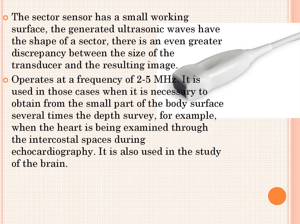
The sector sensor has a small workingsurface, the generated ultrasonic waves have
the shape of a sector, there is an even greater
discrepancy between the size of the
transducer and the resulting image.
Operates at a frequency of 2-5 MHz. It is
used in those cases when it is necessary to
obtain from the small part of the body surface
several times the depth survey, for example,
when the heart is being examined through
the intercostal spaces during
echocardiography. It is also used in the study
of the brain.
21.
The main types of sensors and directions of propagation ofultrasonic waves generated by them: a - linear; b - convective; in
- sector
22.
So, the whole process of ultrasonic scanning can be divided intothe following stages:
generation of ultrasonic waves (reverse piezoelectric effect);
penetration of ultrasonic waves into tissues;
interaction of ultrasound with tissues, reflection from interfaces of
media in the form of different echo strength;
transformation of reflected signals into electrical signals (direct
piezoelectric effect);
visualization of electrical signals by means of various types of
registration of reflected signals or image scanning.
23.
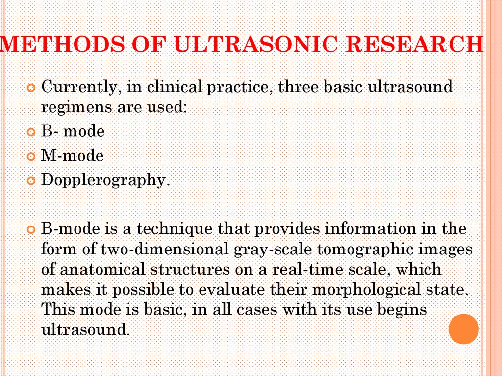
METHODS OF ULTRASONIC RESEARCHCurrently, in clinical practice, three basic ultrasound
regimens are used:
B- mode
M-mode
Dopplerography.
B-mode is a technique that provides information in the
form of two-dimensional gray-scale tomographic images
of anatomical structures on a real-time scale, which
makes it possible to evaluate their morphological state.
This mode is basic, in all cases with its use begins
ultrasound.
24.
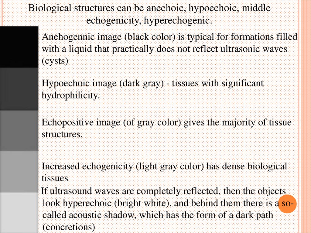
Biological structures can be anechoic, hypoechoic, middleechogenicity, hyperechogenic.
Anehogennic image (black color) is typical for formations filled
with a liquid that practically does not reflect ultrasonic waves
(cysts)
Hypoechoic image (dark gray) - tissues with significant
hydrophilicity.
Echopositive image (of gray color) gives the majority of tissue
structures.
Increased echogenicity (light gray color) has dense biological
tissues
If ultrasound waves are completely reflected, then the objects
look hyperechoic (bright white), and behind them there is a socalled acoustic shadow, which has the form of a dark path
(concretions)
25.
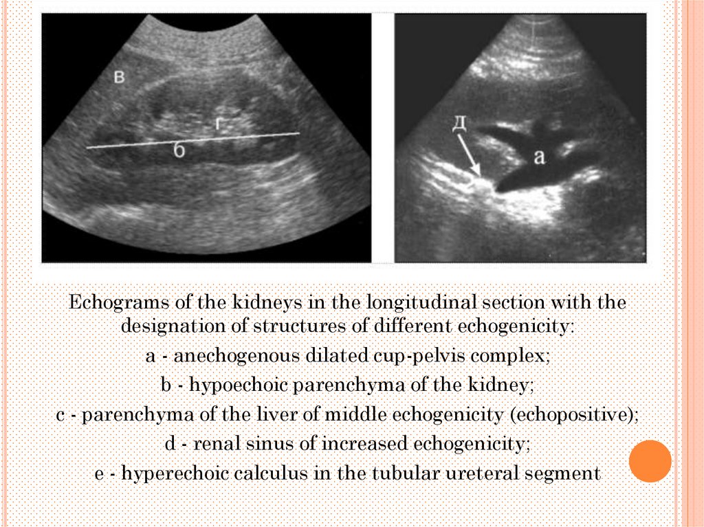
Echograms of the kidneys in the longitudinal section with thedesignation of structures of different echogenicity:
a - anechogenous dilated cup-pelvis complex;
b - hypoechoic parenchyma of the kidney;
c - parenchyma of the liver of middle echogenicity (echopositive);
d - renal sinus of increased echogenicity;
e - hyperechoic calculus in the tubular ureteral segment
26.
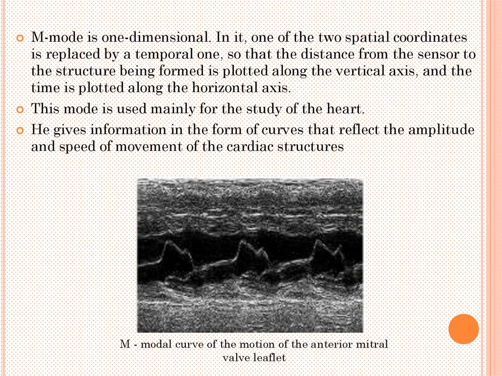
M-mode is one-dimensional. In it, one of the two spatial coordinatesis replaced by a temporal one, so that the distance from the sensor to
the structure being formed is plotted along the vertical axis, and the
time is plotted along the horizontal axis.
This mode is used mainly for the study of the heart.
He gives information in the form of curves that reflect the amplitude
and speed of movement of the cardiac structures
M - modal curve of the motion of the anterior mitral
valve leaflet
27.
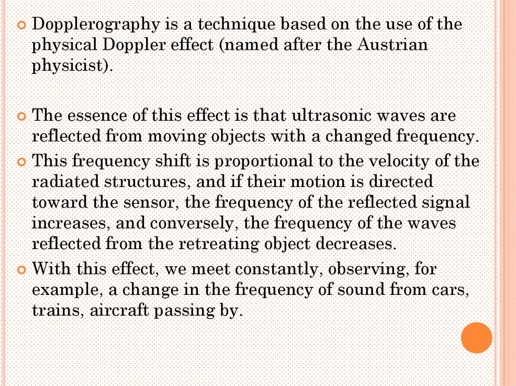
Dopplerography is a technique based on the use of thephysical Doppler effect (named after the Austrian
physicist).
The essence of this effect is that ultrasonic waves are
reflected from moving objects with a changed frequency.
This frequency shift is proportional to the velocity of the
radiated structures, and if their motion is directed
toward the sensor, the frequency of the reflected signal
increases, and conversely, the frequency of the waves
reflected from the retreating object decreases.
With this effect, we meet constantly, observing, for
example, a change in the frequency of sound from cars,
trains, aircraft passing by.
28.
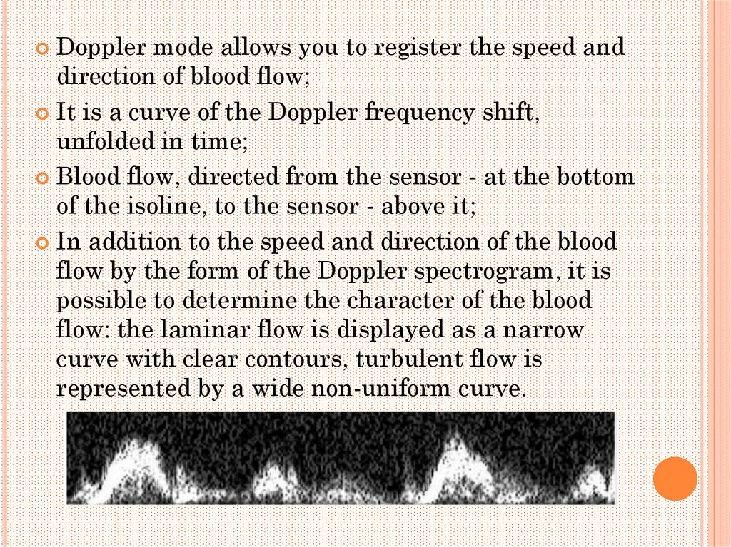
Doppler mode allows you to register the speed anddirection of blood flow;
It is a curve of the Doppler frequency shift,
unfolded in time;
Blood flow, directed from the sensor - at the bottom
of the isoline, to the sensor - above it;
In addition to the speed and direction of the blood
flow by the form of the Doppler spectrogram, it is
possible to determine the character of the blood
flow: the laminar flow is displayed as a narrow
curve with clear contours, turbulent flow is
represented by a wide non-uniform curve.
29.
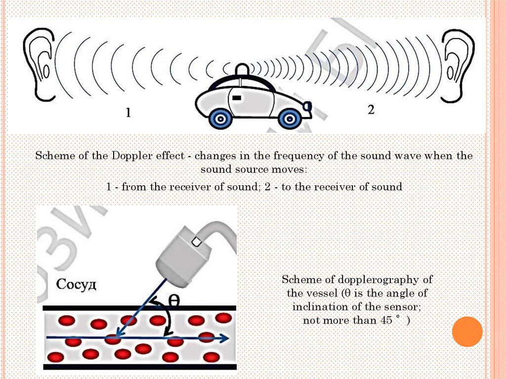
Scheme of the Doppler effect - changes in the frequency of the sound wave when thesound source moves:
1 - from the receiver of sound; 2 - to the receiver of sound
Scheme of dopplerography of
the vessel (θ is the angle of
inclination of the sensor;
not more than 45 °)
30.
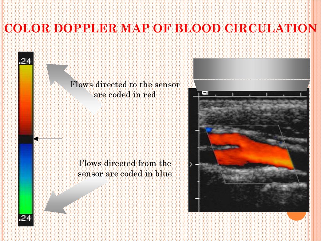
COLOR DOPPLER MAP OF BLOOD CIRCULATIONFlows directed to the sensor
are coded in red
Flows directed from the
sensor are coded in blue
31.
Elastography (sonoelastography) - ultrasound method, which isbased on differential diagnosis of malignant neoplasms based on
changes in their density and rigidity.
Sonoelastography allows the evaluation of tissue stiffness in real
time using soft pressure, performed by a standard ultrasonic
sensor.
Computerized color scale displays the degree of rigidity that
corresponds to a certain color (blue color - rigid structures, red
and green - soft tissues).
The main organs under investigation are the prostate gland,
bladder, uterus, ovaries, liver, breast, lymph nodes, soft tissues.
32.
INDICATIONS FOR USE OF USHead
1. Investigation of the brain in young children, mainly if
there is a suspicion of an inborn violation of its
development.
2. Investigation of cerebral vessels in order to establish
the causes of cerebral circulation disorders and to
evaluate the efficiency of performed operations on
vessels.
3. Investigation of the eyes for the diagnosis of various
diseases and injuries (tumors, retinal detachment,
intraocular hemorrhages, foreign bodies).
4. Investigation of salivary glands for evaluation of their
morphological state.
5. Intraoperative control of the total removal of brain
tumors.
33.
INDICATIONS FOR USE OF USNeck
1. Study of carotid and vertebral arteries:
- prolonged, often recurring severe headaches;
- Frequent fainting;
- clinical signs of cerebral circulation disorders;
- clinical syndrome of subclavian stealing (stenosis or occlusion of
the brachiocephalic trunk and subclavian artery);
- mechanical trauma (vascular damage, hematoma).
2. Thyroid examination:
- Any suspicion of her illness;
3. Investigation of lymph nodes:
- Suspicion of their metastatic damage in the presence of a
malignant tumor of any organ;
- Lymphoma of any localization.
4. Inorganic neoplasm of the neck (tumors, cysts).
34.
Chest1. Heart examination:
- Diagnosis of congenital heart disease;
- Diagnosis of acquired heart defects;
- a quantitative assessment of the functional state of the heart (global and
regional systolic contractility, diastolic filling);
- evaluation of the morphological state and function of intracardial
structures;
- detection and establishment of the degree of intracardiac hemodynamics
disturbances (pathological shunting of blood, regurgitating flow in case of
insufficiency of the heart valves);
- Diagnosis of hypertrophic myocardiopathy;
- diagnostics of intracardiac thrombi and tumors;
- detection of ischemic myocardial disease;
- determination of fluid in the pericardial cavity;
- Quantitative evaluation of pulmonary arterial hypertension;
- Diagnosis of heart damage in case of mechanical trauma of the chest
(bruises, ruptures of walls, partitions, chords, leaflets);
- Evaluation of the radicality and efficiency of heart operations
35.
Chest2. Examination of the respiratory and mediastinal organs:
- determination of fluid in the pleural cavities;
- specification of the lesion of the chest wall and pleura;
- differentiation of tissue and cystic neoplasms of the mediastinum;
- evaluation of the mediastinal lymph nodes;
- diagnosis of thromboembolism of the trunk and main branches of
the pulmonary artery.
3. Breast examination:
- specification of uncertain radiographic data;
- differentiation of cysts and tissue formations revealed by
palpation or x-ray mammography;
- evaluation of seals in the mammary gland of unclear etiology;
- assessment of the condition of the mammary glands with
increasing axillary, sub- and supraclavicular lymph nodes;
- evaluation of the state of silicone prostheses of mammary glands;
- puncture biopsies of formations under the supervision of
ultrasound.
36.
Stomach1. Investigation of the parenchymal organs of the digestive system
(liver, pancreas):
- diagnosis of focal and diffuse diseases (tumors, cysts, inflammatory
processes);
- diagnostics of injuries in case of mechanical injury of the abdomen;
- detection of metastatic liver damage in malignant tumors of any
location;
- Diagnosis of portal hypertension.
2. Investigation of the biliary tract and gallbladder:
- Diagnosis of cholelithiasis with assessment of the state of the
biliary tract and the definition of concrements in them;
- clarification of the nature and severity of morphological changes in
acute and chronic cholecystitis;
- establishment of the nature of postcholecystectomy syndrome.
37.
Stomach4. Research of the intestine:
- diagnosis of intestinal obstruction;
- assessment of local prevalence of colorectal cancer;
- Diagnosis of acute appendicitis.
5. Study of the abdominal cavity:
- Diagnosis of diffuse peritonitis;
- diagnostics of intraperitoneal non-organic abscesses;
- differentiation of intraperitoneal abscesses with inflammatory
infiltrates.
38.
6. Study of the kidneys and upper urinary tract:- Diagnosis of various diseases and evaluation of the nature and
severity of existing morphological changes;
- assessment of local prevalence of malignant tumors of the
kidneys;
- changes in urinalysis that persist for more than 2 months;
- establishing the causes of hematuria, anuria;
- differential diagnosis of renal colic and other acute abdominal
diseases (acute cholecystitis, acute appendicitis, intestinal
obstruction);
- clinical signs of symptomatic arterial hypertension;
- Diagnosis of injuries in case of mechanical trauma to the
abdomen and lumbar region.
39.
7. Investigation of lymph nodes:- detection of their metastatic lesion in malignant
tumors of the abdominal and pelvic organs;
- Lymphoma of any localization.
8. Study of the abdominal aorta and inferior vena cava:
- diagnosis of aneurysms of the abdominal aorta;
- detection of stenoses and occlusions;
- detection of phlebotrombosis of the inferior vena cava.
40.
Pelvis1. Study of the lower urinary tract (distal part of the ureters,
bladder):
- Diagnosis of various diseases;
- assessment of local prevalence of malignant tumors;
- determination of residual urine in the bladder with infravesical
obstruction.
2. Investigation of internal genital organs in men (prostate, seminal
vesicles):
- Diagnosis of various diseases;
- assessment of local prevalence of malignant tumors;
- determination of the stage of benign prostatic hyperplasia.
41.
Pelvis3. Study of internal genital organs in women:
- Diagnosis of various diseases;
- the establishment of causes of infertility;
- determination of the gestational age;
- control over the course of pregnancy;
- determination of the sex of the fetus;
- determination of the expected body weight and length of the fetus;
- determination of the functional state ("biophysical profile") of the
fetus;
- Diagnosis of ectopic pregnancy;
- diagnosis of intrauterine fetal death;
- Diagnosis of congenital malformations and fetal diseases.
42.
Spine1. Diagnosis of degenerative-dystrophic lesions.
2. Diagnosis of damage to soft tissue structures of the spine during mechanical trauma.
3. Diagnosis of birth injuries and their consequences in newborns and children of the first year
of life.
Limbs
1. Diagnosis of damage to muscles, tendons, ligaments.
2. Diagnosis of diseases and injuries of extra- and intra-articular structures.
3. Diagnosis of inflammatory and neoplastic diseases of bones and soft tissues.
4. Diagnosis of congenital disorders of limb development (congenital dislocation of the hip,
deformities of the foot, incomplete muscles).
Peripheral blood vessels
1. Diagnosis of arterial aneurysms.
2. Diagnosis of arteriovenous anastomoses.
3. Diagnosis of thrombosis and embolism.
4. Diagnosis of stenosis and occlusion.
5. Diagnosis of chronic venous insufficiency.
6. Diagnosis of vascular injuries in case of mechanical trauma.
In general, the ultrasound method has become an integral part of the clinical examination of
patients, and its diagnostic capabilities continue to expand
43.
Limbs1. Diagnosis of damage to muscles, tendons, ligaments.
2. Diagnosis of diseases and injuries of extra- and intra-articular
structures.
3. Diagnosis of inflammatory and neoplastic diseases of bones and soft
tissues.
4. Diagnosis of congenital disorders of limb development (congenital
dislocation of the hip, deformities of the foot, incomplete muscles).
Peripheral blood vessels
1. Diagnosis of arterial aneurysms.
2. Diagnosis of arteriovenous anastomoses.
3. Diagnosis of thrombosis and embolism.
4. Diagnosis of stenosis and occlusion.
5. Diagnosis of chronic venous insufficiency.
6. Diagnosis of vascular injuries in case of mechanical trauma.