


")


 medicine
 english
Similar presentations:


Doppler ultrasound
1. Doppler ultrasound
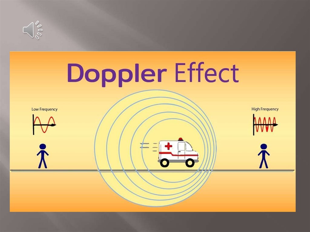
Dr. Badira Al Qudah2. Doppler effect
We are all aware that the pitch of an ambulancesiren changes as we stop and listen to it as it
drives by.
The frequency that reaches you is higher as the
ambulance approaches and lower as the
ambulance passes by.
This is a consequence of the Doppler effect.
3.
4. Physics
Ultrasound images are formed by reflectedechoes. These waves have an amplitude
(strength) and a frequency, which is equal to
the frequency of the emitted wave, if the tissue
is static. Tissue movement (e.g. blood)
promotes a frequency shift (Doppler shift) in
the reflected echoes.
5. Application of Doppler
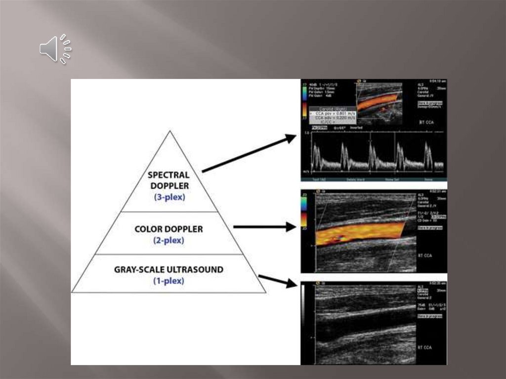
Three basic levels of US can be performed, with each leveladding information to the preceding level.
At the first level is the traditional standard brightness
mode (B-mode) gray-scale examination, in which no
Doppler is used.
The second level superimposes a color Doppler
interrogation region of interest. This level produces an
image that shows blood flow in vessels.
The third level superimposes a small interrogation
region, called a sample volume, over a vessel of
interest. Targeted interrogation of the vessel produces
a spectral Doppler waveform.
6.
7.
-The Doppler effect in diagnostic imaging can
be used to study blood flow, for example, and
provides the operator with three pieces of
information to determine:
Presence or absence of flow
Direction of blood flow
Velocity of blood flow.
8. Doppler shift
Blood flow moving towards the transducer producespositive Doppler shifted signals and conversely blood
flow moving away from the transducer produces negative
Doppler shifted signals
9. Angle of insonation (Doppler angle)
The angle of insonation is very important.The angle of insonation is the angle between
the transducer and the vessel being studied.
The angle of insonation should be between 45°
and 60°.
The velocity measurements become unreliable
with angles more than 60°.
10.
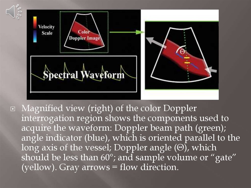
Magnified view (right) of the color Dopplerinterrogation region shows the components used to
acquire the waveform: Doppler beam path (green);
angle indicator (blue), which is oriented parallel to the
long axis of the vessel; Doppler angle (Θ), which
should be less than 60°; and sample volume or “gate”
(yellow). Gray arrows = flow direction.
11. Doppler US
is an application of diagnosticultrasound used to detect moving blood cells or
other moving structures and measure their
direction and speed of movement.
The Doppler effect is used to evaluate
movement by measuring changes in frequency
of the echoes reflected from moving structures.
12.
In many instances, Doppler ultrasound hasreplaced x-ray methods such as angiography,
as a method to evaluate blood vessels and
blood flow.
Doppler ultrasound permits real-time viewing
of blood flow that cannot be obtained by other
methods.
Doppler ultrasound has proved a helpful in all
areas of ultrasound, aiding in the evaluation of
the major arteries and veins of the body, the
heart, and in obstetrics for fetal monitoring.
13.
Types of Doppler ultrasound include:Color Doppler
Power Doppler
Spectral Doppler
14. Color Doppler
uses a computer to convertthe Doppler measurements into an array of colors.
Colour Doppler imaging colour codes Doppler
shift information and superimposes that
information over a B-mode image
Color Doppler displays blood flow toward the
transducer as red and blood flow away from the
transducer as blue.
This color visualization when combined with a
standard ultrasound picture of a blood vessel
shows the speed and direction of blood flow
through the vessel.
15. Color Doppler
16. Power Doppler
is a technique that uses theamplitude of Doppler signal to detect moving
matter.
It is an ultrasound technique that is used to obtain
images that are difficult or impossible to obtain
using standard color Doppler and to provide
greater detail of blood flow, especially in vessels
that are located inside organs.
Power Doppler is more sensitive than color
Doppler for the detection and demonstration of
blood flow, but provides no information about the
direction of flow.
Color and spectral Doppler both reveal the
direction of blood flow which can be valuable
information.
17. Power Doppler
--
-
Power Doppler:
is independent of velocity and direction of
flow.
is independent of angle, allowing detection of
smaller velocities than color Doppler,
facilitating examinations in certain technically
challenging clinical setting
has higher sensitivity than color Doppler
18. Spectral Doppler
Instead of displaying Doppler measurementsvisually as in the color and power
Doppler methods, spectral Doppler displays
the blood flow measurements graphically,
displaying flow velocities recorded over time.
Spectral analysis of Doppler signal contains
both frequency and amplitude information.
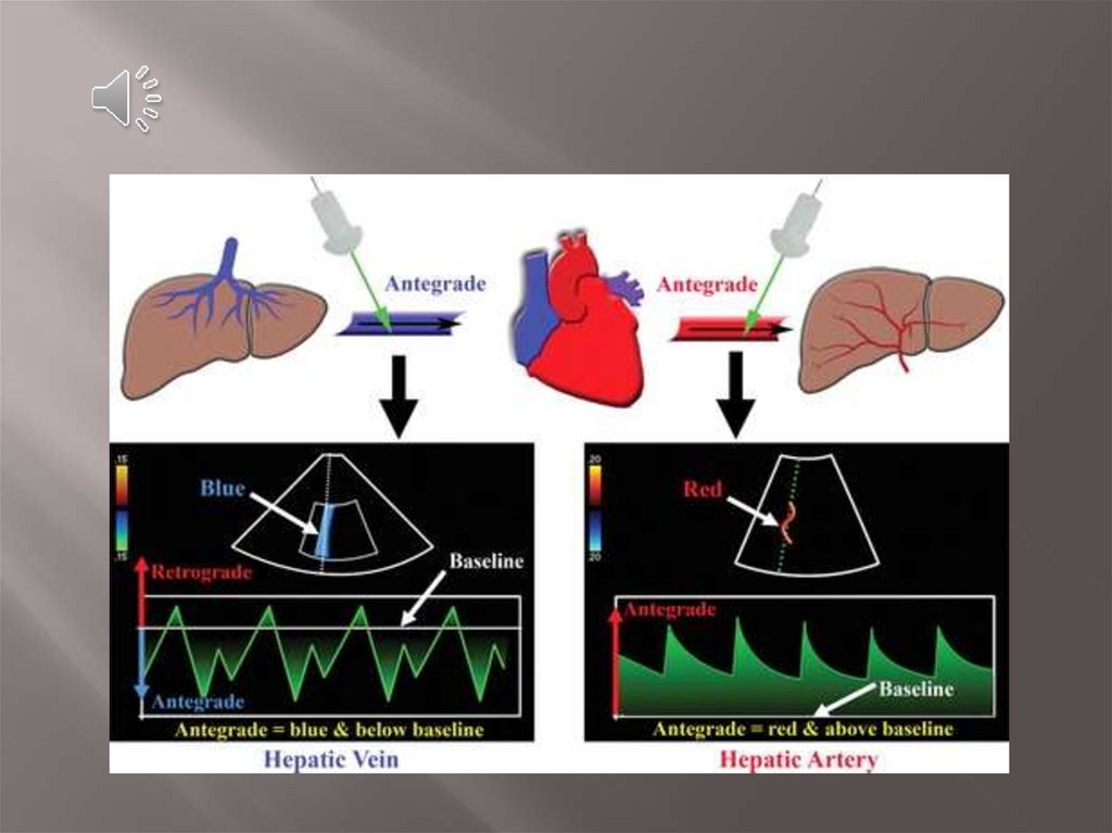
At spectral Doppler, blood flow toward the
transducer is displayed above the baseline and
blood flow away from the transducer is
displayed below the baseline
19.
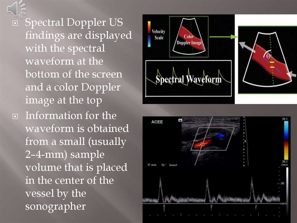
Spectral Doppler USfindings are displayed
with the spectral
waveform at the
bottom of the screen
and a color Doppler
image at the top
Information for the
waveform is obtained
from a small (usually
2–4-mm) sample
volume that is placed
in the center of the
vessel by the
sonographer
20.
21. Introduction to Doppler ultrasound
https://www.youtube.com/watch?v=tQn8jKtwk6o
22. References
https://radiologykey.com/physicalprinciples-of-doppler-ultrasoundOptimizing Doppler and Color Flow US:
Application to Hepatic Sonography,Jonathan
B. Kruskal
Doppler US of the Liver Made Simple, Dean
Alexander McNaughton, Monzer M. AbuYousef, gastrointestinal imaging
Radiologyinfo.org