


 medicine
Similar presentations:


Radiation methods for studying the organs of the cardiovascular system in childhood
1. Radiation methods for studying the organs of the cardiovascular system in childhood.
Checked by:Done by:
2. USM
• The method of choice in the diagnosis ofpathological changes in the cardiovascular
system in children is ultrasound.
Ultrasound examination of the heart includes
two methods:
• echocardiography
Doppler study.
3.
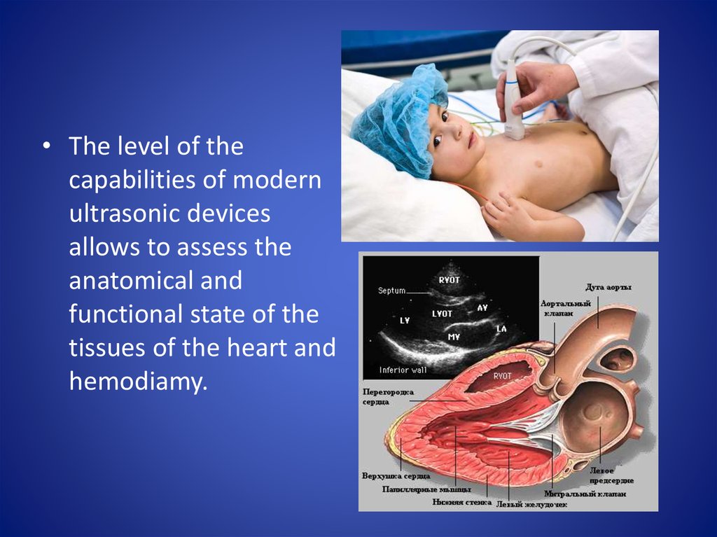
• The level of thecapabilities of modern
ultrasonic devices
allows to assess the
anatomical and
functional state of the
tissues of the heart and
hemodiamy.
4.
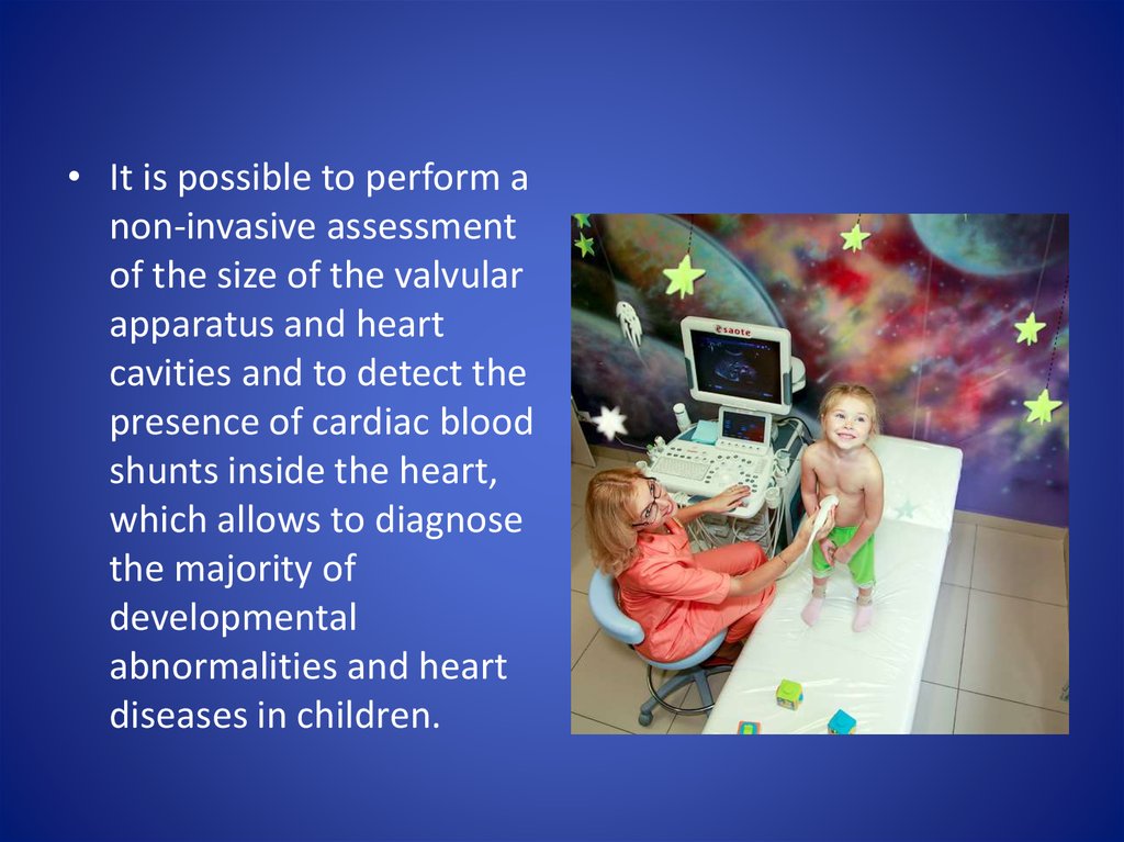
• It is possible to perform anon-invasive assessment
of the size of the valvular
apparatus and heart
cavities and to detect the
presence of cardiac blood
shunts inside the heart,
which allows to diagnose
the majority of
developmental
abnormalities and heart
diseases in children.
5.
The main indications for Echocardiography are:- auscultative picture (noise);
- complaints of the child for fatigue, dyspnea, pain in the chest;
- Cyanosis, pallor;
- bad weight gain;
- arterial hypertension;
- frequent colds;
- cough without signs of respiratory infection;
- changes on the roentgenogram: atypical configuration of the heart, atypical
pulmonary pattern, etc.
6. The main scanning modes for echocardiography are:
- B-mode: visualization ofthe heart in real time from
various accesses. Used to
visually assess the
anatomy of the heart, its
location, contractions, the
presence of defects,
pathological inclusions
• - M-mode is usually in
modern devices is
switched in parallel to
the B-mode. This is a
one-dimensional scan in
real time. As accurately
as possible, it allows
measurements of
various anatomical
structures.
7.
EchoCG in B-mode: 1 - cavity of the right ventricle; 2 - pulmonaryartery trunk:
a - two defects of the interventricular septum (arrows);
b - a significant amount of purulent effusion in the pericardium;
в - a hypoplasia of a trunk of a pulmonary artery (the right ventricle
is expanded)
8.
EchoCG in M-mode:a - decreased contractility of the left ventricle (between the
arrows - interventricular septum);
b - early carditis, severe myocardial hypertrophy, ventricular
cavity
sharply reduced (compare with the image of "a");
в - movements of the mitral valve leaf (arrow)
9.
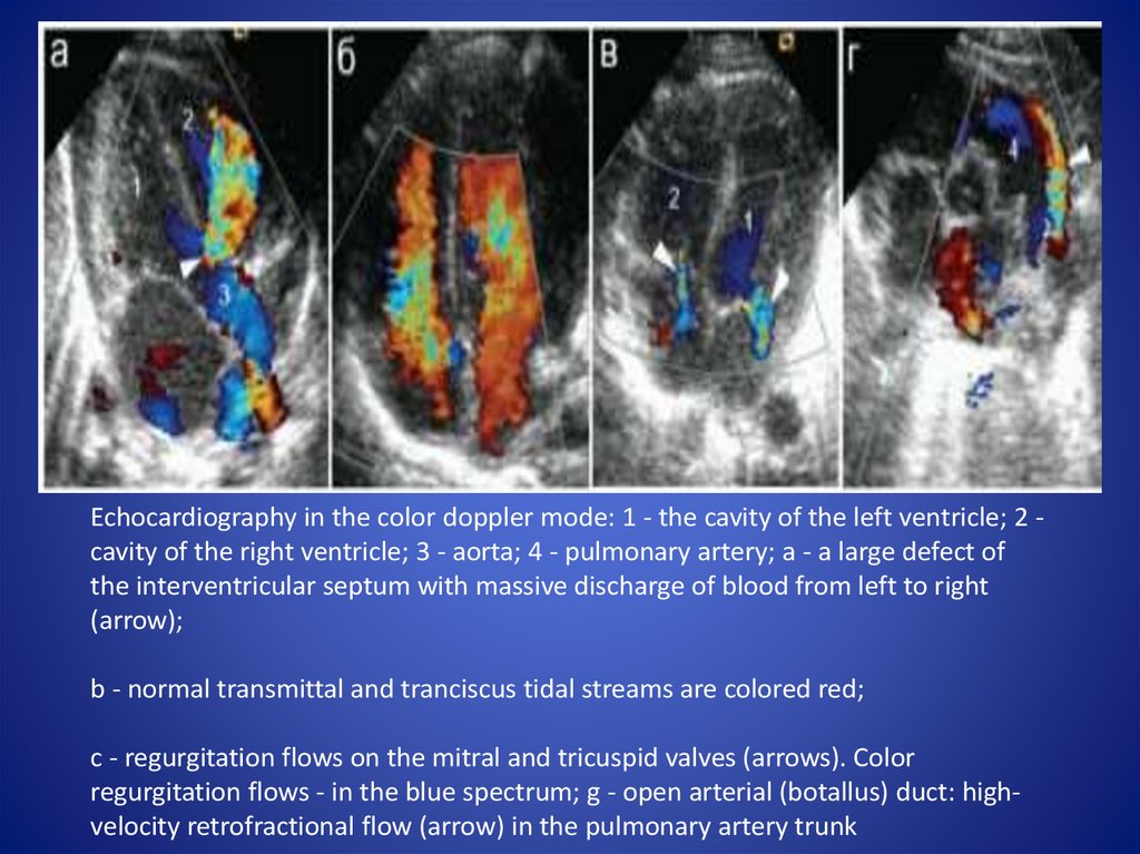
Echocardiography in the color doppler mode: 1 - the cavity of the left ventricle; 2 cavity of the right ventricle; 3 - aorta; 4 - pulmonary artery; a - a large defect ofthe interventricular septum with massive discharge of blood from left to right
(arrow);
b - normal transmittal and tranciscus tidal streams are colored red;
c - regurgitation flows on the mitral and tricuspid valves (arrows). Color
regurgitation flows - in the blue spectrum; g - open arterial (botallus) duct: highvelocity retrofractional flow (arrow) in the pulmonary artery trunk
10.
Echocardiography in the regime of constant wave doppler (arrows showthe components of Doppler curves characterizing the defect): a - open
arterial (botalla) duct; b - stenosis of the pulmonary artery; c regurgitation on the tricuspid valve
11. x-ray method
• Historically, the x-raymethod belongs to the
precedence in the diagnosis
of congenital heart defects
on the basis of both an
analysis of the actual
position and configuration
of the heart and a
pulmonary picture that
allows one to judge the
presence or absence of
pathological changes from
the pulmonary blood flow.
12.
• With the review of radiography can be identifiedonly such views that lead to a change in the
structure of the heart and / or pathology of the
small circle of blood circulation. The main
advantages of the method are the possibility of
its implementation in almost any medical
institution and the simultaneous receipt of
information on both the structure of the heart
and the state of the lungs and the small
circulation
13.
Normal radiographic anatomy of the heart in a direct projection:a - arches of the heart along the right and left contours;
b - measurements for calculation of cardiothoracic index and
Moore's index
14.
• On the right contour, 2 arcs are distinguished: the upperone is formed by the ascending aorta or the superior
hollow vein (1), the lower one by the lateral wall of the
right atrium (2). Between them, in the middle of the height
of the heart shadow, is the atriovasal angle. Shifting it
upwards indicates an increase in the right atrium,
downward - about the expansion and lengthening of the
ascending aorta.
• On the left contour of the heart, 4 arcs are visualized: the
first (1) - the arch of the aorta; the second (2) - the arc of
the pulmonary artery; the third arc (3) - the eye of the left
atrium; the fourth arch (4) is the lateral wall of the left
ventricle, which should not protrude into the left
pulmonary field behind the median-clavicular line (Figure
7.2.2).
15.
• To determine the size of the heart cardiothoracic index is used - theratio of the diameter of the heart to the internal diameter of the
chest (at the level of the right dome of the diaphragm):
• cardiothoracic index = (a + b): with x 100% and is normally no more
than 50%.
• The degree of expansion of the pulmonary artery is determined by
the Moore index:
• d: 1 / 2c
• and is normally no more than 0.25.
16.
X-ray signs of pulmonary edema in children: a - newborn,arrows show "butterfly wings"; b - child 2 years old
17.
X-ray signs of heart defects with normovolemia of the small circle ofblood circulation:
a - normovolemia of the small circle, we determine the usuras along the
lower edge of the VII and VIII ribs on the right (arrows) in a child with
coarctation of the aorta; b - valve stenosis of the pulmonary artery
(swelling of the pulmonary arterial contour is indicated by an arrow)
18. Angiocardiography
• Congenital abnormalities of the aortic arch andpulmonary bifurcation may occur in isolation or in
combination with other AMS, and also be accompanied
by symptoms of dysphagia and tracheal obstruction.
Diagnosis of these anomalies is based on the detection
of deformation of the contrasted esophagus, which is
an indirect sign of the presence of an aberrant or
abnormal vessel passing through the mediastinum. For
this purpose, a special examination (angiocardiography
with cardiac catheterization) is carried out, designed to
accurately diagnose these conditions.
19.
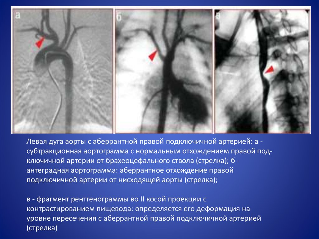
Левая дуга аорты с аберрантной правой подключичной артерией: а субтракционная аортограмма с нормальным отхождением правой подключичной артерии от брахеоцефального ствола (стрелка); б антеградная аортограмма: аберрантное отхождение правойподключичной артерии от нисходящей аорты (стрелка);
в - фрагмент рентгенограммы во II косой проекции с
контрастированием пищевода: определяется его деформация на
уровне пересечения с аберрантной правой подключичной артерией
(стрелка)
20. СT and MRI
• Computer tomography is performed after surgery forheart defects, for visualizing the noncompact
myocardium and coronary arteries.
• Magnetic resonance imaging in children is used to
diagnose cardiomyopathies and myocarditis and
associated fibrotic changes in the myocardium and to
assess the presence of effusion in pericarditis.
• Computer tomography and magnetic resonance
imaging of the heart are used to diagnose and assess
the size of tumors. The final answer is biopsy.