


")


 of rheumatic fever.")
 of rheumatic fever.")
 of rheumatic fever.")
 of rheumatic fever.")
 of rheumatic fever.")


 medicine
Similar presentations:


The anatomical and physiological particularities of cardiovascular system in children and their clinical importance
1.
The anatomical andphysiological
particularities of
cardiovascular system
in children and their
clinical importance.
Outline of the lecture
1/ Embryogenesis, Embryopathyes.
Fethophaties
2/ What is the fetus blood circulation
about?
3/ Morphological and functional
particularities of the heart and blood
vessels in children.
4/ The clinical study.
5/ The Semiotics of the commonest
2.
The heart and largeblood vessels appears
at the 3-rd week of the
embrionic phase. The
first contractions of
two chambers
embryonic heart occur
at the 4-th week of the
embryogenesis. The
heart sounds can be
heared through the
mother`s abdominal
wall since the fourth
month of gestation.
3. Briefly the process of heart and large blood vessels embryogenesis can be described as a complex process of yolk sack and umbilical vessels interactions forming two tube-shaped hearts. After that thay merge forming the primitive embryonic heart.
Briefly theprocess of heart
and large blood
vessels
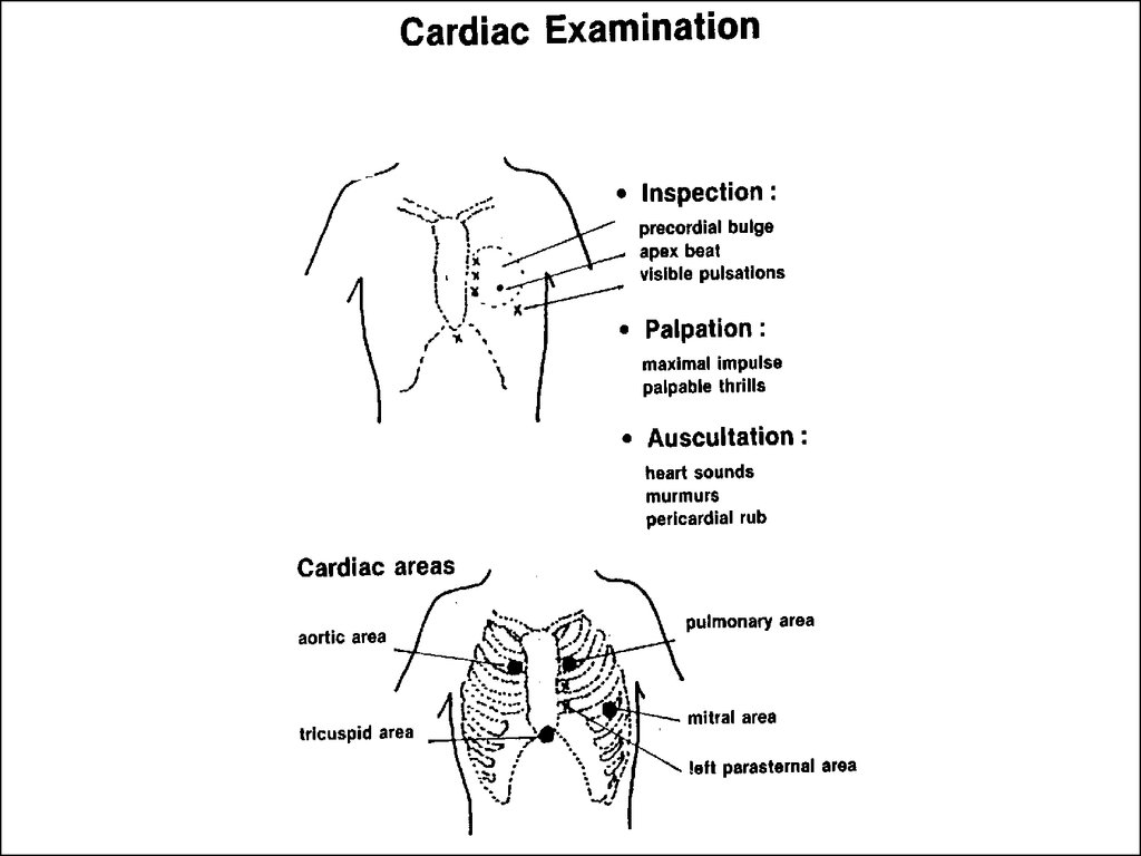
embryogenesis
can be described
as a complex
process of yolk
sack and
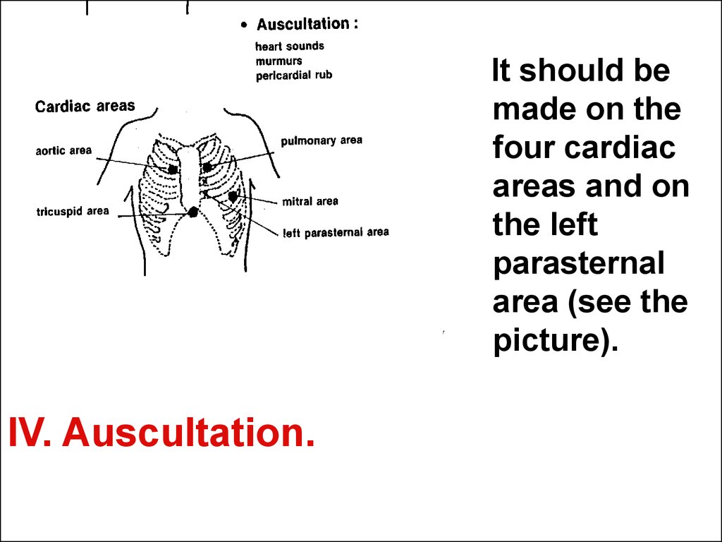
umbilical vessels
interactions
forming two tubeshaped hearts.
After that thay
merge forming
the primitive
embryonic heart.
4.
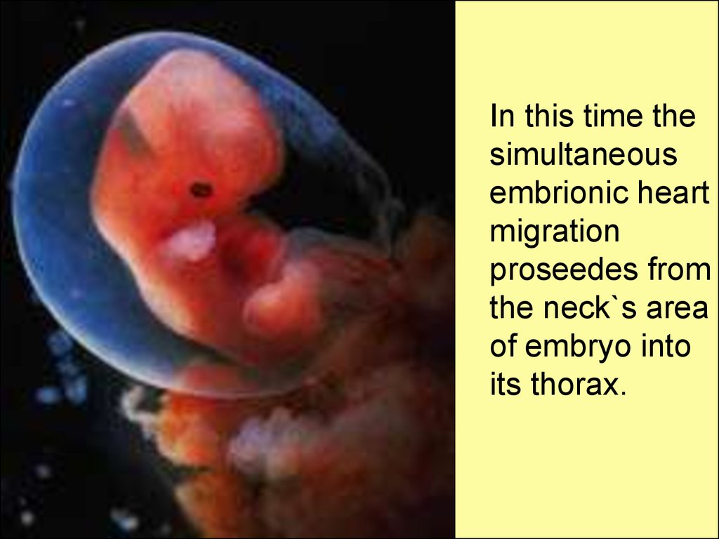
In this time thesimultaneous
embrionic heart
migration
proseedes from
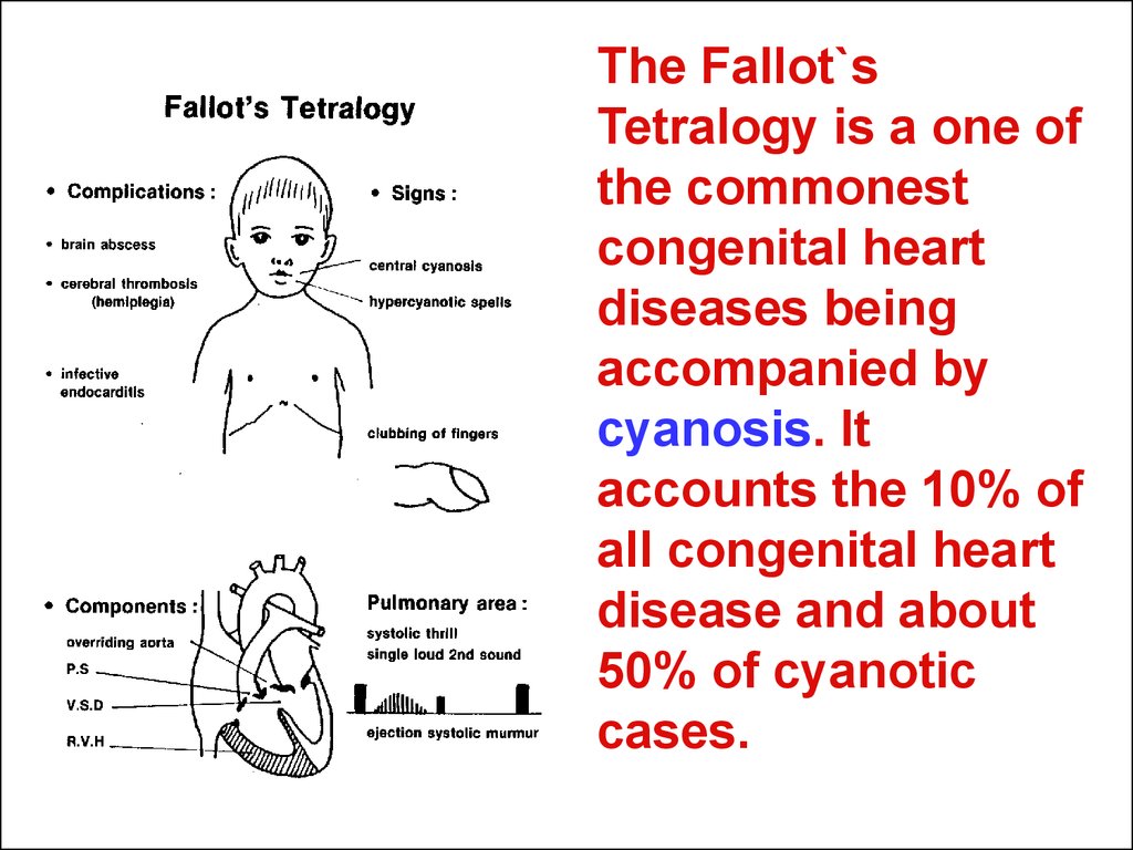
the neck`s area
of embryo into
its thorax.
5. Heart congenital abnormalities
• It is important to confess that embryo heartduring at list the 1-st month of life is
staying in raised risk to get a damage due
to teratogenic (causing congenital
abnormalities) factors.
• The viruses are the most common
pathogens damaging heart`s growing and
differentiation and leading to congenital
heart disease.
6. Embryopathyes.
blood vessels transpositionAorta
Right
heart
Pulmonary
artery
Left
heart
Ventriculum
septum
deffect
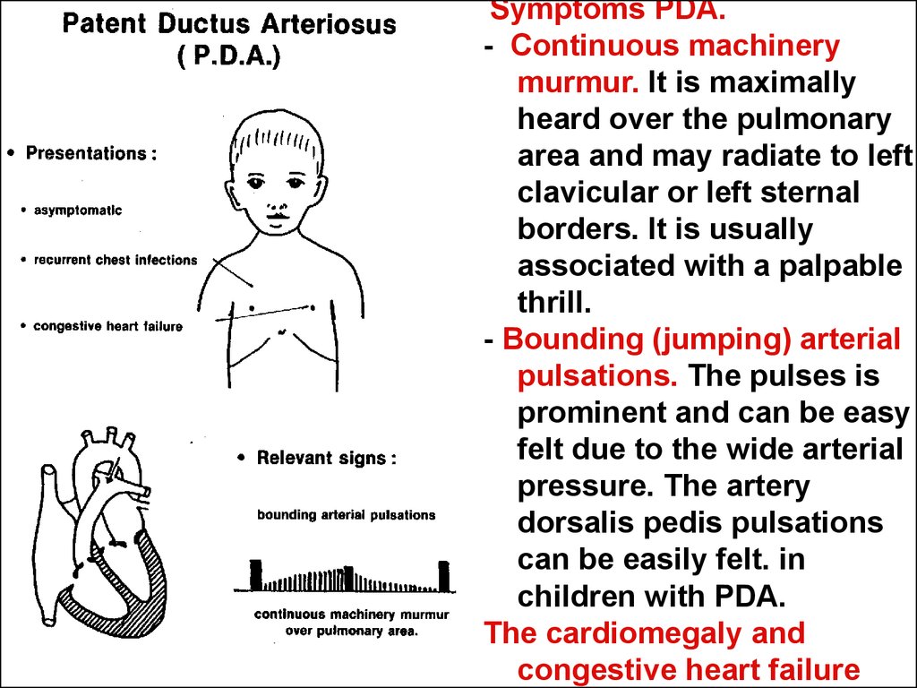
• Develop
within first 9
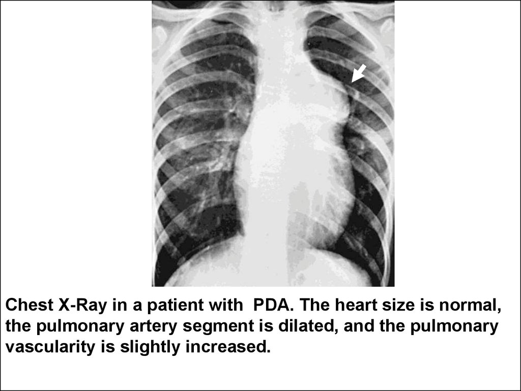
weeks of
gastation
• Are usually
severe
diseases
• Show huge
anatomical
abnormalitie
s
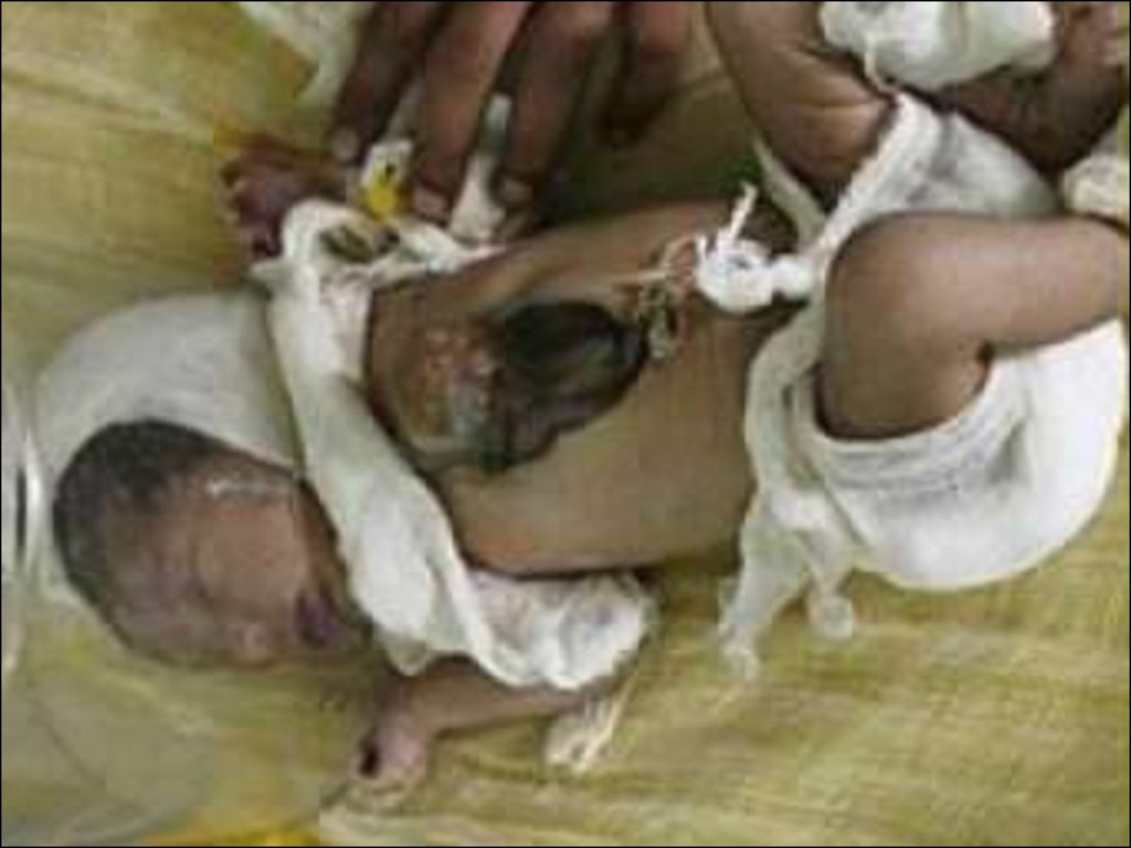
7. Heart ecthopia
Newborn male withthoracoabdominal ectopia
cordis (Cantrell
pentalogy). Flaring of the
lower thoracic cavity is
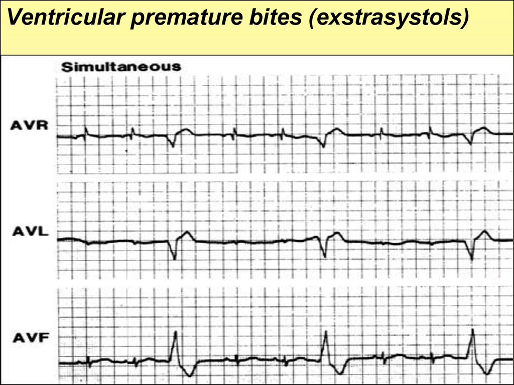
present with a large
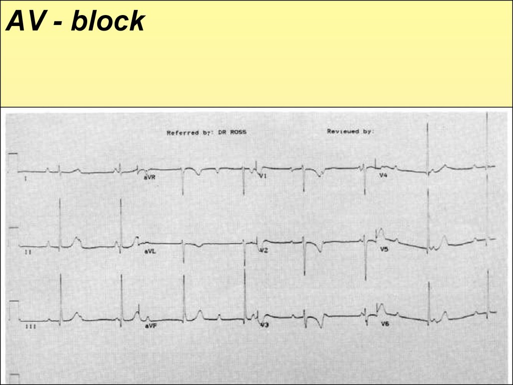
epigastric omphalocele.
The transverse septum of
the diaphragm and the
inferior portion of the
pericardium were absent.
The patient also had
tetralogy of Fallot. (From
Shamberger R, Welch K:
Chest wall deformities.
Ashcraft K, Holder T
(eds): Pediatric Surgery,
2nd ed. Philadelphia, WB
Saunders, 1993, p 158.)
8.
9. dextrocardy
10. Ultrasound investigation in utero
• By means of ultrasound investigationmethod it is possible to define embryo
and fetus heart`s contractions, to consider
a heart rate, to assess the heart and its
chambers sizes, shape and even some
abnormities that allows to required surgical
handling for children immediately after
delivery.
11. Fethophaties
• The 3-rd month old normal fetus has analready wholly formed heart.
• If congenital heart disease starts at this time
it`ll be less severe and easier in subject for
surgical correction.
• This sort of heart desease pertaines to
fethophaties.
• A clinical examples of fethophaties are:
- the patient ductus arteriosus (which
matches the aorta and pulmonary arteria);
- open foramen ovale linked right and left
atriums.
12.
The existence of Fethophaties may byexplained from position of the fetus
blood circulation.
13.
What is the fetus blood circulationabout?
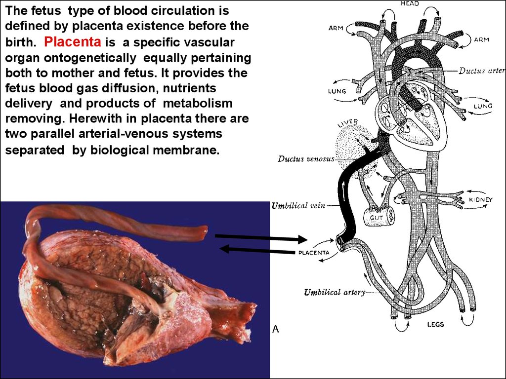
Plan of the human
circulation before
birth. Black shading
indicates more
oxygenated blood, and
arrows indicate the
direction of flow.
(From Rudolph AM:
Congenital Diseases of
the Heart. Chicago,
Year Book Medical
Publishers, 1974.)
14.
The fetus type of blood circulation isdefined by placenta existence before the
birth. Placenta is a specific vascular
organ ontogenetically equally pertaining
both to mother and fetus. It provides the
fetus blood gas diffusion, nutrients
delivery and products of metabolism
removing. Herewith in placenta there are
two parallel arterial-venous systems
separated by biological membrane.
15.
The pump function of fetusheart and two arteries
connected with fetus aorta in
place of its fission in low
abdomen provide the fetal
placental blood circulation.
This two arteries come out
through umbilical ring, reach
placenta and inside it divide
onto capillary network. From
here the blood enriched by
nutritive materials and oxygen
required for fetus development
comes back to fetus body by
means of umbilical vein. Two
arteries and one vein, thereby,
form the umbilical cord of the
fetus.
16.
After delivery theumbilical cord crossing
(cut) usually has to be
done. Immediately after
the umbilical cord must
be examined for
congenital vessels
abnormalities. In normal
umbilical cord the
umbilical vein looks like a
single big usually mildly
bloody vessel. Also there
are two contracted
vessels which must be
defined as an umbilical
arteries.
A
A
V
AA
A
V
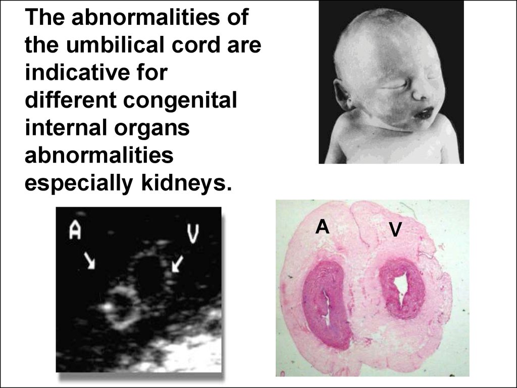
17.
The abnormalities ofthe umbilical cord are
indicative for
different congenital
internal organs
abnormalities
especially kidneys.
A
V
18.
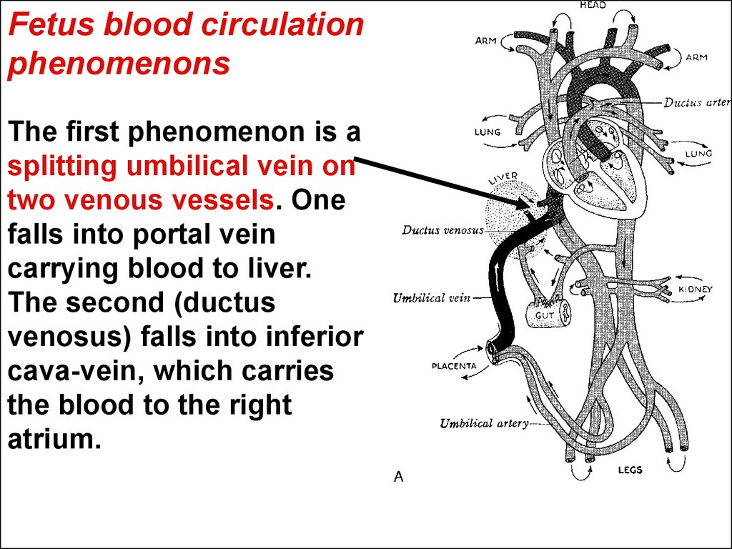
Fetus blood circulationphenomenons
The first phenomenon is a
splitting umbilical vein on
two venous vessels. One
falls into portal vein
carrying blood to liver.
The second (ductus
venosus) falls into inferior
cava-vein, which carries
the blood to the right
atrium.
19.
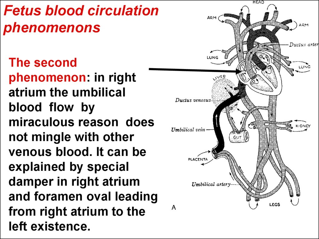
Fetus blood circulationphenomenons
The second
phenomenon: in right
atrium the umbilical
blood flow by
miraculous reason does
not mingle with other
venous blood. It can be
explained by special
damper in right atrium
and foramen oval leading
from right atrium to the
left existence.
20.
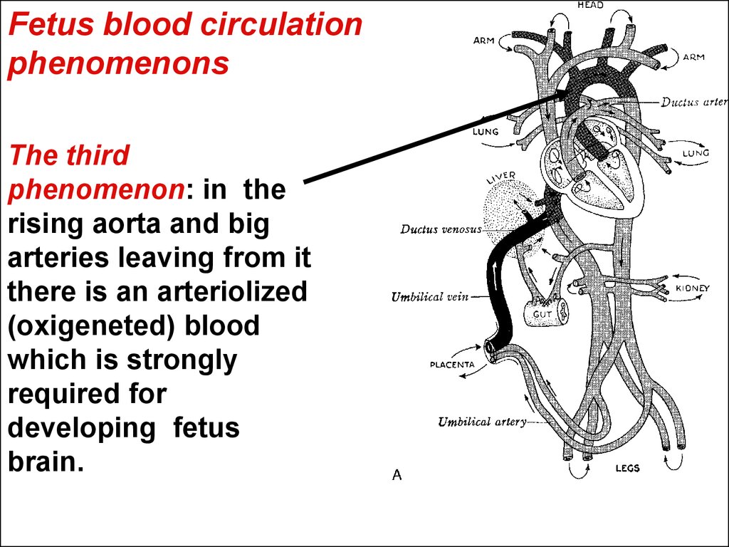
Fetus blood circulationphenomenons
The third
phenomenon: in the
rising aorta and big
arteries leaving from it
there is an arteriolized
(oxigeneted) blood
which is strongly
required for
developing fetus
brain.
21.
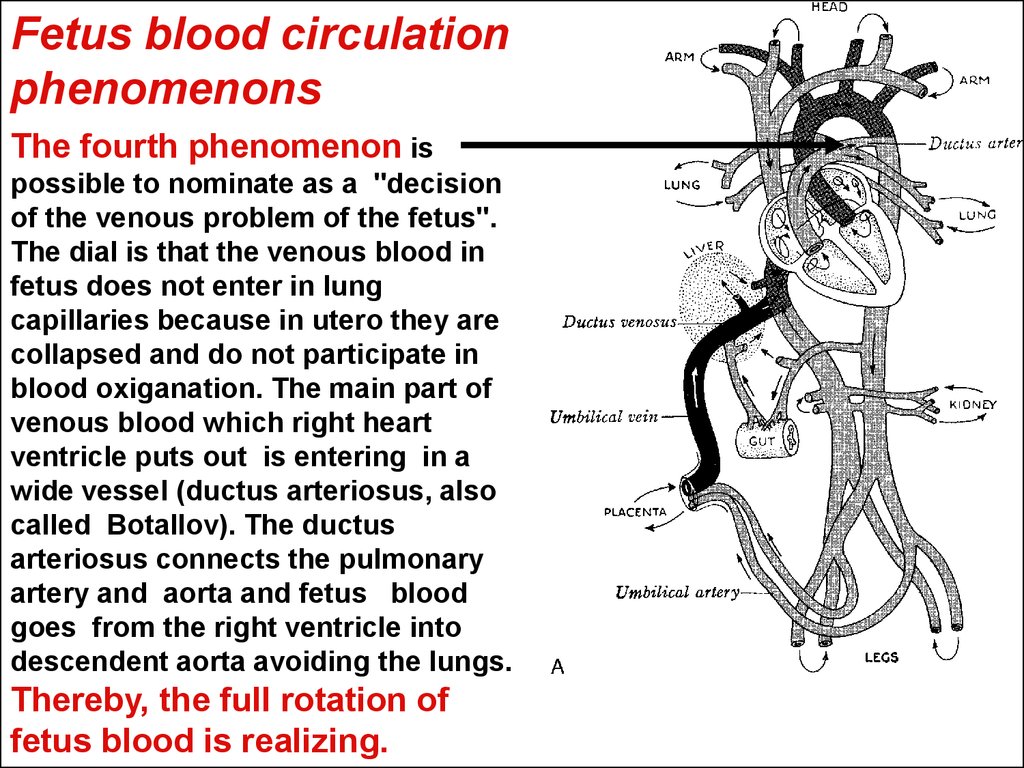
Fetus blood circulationphenomenons
The fourth phenomenon is
possible to nominate as a "decision
of the venous problem of the fetus".
The dial is that the venous blood in
fetus does not enter in lung
capillaries because in utero they are
collapsed and do not participate in
blood oxiganation. The main part of
venous blood which right heart
ventricle puts out is entering in a
wide vessel (ductus arteriosus, also
called Botallov). The ductus
arteriosus connects the pulmonary
artery and aorta and fetus blood
goes from the right ventricle into
descendent aorta avoiding the lungs.
Thereby, the full rotation of
fetus blood is realizing.
22.
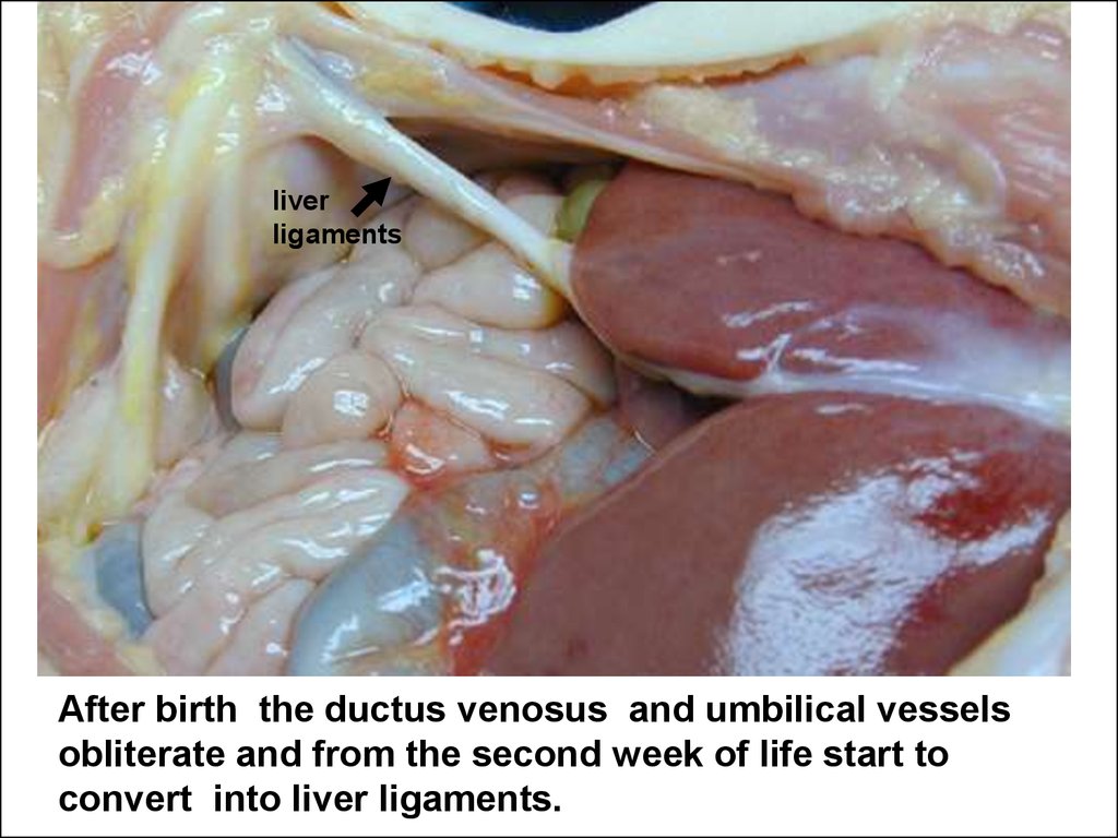
liverligaments
After birth the ductus venosus and umbilical vessels
obliterate and from the second week of life start to
convert into liver ligaments.
23.
The ductus arteriosus and foramen ovale close for severalseconds or minutes after birth. Their complete obliteration
occurs for 6-8 weeks later. But this process can be delayed.
Some times it happens that they never close because of their
innate big anatomical size or more often due to high blood
pressure in pulmonary artery system, for instance when the
newborn is sick with severe pneumonia.
24.
Morphological and functional particularitiesof the heart and blood vessels in children.
25. The heart size
The heart of the fetus or newborn iscomparatively greater that one in older children
and forms nearly 1% from mass of the body. In
children aged 1 yr and older it is approximately
0,5%. All the time the left ventricle mass is
bigger than the right one. But in very small
children the electric and mechanical activities
prevalence of the right ventricle must be
emphasized. This fact can be explaned by fetal
blood circulation.
26. The myocardial infarction in children is a casuistry
• Heart coronary arteries before age two aredistributed in children on splinting type. In
children aged 2 yr the coronary arteries are
distributed on mixed and after 10 years on
adult type. It means the main arterial branches
existence. According to this anatomical feature
the child heart is not predisposed to ischemic
heart attack like adult heart. In this conditions
the myocardial infarction in children is a
casuistry.
27. The myocardium.
In small children the heart myocytes are fine, havenot transverse lines and contain big amount of
nucklear substance. During the first two years of
the child life the intensive growing and
differentiation of myocardium occur. The muscular
filaments become to be more thick and strong. In
10 year old child the heart in its histology
corresponds to such one as in adult persone. A
little bit later in 14-15 year old children the
histological development of heart conductive
system forme definitively from specialized
cardiomyocytes losted contractive activity.
28. The nerviouse system supplementation of the heart
is realized through surface and deep plexuses combinationformed by nervus vagus and sympathic nerve filaments
contacting with sinus and atrio-ventricular ganglions of the
heart rate pasmacer center. The vagus nervial branches
pertaining to the parasympathetic nervous system finish
their development and myelinization in children aged of 34 years. Before this age the heart rate is defined by
unilateral activity of sympathic division of nervious
system. This fact explains comparatively high rate of heart
rhythm in smoll children. In age 4-5 after increasing
parasympathic activities the pulse rate is getting low. In
this period in well-children the physiological phenomenon
well known as respiratory arrhythmia characterized by
some lengthened intervals between heart bites also
appears.
29. The premature heart bites
• The premature heart bites(extrasystoles) if they are not occurring
often also are characteristic for
children.
30. The heart rate
• In children the heart rate is verychangeable due to different
physiological and pathological
influences. The heart rate changes
according to physical and emotional
activity, due to stimuli from internal
organs, CNS receptors and
corresponding reflexes.
31. Characterizing anatomical and physiological particularities of children heart
it is necessary to underline its high levelof endurance, ability to execute big volume
of work, possibility without harm vastly to
enlarge the heart rate. The inherent for
children low arterial pressure which is
conditioned by small volume of heart output and low arterial vessels resistance is
also considered as one of the age
depending factor protecting a child from
heart and vascular attackes vastly
characteristic for adult persons.
32. The vessels
conduct and distribute the blood onorgan and tissues. The acute
difficiency of blood circulation leads to
the shock. The shock is an universal
pathophysiological reaction of the
organism in toto connected with
significant low blood circulation in the
capillaries.
33. By origin the shock can be
• cardiogenic connected with low heart out-putlooks like a deficiency of pump work forcing
blood in vessels. This type of shock is rare in
children.
• distributive (anafilactic) shock occures when the
big amount of blood agglomerates in extended
venous riverbed especially in abdominal cavity.
At the same moment the capillary blood flow in
others organs becomes vastly ripoffed.
• hipovolemic shock develops due to blood
circulating volume loss. In children the typical
condition leading to this shock type is dehydration
state (for instance, in diarrhea).
• infectious toxic type of shock develops due to
precapillary sphincters spasm of areterioles, when
the blood can not enter in capillaries.
34.
The clinical study ofcardiovascular system.
35. The cardiovascular dependent complaints
• in children are very unspecific. Especially they aredoubtful when the child claims on the heart pain.
Unlike as in adult patientes in children the
complains on heart pain are often formed as
conscious or unconscious aggravation when a
child repeats the wide-spread complaints of
adults, his or her relatives. The good appiarence
of a well child can help to differentiate the
exaggeration from real disease.
• If complaint on a heart pain really exist it as a
rule acompanias with palpitations, palor, fear and
other serious signs. In this situation it is
necessary immediately to exert all efforts to install
the reason of the chest or heart pain and render
the aid. Delay in this condition can be dangerous
for a child!
36. The physical load intolerance complaint
• In children the big clinical importance indiagnostics of cardiovascular diseases has
a complaint of insufficient tolerance of the
physical load (for instance, then the child
rising on stairway has to have some
minutes to rest) The physical load
intolerance serves one of the most
objective signs of heart failure.
37. The heart rate, arterial blood pressure
The clinical characteristics ofcardiovascular system pertain to
vitality-important signs of the human
body. So all physicians have to
possess the concrete knowledge about
the age dependent normal heart rate
(frequency of pulse) and value of
arterial blood pressure.
38. The normal frequency of pulse is changing depending on age:
• - in newborns 120 - 140 per minute(immediately after the birth it is 100-160 per
minute),
• - in infants and late infants (1-2 yrs.) - 110100 at minute,
• - in toddlers and preschoolers (3-7 years) 100-90 at minute,
• - in school` children and teens - 80 per
minute and less.
39. Pulse count
• An important practical remark consistsin necessary to create for the patient a
standard reference conditions before to
count the pulse frequency. The child
must not be motor and emotional
excited. This conditions can be
established before usual morning
round when in the hospital all patients
have to stay in beds.
40. Pulse count
• The pulse rate also depends on the bodytemperature. In fever the pulse usually is
getting more frequent. After normalizing of
the body temperature the pulse rate returns
to the normal level. In other events the
tachycardia (the heart rate measured for
minute is over normal level) in children
has to be evalueted as a sign of shock or
heart failure.
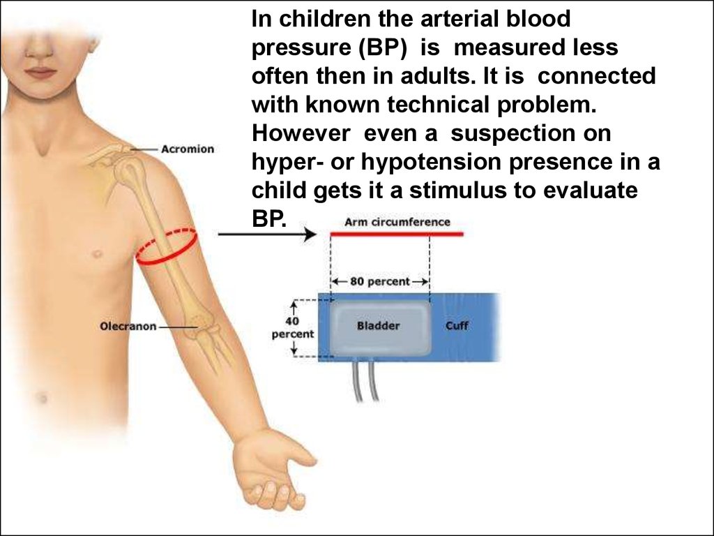
41.
In children the arterial bloodpressure (BP) is measured less
often then in adults. It is connected
with known technical problem.
However even a suspection on
hyper- or hypotension presence in a
child gets it a stimulus to evaluate
BP.
42. The Normal values of arterial blood pressure
• depend on the child age and should beapproximately considered:
• in newborns 80/50 mm Hg,
• in infants (up to 1 year) - 85/55 mm,
• in children adult then 2 years - 90/60мм.
43.
Current cardiac examination ismade in 4 steps.
44.
45. I. The inspection of heart area allows to reveal following symptoms:
• Precordial bulge, which isindicative for significant
increasing of heart in size.
• Visible pulsations. Their
localization has a
diagnostic relevance:
a) Exaggerated apical
pulsations is indicative for
left ventricular
hypertrophy (LVH).
b) Left parasternal and
epigastric pulsations can
denote right ventricular
hypertrophy (RVH).
c) Pulmonary arteria
pulsations (over left
second intercostal space)
are present in pulmonary
hypertension (raised
blood pressure in
pulmonary artery).
46. II. Palpation is important for determination:
• Apex beat. It isshifted downwards
and outwards in LVH
and outwards in
RVH.
• The maximal
impulse is apical in
LVH and left
parasternal in RVH.
• Palpable thrills
(flutter) are
sensations of
vibration felt by
hand of the
physician. They
accompany usually
the significant
organic murmurs.
47. III. Percussion.
The percussion of cardiac borders is
obsolete now in modern cardiology. It can
not easily detect cardiac enlargement or
to differentiate between left and right
hypertrophy. The inspection and palpation
are clinically more informative. The
percussion of heart borders can be useful
in hydropericardium evoluation. In this
case the conclusion that “the heart is
dramatically enlarged and its borders are
extended so it is difficult to define them"
could be done.
48.
It should bemade on the
four cardiac
areas and on
the left
parasternal
area (see the
picture).
IV. Auscultation.
49. The clinical comment of auscultation shold include next characteristics:
The heart sounds.• Normal heart sounds can be marked like the abbreviation S1+S2
(sound 1 and sound 2).
• The heart sounds can be splinted or reduplicated. It means a broad
gap existents between dubbed sound. For example the heart sounds
formula can be written as S1+S2+S2 and it is serious application for
congenital heart disease as atrial septal defect existence.
• When one tone is heard as louder then other it can be noted as the
fixed sound: s1+S2, for instance. This tune over aortal area is typical
for arterial hypertension.
• If the additional sounds are listened they have to be marked as s3
and even s4. As a rule they are not too much loud.
• Muffled (weak) sounds (s1+s2) with tachycardia suggests myocarditis
(inflammation disease of heart muscle).
• Distant sounds with quiet precordium suggests pericardial effusion.
• The second sound (S2) on pulmonary area is a essentially useful for
diagnostics noncyanotic congenital heart disease (see latter).
50. The clinical comment of auscultation shold include next characteristics:
Murmurs. Their comment should include
the next points.
Duration and relationship with cardiac
phases: systolic, diastolic or continuous.
Intensity: faint or loud, graded from 1 to 6
degree by Styll.
Character: soft, harsh or rumbling.
Location: area of maximal intensity.
Propagation: on body back, according
blood flow etc.
51. The clinical comment of auscultation shold include next characteristics:
• Pericardial rub. It is a friction soundheard in pericarditis.
52.
The heart sounds can be visualized in paper orelectronic screen by phonocardiography
method. Phonocardiogram allows to study the
frequency of acoustic waves forming heart
sounds and cordial murmurs, abnormal
fluctuations corresponding to splitting or
additional sounds and other parameters.
53.
For successful diagnostics ofcardiovascular diseases in
children it is necessary to
master several acceptances of
clinical study and to know
important cardiological signs
and syndromes.
54. Cyanosis
is defined as skin or mucosa bluishness.• Acute cyanosis with respiratory distress is
observed in respiratory or heart failure.
• Chronic cyanosis is moistly due to
congenital cyanotic heart disease.
• In both events the capillary blood is
deficient to oxygen and this way has a dark
color.
55. A femoral pulsation
or palpation of the pulse on hip arteryis important sign.
Weak or absent pulsation suggests
coarctation (narrowing) of the aorta.
56. The syndrome of congestive heart failure
is formed from specific signs.It can be acute or chronic.
For understanding the developmental
mechanisms of this important
syndrome it is necessary to consider
the heart as pump executing blood
pumping from venous riverbed into
arterial.
57. In acute failure, the cardinal triad is tachycardia, tachypnea and enlarged and tender liver.
1. Tachycardia (high heart rate): The sickheart tries to compensate its pumping
insufficiency by frequency of own efforts.
Pulse is rised.
• 2. Tachypnea (breathing frequency
increasing): The organism tries to rise blood
oxygen because in conditions of slowly
blood circulation there is tissues hypoxia.
• 3. Enlarged and tender liver due to venous
blood sequestration in it in conditions when
the diastolic heart function is insufficient.
58. In chronic failure:
• 1. Exertional dyspnea is present. Thebreathlessness appears in response to
physical load, for instance, the rising
on floors is getting difficult for the
child.
• 2. Other features as engorged neck
veins, hepatomegaly and edema of
lower limbs are presented.
59.
The Semiotics of the commonestdiseases of cardiovascular
system in children.
60.
The Fallot`sTetralogy is a one of
the commonest
congenital heart
diseases being
accompanied by
cyanosis. It
accounts the 10% of
all congenital heart
disease and about
50% of cyanotic
cases.
61.
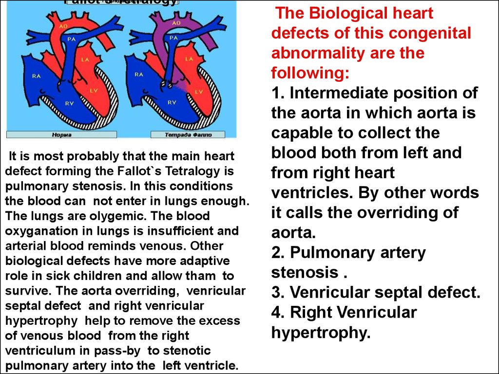
Fallot`s TetralogyIt is most probably that the main heart
defect forming the Fallot`s Tetralogy is
pulmonary stenosis. In this conditions
the blood can not enter in lungs enough.
The lungs are olygemic. The blood
oxyganation in lungs is insufficient and
arterial blood reminds venous. Other
biological defects have more adaptive
role in sick children and allow tham to
survive. The aorta overriding, venricular
septal defect and right venricular
hypertrophy help to remove the excess
of venous blood from the right
ventriculum in pass-by to stenotic
pulmonary artery into the left ventricle.
The Biological heart
defects of this congenital
abnormality are the
following:
1. Intermediate position of
the aorta in which aorta is
capable to collect the
blood both from left and
from right heart
ventricles. By other words
it calls the overriding of
aorta.
2. Pulmonary artery
stenosis .
3. Venricular septal defect.
4. Right Venricular
hypertrophy.
62. The semiotics reflect pathophysiological changes occurring in this congenital heart disease.
• In advanced stage of disease in children withFallot`s Tetralogy always there is the central
cyanosis. It is a bluish discoloration mostly seen
in lips, tongue, mucosa membranes of oral cavity
and fingernails.
• The onset of cyanosis is usually delayed to 1 – 2
months after birth. In early cases it reveals only
during physical exertion (for example, crying or
mother`s breast sucking) and appears near the
mouth (this is so-called circumoral cyanosis) and
eyes (circumocular cyanosis).
63. High hemoglobin
• The compensaroty hemoglobin anderythrocytes elevation reachs the level
nearly double exceeding the normal.
The polycytemic blood is characterized
by high viscosity. In this conditions in
young patient with Fallot`s tetralogy the
hypercyanotic spells appear.
64. Hypercyanotic spells
They are attacks of deep cyanosis and respiratory distresswhich may by precipitated by crying or infection. In this
conditions the high polycytemic blood viscosity provoke
severe disturbance of blood circulation in lungs. It leads to
sudden hypoxemic attacks look as hypercyanotic spells.
Mild attacks (for minutes) are followed by weakness and
sleep, while severe attacks (for hours) may progress to
convulsions and unconsciousness. It is characteristic that
children aged above 2 years are trained to fight with
beginning of the spell, sitting squat. In this position with
heavy flexed hips and knees the squeezing of lower limbs
large arteries reduces aortal out-put and directs more
blood in pulmonary artery. It provides pulmonary
oxygenation improve. Seems, the nature itself prompts to
physician how to help to the patient with hypercyanotic
spell.
65.
The othersuggestive
clinical
features:
Clubbing of
fingers. It is
usually
observed after
the age of 1-2
years and the
clubbing looks
blue.
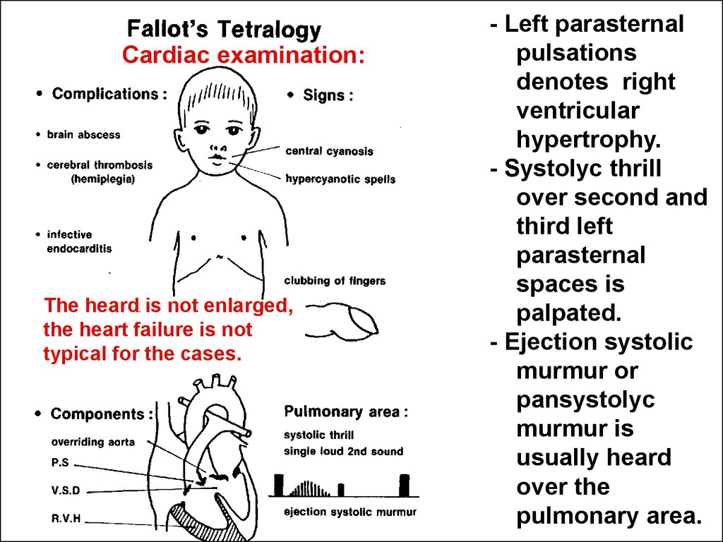
66.
Cardiac examination:The heard is not enlarged,
the heart failure is not
typical for the cases.
- Left parasternal
pulsations
denotes right
ventricular
hypertrophy.
- Systolyc thrill
over second and
third left
parasternal
spaces is
palpated.
- Ejection systolic
murmur or
pansystolyc
murmur is
usually heard
over the
pulmonary area.
67.
The additional clinical investigations.Chest X-ray shows pulmonary oligemia, normal sized heart,
prominent right ventricle with uplifted apex (“boot shaped”
heart or “duck sitting on the water”).
Roentgenogram of an
8-yr-old boy with
tetralogy of Fallot.
Note the normal heart
size, some elevation of
the cardiac apex,
concavity in the region
of the main pulmonary
artery, right aortic
arch, and diminished
pulmonary vascularity.
68. The guidelines of care for the sick children.
• When the hypercyanotic spell happens it isnecessary to becalm a child and put him or
her on "frog” on belly position with flexed
hips and knees brought to the bosom until
the condition will improve. The moistened
oxygen from mask also can be helpful.
• Other important element of the permanent
care is reasonable overdrinking in a sick
child. The patients with Fallot`s Tetralogy
must use more fluids because it
counteracts with the high blood viscosity.
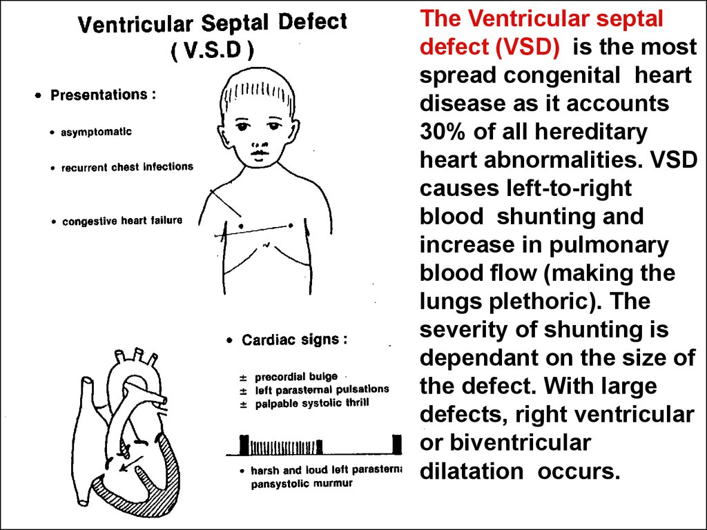
69.
The Ventricular septaldefect (VSD) is the most
spread congenital heart
disease as it accounts
30% of all hereditary
heart abnormalities. VSD
causes left-to-right
blood shunting and
increase in pulmonary
blood flow (making the
lungs plethoric). The
severity of shunting is
dependant on the size of
the defect. With large
defects, right ventricular
or biventricular
dilatation occurs.
70.
Patients with VSD are frequently asymptomatic andthe condition is accidentally discovers on routine
cardiac examination. In severe cases the disease
revels on recurrent chest infections due to plethoric
lungs. In this cases the pneumonia is complicated
with congestive heart failure. So this condition in a
young child can be the main presentation of VSD
In other hand the children with a small VSD (so
called the Rogee`s disease) do not show signs of
severe heart disease and develop well. The small
defects of ventricular septum (about 60% of cases)
are capable to close spontaneously within 2 – 4
years making joy for parents, patient and physician
in charge.
71.
Healthy!Murmur,
VSD!
72. Semiotics of V.S.D.
• Characteristic murmur is pansystolic, loud, harsh,and left parasternal mainly over 3rd and 4 rd left
parasternal spaces localized. This murmur should
be clinically differentiated from other causes of
pansystolic murmurs especially mitral
incompetence (maximum intensity is heard over
mitral area) and tricuspid incompetence
(maximum intensity is heard over tricuspid area).
Both conditions are much less commoner than
V.S.D and are characteristic for chronic rheumatic
heart disease in school aged children.
• Cardiomegaly occurs with moderate and large
sized V.S.D. It can be detected clinically by the
precordial bulge and the left parasternal
pulsations.
• Congestive heart failure (CHF) also occurs with
moderate and large sized
73. The manifestations of CHF may by chronic or acute.
• Chronic CHF appears gradually in the form of dispneaduring breast feeding and can lead the children to growth
delay. The Explanation is that the nursing provokes in
baby a physical effort (especially sucking) but any
physical efforts in congestive heart failure are bad
tolerated. The other explanation is concluded that each
breast feeding is a water load. In this conditions the infants
with CHF can instinctively avoid to enlarge the blood
circulating volume and worse the diastolic function of
insufficient heart.
• In advanced cases the dispnea becomes evident at rest
and other manifestations of chronic failure as engorged
pulsating neck veins, hepatomegaly and edema of lover
limbs appear.
• b) Acute CNF is usually precipitated by chest infection.
Diagnosis depends on the presence of the clinical triad of
tachycardia, tachypnea and tender liver. In severe cases
chest retractions and bilateral crepitations as
manifestations of pulmonary edema may appear.
74. Other diagnostic approaches
• Diagnostic echocardiographydemonstrates the septal defect, its size
as well as the degree of cardiac
enlargement
• Chest X-ray shows variable degree of
cardiomegaly and typical pattern of
pulmonary plethora.
75.
Presence of cardiomegaly orcongestive heart failure in
infancy is an indication for early
surgical intervention in VSD.
76.
A) Preoperative roentgenogram in a ventricular septal defect with alarge left-to-right shunt and pulmonary hypertension. Significant
cardiomegaly, prominence of the pulmonary arterial trunk, and
pulmonary overcirculation are evident.
B) Three years after surgical closure of the defect. There is a marked
decrease in the heart size, and the pulmonary vasculature is
77. The guidelines of care for sick children.
The children sufferring from VSD with chronic
congestive heart failure need a patient care providing
sufficient feeding supply especially nursing. All
anthropometrics like weight and length in sick children
mast be controlled attentively.
The children are predisposed to respiratory infections.
That is way the contacts between chronic and acute sick
patients have to be restricted.
Even for children with small size of VSD the prevention
of bacterial endocarditis mast be provided very early. The
kids aged 2 years should be accustomed to use toothbrush
that reduces the risk of toothcaries, odontogenic
bacteriemia and septic endocaditis localized in VSD area.
78. The atrial septal defect (ASD)
• The atrial septal defect (ASD) is other one of the widespread innate heart diseases as it accounts 30% of allhereditary heart abnormalities.
• Atrial septal defect (or ostium secundun defect) causes
left-to-right at the atrial level. The blood abnormally enters
from the left atrium to the right throught the abnormal
foramen overloading right intraventricular volume and
blood volum in pulmonary vessels riverbed.
• Patients with ASD are frequently asymptomatic and the
condition can be accidentally discovered on routine
cardiac examination. In severe cases the disease reveals
on recurrent chest infections due to plethoric lungs. In this
cases the sudden pneumonia is complicated with
congestive heart failure. So this condition in a young child
can be the main presentation of ASD.
79. Symptoms of ASD
1. The systolic murmur over the pulmonary area is usuallyheard. It is not very loud (grade 1-2 by Styll) and seldom
accompanied by thrill felt in palpation of heart area.
2. Abnormal second sound. A broadly (widely) split (forked)
and fixed (exaggerated) second sound (S1+S2+S2) on
pulmonary area is the most characteristic finding of ASD
This important clinical phenomenon appears due to
insimultaneouse closing aorta`s (first component of sound 2)
and pulmonary artery valves (second component of sound 2)
in conditions of high blood pressure in right ventricle.
3. The leftside parasternal pulsations is defined over the
chest and denotes a right ventricular hypertrophy.
4.
Diagnostic echocardiography demonstrates the defect
and degree of righr atrial and right ventricular enlargement.
5. Chest X-ray shows variable degree of right atrium, right
ventricle or common cardiomegaly and tipical pattern of
pulmonary plethora.
80.
81. The other causes of ejection systolic murmur
1. Innocent (functional, non-malignant) systolic murmur isfaint, soft and short. It is commonly heard in up to 50% of
well children. This murmur should not be confused with the
significant pathological murmurs of congenital heart disease.
2. Hemic murmur. It is soft murmur maximally heard over
pulmonary area. It changes in character with changes in
body position and not associated with a thrill. A symptoms of
severe anemia are usually presented, especially a skin pallor
and Hb < 100 g/l.
3. Pulmonary stenosis (PS) murmur is systolic and
maximally heard over the pulmonary area. It is commonly
associated with a palpable thrill. The second sound is split
with weak even inaudible pulmonary component (S1+S2+s2).
Sygnificant right ventricular hypertrophy is usually present.
4. Aortic stenosis (A.S.) provokes the maximum intensity of
the ejection systolic murmur over aortic area. The murmur is
rough and loud and commonly associated with palpable
thrill. It usually propagates to the neck. Left ventricle
hypertrophy is usually present.
82. The guidelines of care for sick children with ASD.
They are the same as discussing before in patientswith VSD. The children sufferring from A.S.D. with
chronic congestive heart failure need a patient
care providing sufficient feeding supply especially
nursing. The children with A.S.D. are
predisponded to respiratory infections. That is
way a contacts between chronic and acute sick
patients have to be restricted. Also the prevention
of bacterial endocarditis mast be provided very
early. The kids aged after 2 years should be
accustomed to use toothbrush that reduces the
risk of toothcaries, odontogenic bacteriemia and
septic endocaditis localized in ASD area.
83.
Symptoms PDA.- Continuous machinery
murmur. It is maximally
heard over the pulmonary
area and may radiate to left
clavicular or left sternal
borders. It is usually
associated with a palpable
thrill.
- Bounding (jumping) arterial
pulsations. The pulses is
prominent and can be easy
felt due to the wide arterial
pressure. The artery
dorsalis pedis pulsations
can be easily felt. in
children with PDA.
The cardiomegaly and
congestive heart failure
84. Other diagnostic approaches
Echcardiography reveals increased left
atrial and left ventricular sizes.
UltraSound-Scanning from the
suprasternal notch can visualize the
ductus.
For confirming diagnosis PDA some times
the contrast aortography is
recommended.
85.
Chest X-Ray in a patient with PDA. The heart size is normal,the pulmonary artery segment is dilated, and the pulmonary
vascularity is slightly increased.
86.
The Rheumatism (rheumatic fever) is anautoimmune disease, which develops as
complication of streptococcal infections
(pharyngitis or scarlet fever), caused by betahaemolytic streptococcus of group A
87. Jones`s criteria
• 5 Jones`s criterias (or clinical manifestations) aregenerally used for diagnosis of rheumatic fever.
• The criteria`s using is based on two rules.
• For diagnosis it is necessary the presence of 2
major criterias (or major clinical manifestations) or
one major and 2 minor manifestations.
• There is an evidence of recent streptococcal
infection (case history, positive ASL-O test
showing antistreptococcal antybodies in serum
etc.).
88. Major criteria (Jones`s criteria or manifestations) of rheumatic fever.
• 1/ Polyarthritis occurs in 75% of cases. Therheumatic arthritis is typically multiple and
affects mainly the large joints as knees,
ankles, elbows and wrists. The inflamed
joints are usually swollen, red, hot and
tender with limitation of movements. The
rheumatic arthritis is also transient (lasts
less then one week in the affected joint
never produsing destruction or other
complicatons) and migratory (leaves one
joint to affect other).
89. Major criteria (Jones`s criteria or manifestations) of rheumatic fever.
2/ Carditis occurs in 50% of cases. The
next symptoms are characteristic for
carditis.
– Disproportionate tachycardia. It means the
heart rate is bigger as it has to be according
the child age and his or her fever.
– Significant murmur. In common cases it is
systolic murmur localized in apical area of
heart and characteristic for mitral valvulitis
(inflamarory disturbans of mitral valve).
– Pericardial friction rib is tipical for pericarditis
(inflammation of pericardial serosa).
– Congestive heart failure may be also present.
90. Major criteria (Jones`s criteria or manifestations) of rheumatic fever.
• 3/ Reumatic chorea.The chorea is the neurological
manifestation of rheumatic fever. This syndrome is
characteristic for 10% patients suffering from rheumatism.
The condition occurs mainly in school age children and
females are often more affected.
The main features of rheumatic chorea are the following.
• Chorea movements. They are rapid, jerky, purposeless and
nonrhythmic involving mainly the muscles of the face,
trunk and distal extremities. Movements are aggravated by
emotional stress and disappear during sleep. The
condition can be misinterpreted as a conscious intention
to irritate parents or teachers.
• Emotional lability. The patients often appears nervios and
may show crying without apparent reason.
• Muscle hypotonia of variable degree. The child becomes
unable to eat by himself and frequently drops objects. In
severe cases the pseudoparalysis due to muscle hypotonia
can be revealed.
91. Major criteria (Jones`s criteria or manifestations) of rheumatic fever.
• 4/ Erythemamarginatum
(annular
erythema) revels
in 5% children
with acute
rheumatic fever.
It forms wavy
lines or rings of
sharp margins
mainly over the
trunk.
92. Major criteria (Jones`s criteria or manifestations) of rheumatic fever.
• 5/ Subcutaneous nodules. It can befound in 1% children with acute
rheumatic fever. They are a small round
hard and painless nodules felt in the
field over bony prominences.
93.
The Chronic rheumatic heart diseaseis a continuation of the acute
rheumatic fever and is characterized
by inconvertible valvular damage.
94. Recognition of the valvular lesions depends on auscultation of the characteristic murmur.
Mitral incompetence (M.I.).– An apical harsh pansystolic murmur is
present. It always propagates to the
axillaries area and commonly
accompanied with a systolic thrill.
– Simultaneously the left ventricular
hypertrophy (LVH) develops.
95.
Mitral stenosis (M.S.).
An apical mid-diastolic rumbling
murmur is present.
Simultaneously the right ventricular
hypertrophy (RVH) develops.
96.
Aortic incompetence.A soft and bloving diastolic murmur is
heard over aortic area.
The left ventricular hypertrophy (LVH)
develops in severe cases.
Periferal signs of wide pulse pressure are
present. It is so-called water hummer pulse
and elevated systolic with lowered diastolic
blood pressure (for instance, 140/40 mm
Hg).
97.
Aortic stenosis.
A harsh and loud ejection systolic
murmur is heard over aortic area. It
usually propagates to the neck and is
commonly associated with systolic
thrill.
The left ventricular hypertrophy (LVH)
of varios degrees develops in some
cases.
98.
Atrial flatter99.
Ventricular premature bites (exstrasystols)100.
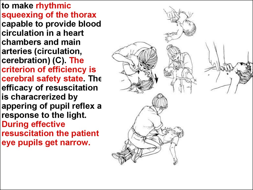
AV - block101. ABC program of cardio-pulmonary and cerebral resuscitation
The sudden cardial arrest for severalminutes leads a child to the clinical death,
which without skilled help ends by biological
death. That is why everybody but medical
staff in the first hand must know the
practical approaches and always has to get
ready to render the cardio-pulmonary and
cerebral resuscitation.
102. The principles of resuscitation are:
to restore airways abilityfor air pass
(A), organize
artificial lung
ventilation
(breathing)
(B),
103.
to make rhythmicsqueexing of the thorax
capable to provide blood
circulation in a heart
chambers and main
arteries (circulation,
cerebration) (C). The
criterion of efficiency is
cerebral safety state. The
efficacy of resuscitation
is characrerized by
appering of pupil reflex at
response to the light.
During effective
resuscitation the patient
eye pupils get narrow.