


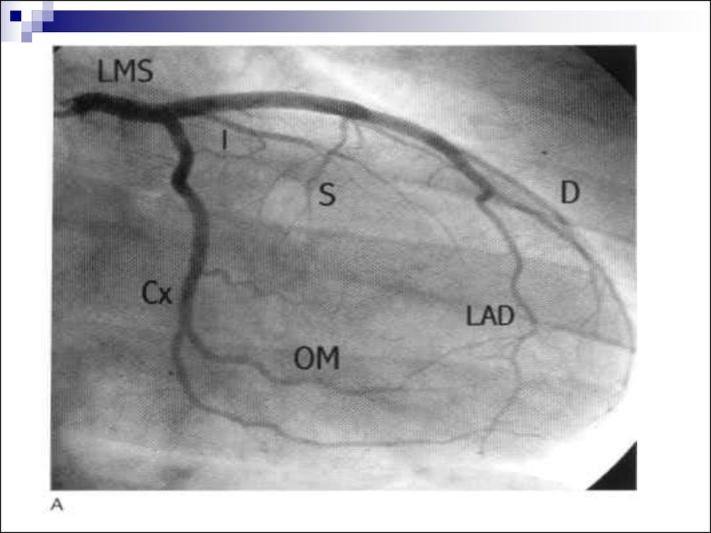
 Coronary arteriogram, same projection and patient as in (A), obtained 1 day later. The stenosis in the left anterior descending coronary artery (arrow) has been reduced after percutaneous balloon angioplasty.")
Aortogram in a patient with acute traumatic aortic injury. The site of injury is the focal outpouching at the insertion of ductus arteriosus (arrow).")


")


 and PA (B) radiographs of the chest in same patient on same day. Note that the cardiac silhouette appears larger on the AP radiograph and may be mistaken for disease if patient position is not considered in the interpretation.")


.")


 medicine
Similar presentations:


The Heart
1. The Heart
2. Imaging of the heart will be considered under the following headings:
1. Simple x-ray2. Screening
3. Cardiac catheterization
4. Angiocardiography
5. Coronary arteriography
6. Ultrasound
7. Isotope scan
8. MRI
3. Simple x-ray
A simple x-ray of the chest is mandatory asthe first imaging investigation in cases of
heart disease, because it yields vital
information concerning of the
1. size of the heart
2. enlargement of individual chambers
3. condition of the lung fields
4. PA view of normal chest. RA, right atrium; RDPA, right descending pulmonary artery; RPA, right main pulmonary artery; SVC, superior vena cava; AA, aortic arch; DA, proximal descending thoracic aorta; LPA, left pulmonary artery; RV, right ventricle.
5. Lateral view of normal chest. RV, right ventricle; RSS, retrosternal clear space; AA, ascending aorta; LPA, left pulmonary artery; RPA, right pulmonary artery en face; IVC, inferior vena cava; LA, left atrium; LV, left ventricle.
6. Screening
Cardiac calcification is seen at screening withan image intensifier than on a simple film.
Calcification is most commonly seen:
in the mitral or aortic valves
but may also be seen in atheromatous coronary
arteries, in the mitral annulus
or in a left atrium containing mural thrombus
7. Echocardiography
1. Echocardiography is a highly versatiletechnique, which is central in cardiological
diagnosis but is operator dependent and
requires considerable experience.
2. Echocardiography is performed from the
transthoracic route using a sector probe.
8.
3. Patient is positioned in a 45 degreesemierect position rotated towards his/her
left side to enhance cardiac contact with
chest wall.
4. Two-dimensional imaging gives direct
information about the anatomy and
physiology of the heart
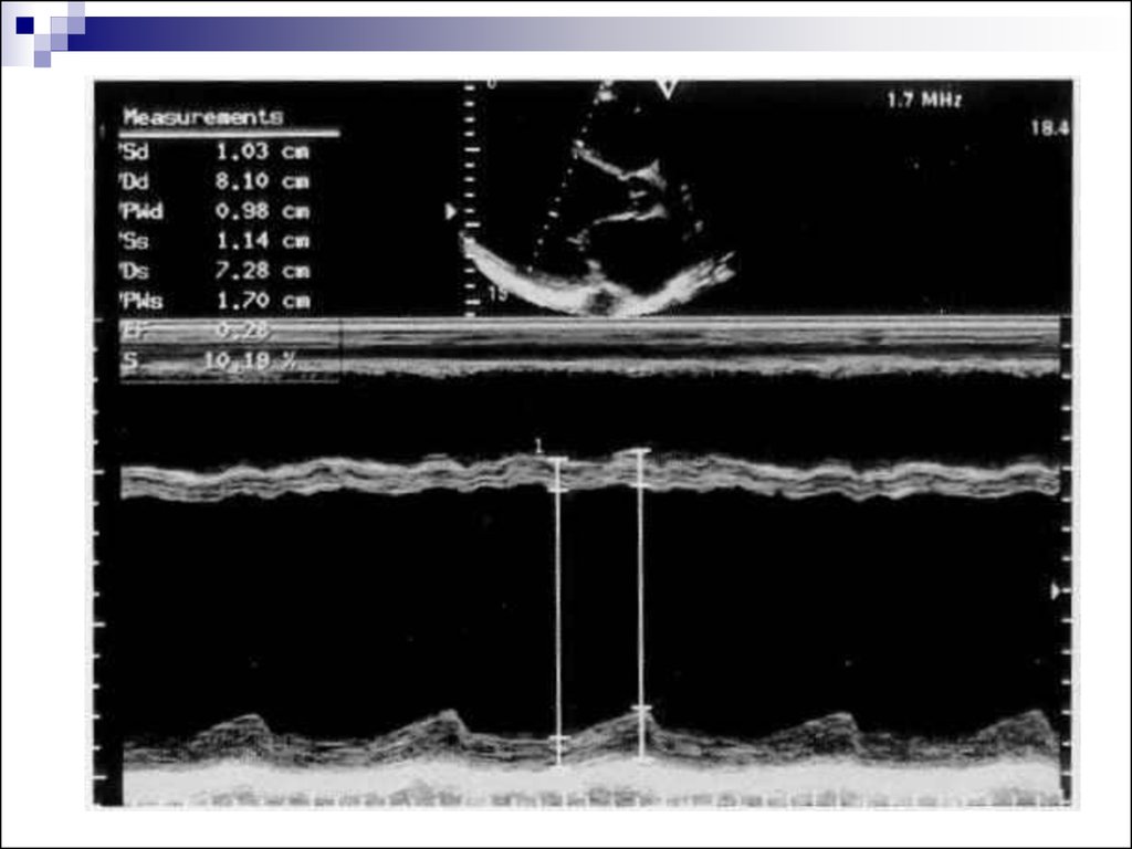
5. M-mode is a one-dimensional
evaluation useful for precise measurement
and timing of cardiac events.
9. Echocardiography an aneurysm of the apex of the left ventricle
10.
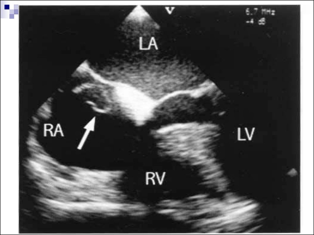
11. Apical four-chamber transthoracic echocardiogram in a patient with hypertrophic cardiomyopathy.
12.
13. Doppler examination
Doppler evaluation allows the study1. of different flow velocities within the
cardiac chambers and in the outflow tracts
2. calculation of the cardiac output,
ejection fraction
14. Apical continuous-wave Doppler trace in a patient with dynamic left ventricular outflow tract obstruction due to hypertrophic cardiomyopathy.
15. Cardiac catheterization
This procedure requires the introduction ofa catheter into the heart and manipulation
of its tip under screen control so as to
enter different chambers of the heart or to
pass through abnormal defects of
communications.
16. Right heart catheterization
This can be performed percutaneously or aftersurgical exposure of a vein in the arm or groin,
and passage of a catheter from there to the
right. The tip is manipulated into the right
ventricle or beyond into the pulmonary artery or
lung fields. If there is an atrial septal defect,
ventricular septal defect, or patent ductus
present, the catheter may be passed to the left
atrium, left ventricle or aorta through the defect.
17.
The site of the catheter tip can beconfirmed by taking pressure recordings
during the investigation and also by taking
blood samples which are examined for
oxygen saturation. The pressure
recordings and oxygen saturation levels
are of vital importance in the diagnosis of
the different forms of congenital heart
disease.
18. Left heart catheterization
The usual technique of left heart catheterizationis for the radiologist to introduce a catheter
percutaneously into the femoral artery and to
pass it under screen control into the aortic arch
and through the aortic valves into the left
ventricle. Pressures are obtained from inside the
ventricle recorded, as is a withdrawal pressure
trace into the aorta.
19. Isotope scanning
Technetium-99m pyrophosphateaccumulates in damaged myocardium
whereas thallium-201 produces a deficient
uptake in territories supplied by occluded
or narrowed arteries. Thallium is most
commonly used as a screening technique
in patients with suspected coronary artery
disease.
20. demonstrates a partially reversible perfusion defect in th interventricular septum and posterior wall of the left ventricle
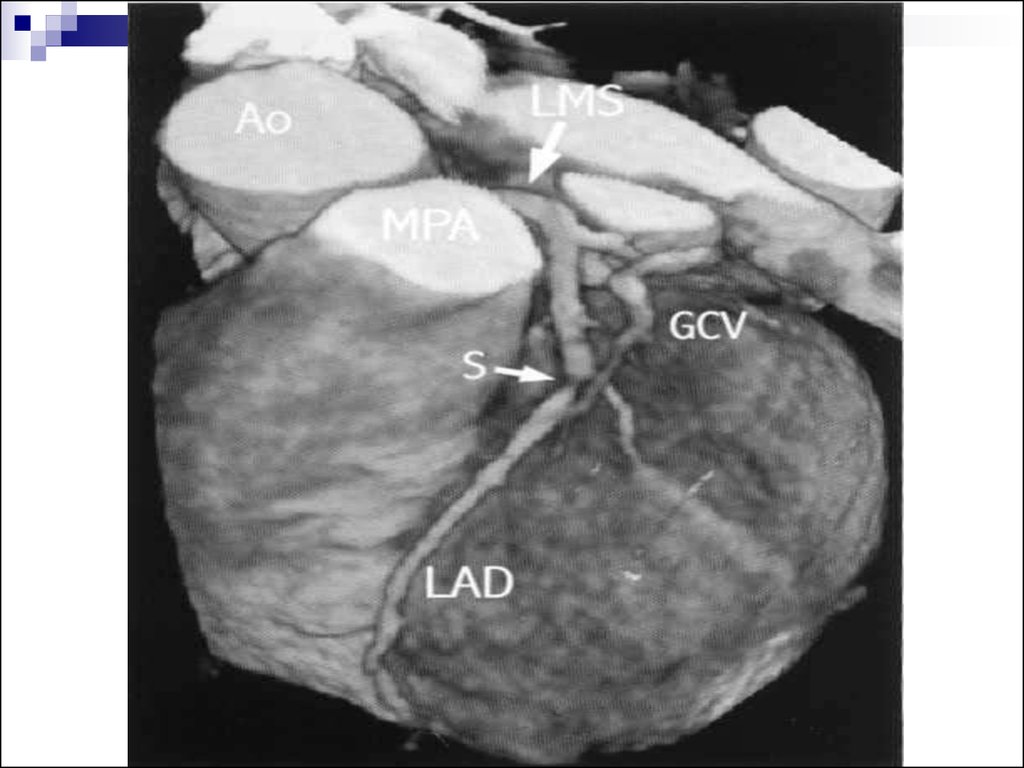
21. CT scan
CT evaluation of the heart is useful for detecting:1. the atherosclerotic disease of the coronary
vessels
2. myocardial calcifications and aneurysmal
3. dilatations and dissection of aorta
4. CT is the investigation of choice for the
evaluation: of cardiac tumors like myxoma, for
pericardial diseases like effusion and
pericardial tumors and dissection of aorta
22. Axial composite image
23.
24. Arteriography
Vascular access is usually obtained usinga percutaneous approach via the femoral
artery. Any major vessel or blood supply to
an organ can be studied by selective
arterial cannulation with contrast injection.
Radial, brachial, axillary or popliteal
arteries can also be punctured
percutaneously, if femoral artery access is
unsuitable. Anatomical detail is excellent;
hemorrhage and arterial thrombus are
recognized rare local complications.
25.
26. (B) Coronary arteriogram, same projection and patient as in (A), obtained 1 day later. The stenosis in the left anterior descending coronary artery (arrow) has been reduced after percutaneous balloon angioplasty.
27. Normal aortogram of transverse arch in patient suspected of having traumatic aortic injury. (B)Aortogram in a patient with acute traumatic aortic injury. The site of injury is the focal outpouching at the insertion of ductus arteriosus (arrow).
28. Intravenous digital subtraction angiography
This technique is utilized to visualize thearterial system by injection of a bolus of
contrast into the superior vena cava. After
passage through the heart and lungs, the
dilute contrast may be imaged in the
arterial circulation by computer
subtraction. Resolution is not as detailed
as conventional arteriography, but can be
an effective investigation in many clinical
situations.
29. MRI
MRI is fast gaining popularity as theinvestigation of choice in most cardiac
pathologies. Assessment of the flow
velocities in different cardiac chambers
and outflow tracts helps in estimating the
ejection fraction, cardiac output.
30.
Perfusion scanning gives the estimationof the surviving and infracted
myocardium following myocardial
infarction.
Cardiac tumors and pericardial diseases
are also better evaluated with MRI.
MRI is the investigation of choice in the
evaluation of congenital heart diseases,
can help in quantifying shunt.
31. MRI image
32. MRI image
33. MRI image
34. Cardiac pulsation
Normally, pulsation on the left border is muchmore prominent than on the right side. During
systole the left border is seen to contract forcibly
and during diastole it moves outwards from
2mm. After left ventricular contraction the
shadow of the pulmonary conus and the aortic
knob bulge forcibly outwards.
On the right side the lower border formed by
right auricle shows a faint contraction of not
more than 1 mm. Pulsation is greater in children
than in adults and increases after exercise.
35. Posterioanterior Projection
the upper right border is formed by:1. the SVC
2. the lower cardiac border is formed by
the RA
36.
the left border has three well-definedsegments:
1. the uppermost is formed by the aortic
arch
2. the main pulmonary artery lies
immediately below the aortic knob
3. LV and the apex (the LA appendage lies
between the pulmonary artery segment
and the LV and is usually not seen as a
separate bulge)
37.
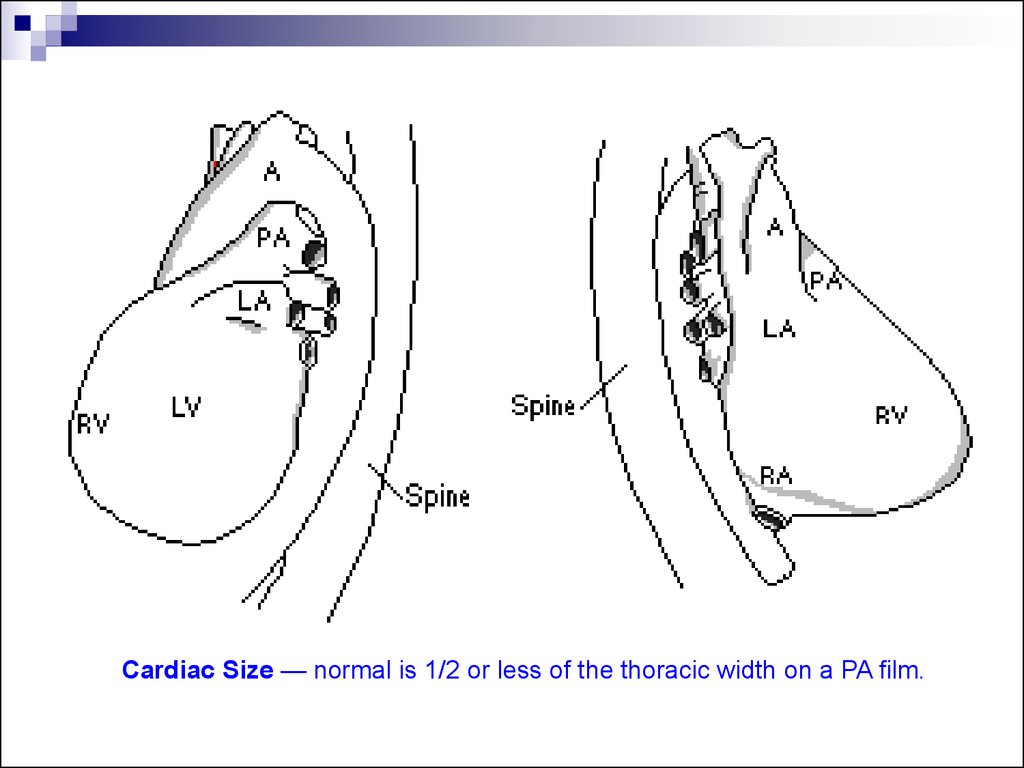
38. Lateral Projection
39.
40.
41.
Cardiac Size — normal is 1/2 or less of the thoracic width on a PA film.42. Technical Factors
• The heart appears larger on AP than PA views.• Film during expiration — simulates pulmonary
edema and the heart appears larger.
• One should check side markers for
dextrocardia.
• One should check the clavicles for angulation.
• Over penetrated films may miss heart failure.
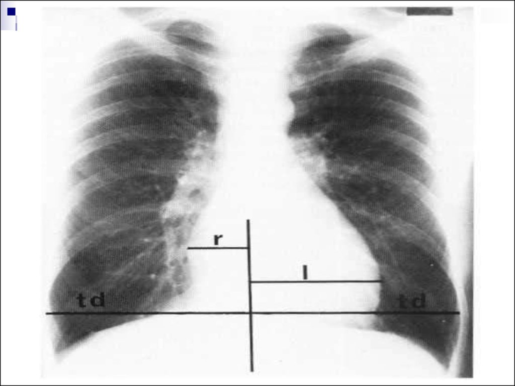
43. Cardiothoracic ratio (CT)
It is a simple method of estimating cardiacenlargement.
Estimation of CT ratio should always be
done in erect PA view.
Normal:
for adults 50%
for neonates 60%
Cardiomegaly is diagnosed on frontal
chest PA radiographs when the CT ratio
exceeds 50%.
44.
45. The causes for increased CT ratio due to nonstandard radiographic techniques include:
poor inspirationsupine position
prone position
AP radiographs, or with a short focus film
distance
46. Expiratory phase on a PA radiograph. Note the low lung volumes, apparent enlargement of the cardiac silhouette, and crowding of bronchovascular structures at the bases. Findings may be misinterpreted as heart failure if analysis of depth of inspiration is
notperformed.
47. AP (A) and PA (B) radiographs of the chest in same patient on same day. Note that the cardiac silhouette appears larger on the AP radiograph and may be mistaken for disease if patient position is not considered in the interpretation.
48. Common causes of cardiomegaly
Valvular heart diseases like mitralstenosis, mitral regurgitation, aortic
regurgitation
Pericardial diseases like pericardial
effusion
Myocardial diseases like ventricular
aneurisms
Congenital cardiac diseases like atria
septal defect, ventricle septal defect
49. Causes of small heart
constrictive pericarditisAddison’s disease
Pulmonary emphysema
50. Enlargement of the heart
It may be general, involving all chambersor eccentric involving one or two chambers
unequally.
51. The common causes of the left ventricular enlargement are:
hypertensionaortic regurgitation
aortic stenosis
coronary arteriosclerosis
acute/chronic nephritis
cardiac aneurism
coarctation of aorta
52.
The left ventricle enlarges to the left andposteriorly and only slightly to the right and
anterioly. Left side of the heart becomes
more globular.
53. Left ventricular enlargement
54. Lateral view shows the left ventricle extending behind the line of the barium-filledoesophagus (arrow).
55. The common causes of right ventricle enlargement are:
mitral stenosiscongestive failure
chronic pulmonary diseases
tricuspid regurgitation
Fallot’s tetralogy
56.
Right ventricle when enlarges, it does soby a broadening of its triangular shape. It
enlarges chiefly to the left and anterioly.
57. Direct signs of right ventricular enlargement are:
upward and outward displacement of theventricular border
elevation of the apex
an upper longer arc above the apex and
a lower shorter arc turning medially
below the apex
58. Indirect signs are:
prominent right atrial borderdilated pulmonary trunk
signs of pulmonary hypertension
59. Gross right ventricular enlargement
60. The common causes of left atrial enlargement are:
ischemic heart diseasemitral stenosis
mitral regurgitation
aortic obstruction and regurgitation
systemic hypertension
left heart tumor
61.
On the anterior view the right atrium formsless than the lower half to the right
mediastinal border in adults.
62. left atrial enlargement
63. The causes of the right atrium enlargement are:
Shunts into right atrium (left ventricular –right atrial shunt, ruptured aortic sinus
into right atrium)
Pulmonary obstruction and regurgitation
Pulmonary arterial hypertension
tricuspid obstruction and regurgitation
Right – sided cardiomyopathy
right atrial tumors
64. Right atrium enlargement
65. Essential hypertension
It is a common cause of cardiacenlargement.
In most cases there is unfolding and
pseudoenlargement of aorta.
The ascending part appears wider and
longer.
The aortic knuckle becomes higher.
66.
Left heart enlargement is common inprolonged hypertension.
The apex lies below the dome of the
diaphragm. Similar findings may be seen
in aortic regurgitation except vigorous
pulsation of the left ventricle.
When failure does occur the heart
enlarges to the left and right in the
transverse diameter greater than the long
diameter.
67.
The pulmonary artery and the conus aresomewhat dilated.
The enlargement hazy outline of the hilar
shadows may precede clinical evidence of
failure and is a useful sign.
68. Chronic nephritis
The heart is enlarged in 80% cases.Marked rounding of the left ventricle is a
conspicuous
Feature in chronic nephritis than in
essential hypertension. Pulmonary edema
occurs.
69. Pericardial effusion
A pericardial effusion is a collection of fluidin the pericardial sac, the fluid being either
serous, blood or lymphatic in origin.
70. Radiological features
Chest film: illustrates a symmetricallyenlarges and globular cardiac shadow only
when there is a significant effusion (>250
ml). Pericardial effusion should be
suspected if there has been a rapid serial
increase in the cardiac shadow, with
normal pulmonary vasculature.
Echocardiography: the investigation of
choice. Effusions are visible as echo-free
areas surrounding the heart.
71.
CT: may also identify the aetiology, e.g.mediastinal malignancy.
MRI: accurate for diagnosis and also
images the chest and mediastinum.
72. Causes
InfectiveUraemia
Posmyocardial infarction
Myxoedema
Malignancy
viral
bacterial
tuberculosis
bronchial and mediastinal tumors with pericardial
invasion
Collagen vascular diseases
systemic lupus erythematosus
rheumatoid arthritis
73. Pericardial effusion
74. Cardiac failure
Cardiac failure is said to be present whentissue demands cannot be adequately
supplied by the heart. It is usually due to
low output from ischaemic heart disease
but, paradoxically, may rarely result from
high output as a consequence of
excessive tissue needs in conditions such
as thyrotoxicosis or Paget”s disease.
75. Radiological features
On a chest x-ray the following may be seen:cardiac enlargement
upper-lobe vascular prominence: from raised
pulmonary venous pressure
pleural effusions: seen as blunting at the
costophrenic angels, but as the effusions
become larger, there is a homogeneous basal
opacity with a concave upper border
76.
interstitial pulmonary oedema: initially,prominence of the upper-lobe and narrowing of
the lower-lobe vessels. As venous pressure
rises, interstitial oedema develops and fluid
accumulates in the interlobular areas with
peripheral septal lines (Kerley “B” lines)
alveolar pulmonary oedema: with further
increases in venous pressure, fluid transgresses
into the alveolar spaces (alveolar shadowing)
with haziness and blurring in the perihilar
regions; in severe cases, pulmonary oedema
develops throughout both lung fields. The outer
thirds of the lungs may be spared, the bilateral
central oedema being described as “bat’s wing”
77. Valvular diseases of heart Mitral stenosis
Mitral stenosis presenting in infancy orearly childhood is due to congenital lesion.
It takes years to develop mitral stenosis
after rheumatic fever. Mitral stenosis
produces a pressure load on the left
atrium and ultimately on the right ventricle.
78. In posterioanterior view
An enlarged left auricle is seen as dense pearshaped opacity lying transversely inside thecardiac shadow.
Double heart shadow in many cases can be
seen to the right of the spine. Left border of the
heart becomes straight and is known as
mitralization.
Small aortic knuckle is caused partly by a true
hypoplasia of aorta and partly by right ventricular
rotation.
79. In right oblique view
The enlargement left auricle bulges backwardsand obliterates the translucent retrocardiac
space.
On barium swallow a bolus passes normally
down to a point just below the left main bronchus
when it seems to halt abruptly. Barium bolus
then fills slowly the lower third of the
oesophagus which is curved sharply backwards.
This sign is more obvious in expiration than in
inspiration.
80.
Elevation of left main bronchus due toenlarged left atrium may be seen.
Horizontally Kerley “B” lines are more
often noted. These lines are usually
persistent. Other more fluid signs such as
mottling, hilar edema and pleural effusion
may develop which disappear on
treatment.
81.
Rheumatic mitral stenosis. This frontal film shows marked enlargement of the82. Mitral regurgitation
Mitral incompetence may result fromfunctional or anatomical disturbance of the
cusps. Familial cases have been reported.
The characteristic signs are mid-systolic
click and a late systolic murmur. There is a
volume and pressure load on the left
ventricle and left atrium and in severe
regurgitation a pressure load on the right
ventricle.
83.
In mild regurgitation heart size may remainnormal.
In late cases, moderate cardiac
enlargement suggests left ventricular
rather than right ventricular enlargement.
Left atrial dilatation is usually obvious.
Gross enlargement of left atrium is noted
in chronic rheumatic regurgitation with
stenosis. Mitral valve calcification is
common.
84. Aortic valve stenosis
In ninety percent it is congenital in origin.Heart is never more than slightly enlarged
unless there is regurgitation.
Left border is often more rounded or longer than
normal with a low apex, a shape characteristic of
left ventricular enlargement.
Poststenotic dilatation of aorta is seen as a
localized bulge to the right above the right
atrium.
Calcification of the valves is almost invariable in
males over the age of 40 years.
85. Aortic regurgitation
Congenital regurgitation is usually due tobicuspid valve whose cusps elongates or
lack support. Aortic regurgitation with
rheumatic heart disease is often
associated with stenosis. In acute
regurgitation following bacterial
endocarditis heart may take many months
to enlarge.
86.
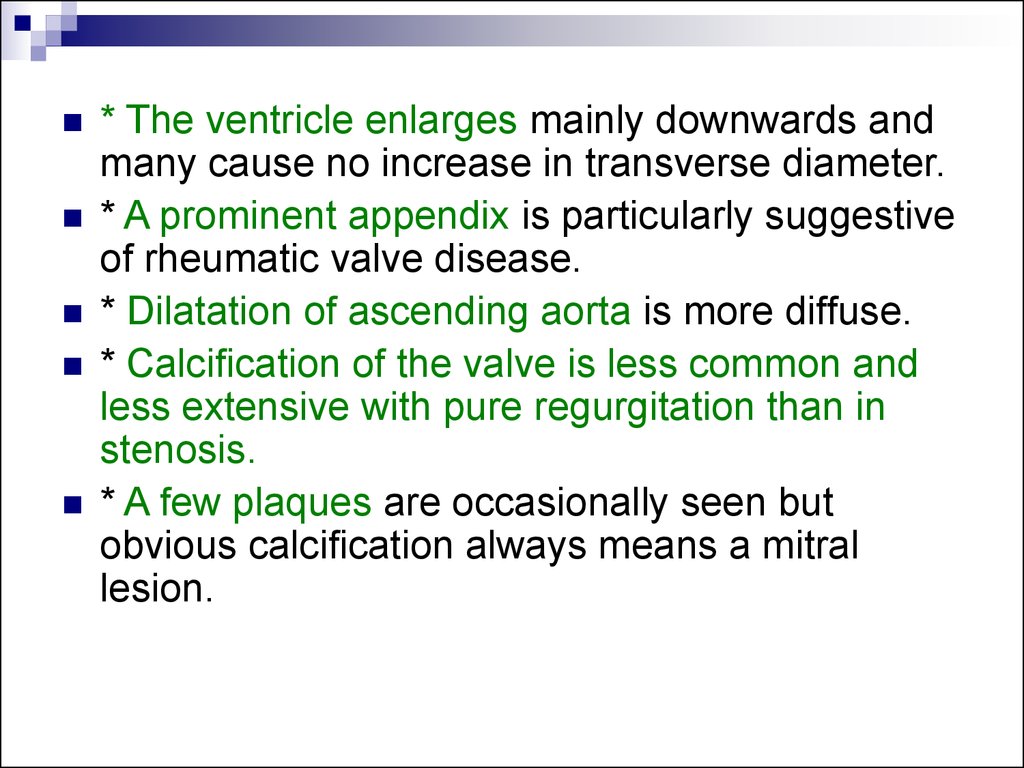
* The ventricle enlarges mainly downwards andmany cause no increase in transverse diameter.
* A prominent appendix is particularly suggestive
of rheumatic valve disease.
* Dilatation of ascending aorta is more diffuse.
* Calcification of the valve is less common and
less extensive with pure regurgitation than in
stenosis.
* A few plaques are occasionally seen but
obvious calcification always means a mitral
lesion.
87. Coarctation of aorta
It is a congenital narrowing of the aortic lumen inthe region of isthmus. If a coarctation presents
after the first year of life, it is usually symptomfree and symptom is discovered due to
hypertension, murmur or an abdominal chest
radiograph. It causes a systolic overload on the
left ventricle with hypertension in the upper part
of the body.
88. X-ray shows:
* enlargement of heart in the early weeks afterbirth and become very large if heart failure is
there
* descending aorta may lie far off to the left off to
the left of the spine
* rib notching is an important finding
* plethora with or without edema suggest a shunt
in addition to coarctation
* in adults aortic knuckle becomes prominent
89. Pulmonary stenosis
Pulmonary valve stenosis is always congenital.The heart is usually normal in size with severe
stenosis but may be slightly enlarged in
childhood as a result of marked hypertrophy of
the right ventricle with elevated apex.
Gross enlargement is seen only with congestive
cardiac failure.
Right atrium appears prominent.
Poststenotic dilatation of pulmonary trunk and or
the left branch occurs in 90% cases.
Pulmonary oligaemia is noted.
90. Pulmonary regurgitation
It may be:congenital
acquired
functional
91.
Isolated pulmonary regurgitation is abenign lesion unless associated with
pulmonary hypertension. The heart and
pulmonary trunk show little or no
enlargement.
Elderly patients on chronicity may develop
congestive failure. When the pulmonary
trunk is large with normal size heart,
idiopathic dilatation is due to pulmonary
regurgitation.
92. Venous hypertension
When there is an increase in resistance to flowbeyond the pulmonary capillaries, pressure rise
in the pulmonary veins with the production of
postcapillary or pulmonary venous hypertension.
i.e. 15 mmhg or more.
Earliest change is dilatation of upper zone
vessels. More often both veins and arteries are
widened, all vessels above the hilum are little
wider than those at lower levels. Vessels may
measure more than 3mm in diameter.
93.
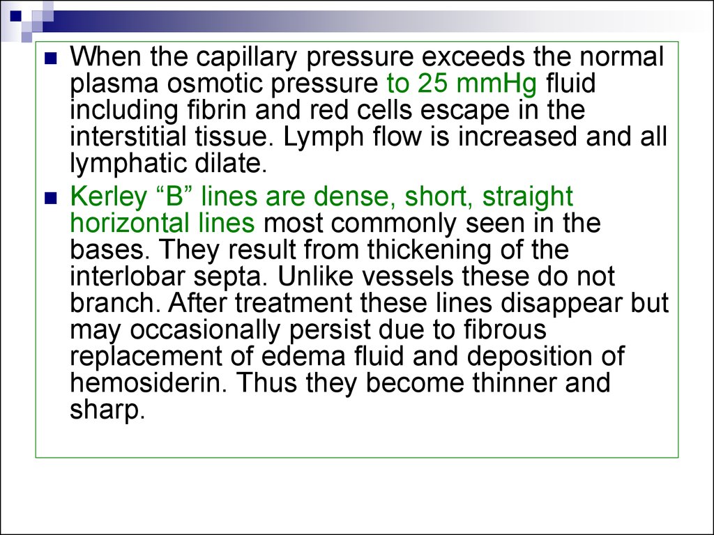
When the capillary pressure exceeds the normalplasma osmotic pressure to 25 mmHg fluid
including fibrin and red cells escape in the
interstitial tissue. Lymph flow is increased and all
lymphatic dilate.
Kerley “B” lines are dense, short, straight
horizontal lines most commonly seen in the
bases. They result from thickening of the
interlobar septa. Unlike vessels these do not
branch. After treatment these lines disappear but
may occasionally persist due to fibrous
replacement of edema fluid and deposition of
hemosiderin. Thus they become thinner and
sharp.
94.
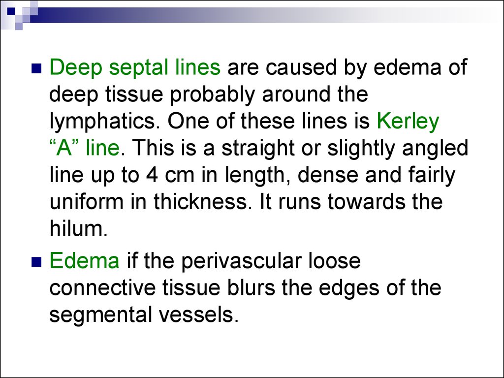
Deep septal lines are caused by edema ofdeep tissue probably around the
lymphatics. One of these lines is Kerley
“A” line. This is a straight or slightly angled
line up to 4 cm in length, dense and fairly
uniform in thickness. It runs towards the
hilum.
Edema if the perivascular loose
connective tissue blurs the edges of the
segmental vessels.
95.
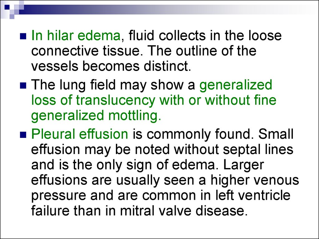
In hilar edema, fluid collects in the looseconnective tissue. The outline of the
vessels becomes distinct.
The lung field may show a generalized
loss of translucency with or without fine
generalized mottling.
Pleural effusion is commonly found. Small
effusion may be noted without septal lines
and is the only sign of edema. Larger
effusions are usually seen a higher venous
pressure and are common in left ventricle
failure than in mitral valve disease.
96.
When the pulmonary venous pressurereaches 30 mmHg, edema fluid may be no
longer contained within the interstitial
tissues but escape into alveoli. X-ray
shows ill-defined semi-confluent lying in
any part of the lung. The commonest
appearance is the “bat’s wing” shadow in
which the edema apparently has a
peripheral distribution. It may be unilateral.
97.
Pulmonary hemosiderosis is due to focaldeposition of hemosiderin. The lung show
diffuse mottling in all zones which may be fine of
course.
Pulmonary ossific nodules are also formed
following organization of intraalveolar edema.
The nodules are dense and irregularly round or
oval and rarely a small central medullary space
may be visible. These vary from 1 to 10 mm
most commonly seen in lower zones. These
increase slowly in number.
98. Fallot’s tetralogy
Consists of:ventricular septal defect
right ventricular outflow tract obstruction
pulmonary stenosis
right ventricular hypertrophy
99. Plain radiograph features:
the heart is usually is not enlarged at birth butmay enlarge later due to biventricular heart
failure
the pulmonary vasculature shows pulmonary
oligemia
the classic “cour en sabot” silhouette is due to
combination of a deeply concave pulmonary bay
and elevation from the diaphragm of slightly
angular cardiac apex due to right ventricular
hypertrophy
the ascending aorta is typically enlarged and
prominent on plain radiograph
100.
101. Ventricular septal defect
is abnormal opening between the twoventricles.
Types:
membranous
muscular
102. Chest radiograph:
left atrium is enlargedassociated hypertrophy of right ventricle
and left ventricle
increased pulmonary vascular markings
(plethora)
103. Atrial septal defect
Atrial septal defect is the abnormalcommunication between the right and the
left atria.
Types:
osteum secondum
osteum primum
104. Chest radiograph:
enlargement of right atrium and rightventricle
pulmonary vascular prominence in lung
field (plethora)
105. Atrial septal defect
106. Cardiac tumors
metastasis from bronchogenic carcinoma,mediastinal tumors, melanoma, and
lymphoma are the most common
malignant lesions of the heart
left atrial myxoma is the most common
primary tumor of the
107. Myxoma:
most common location is left atrium arisingfrom the interatrial septum
in echocardiography, a polypoidal and
mobile mass with heterogeneous
echotexture is seen
on Ct scan, a heterogeneous mass lesion
noted in the left atrium showing
inhomogeneous enhancement