


 medicine
Similar presentations:


Development of the heart
1.
VOLGOGRAD STATE MEDICAL UNIVERSITYDepartment of histology, embryology, cytology
Lecture for the general medicine IInd course
english medium students
Volgograd, 2015
2.
THE OBJECTIVES:Describe the main features of heart development to the fourchambered system.
Describe the development of the pericardium
Describe the development of primary and secondary atrial
septa and the ventricular septum.
Explain the changes occurring in the bulbis cordis and
truncus arteriosus in its transformation from a single to a
double tube.
Describe the developmental aberrations responsible for the
following malformations: patent ductus arteriosus (P.D.A.);
atrial septal defects (A.S.D.) and ventricular septal defects
(V.S.D.); tetralogy of Fallot.
3.
GENERAL PROVISIONS:the CVS is the first system to function in the
embryo,
vascular system appears in the middle of the 3rd
week when the embryo is no longer able to satisfy
its nutritional requirements by diffusion alone,
blood begins to circulate by the end of the 3rd
week.
4.
EndodermEctoderm
Angiogenic
cell cluster
Prechor
dal
plate
Amniotic cavity
Connecting
stalk
EARLY
DEVELOPMENT OF
THE EMBRYO
Allantois
Cloacal
membrane
Middle of the 3rd week – presomite stage, pan-cake appearance of
embryonic disc, intraembryonic endoderm constitutes the roof of
the spherical yolk sac.
Formation of the angiogenic cell clusters: splanchnic mesoderm
gives rise to angioblasts – cells of the mesenchymal origin
condensing into interconnecting cords of cells.
5.
DEVELOPMENT OF THEMESODERM
Notochord
Amniotic
cavity
Ectoderm
Paraxial
mesoderm
Mesoderm
Dorsal
aorta
A
Intermediate
mesoderm
Intercellular
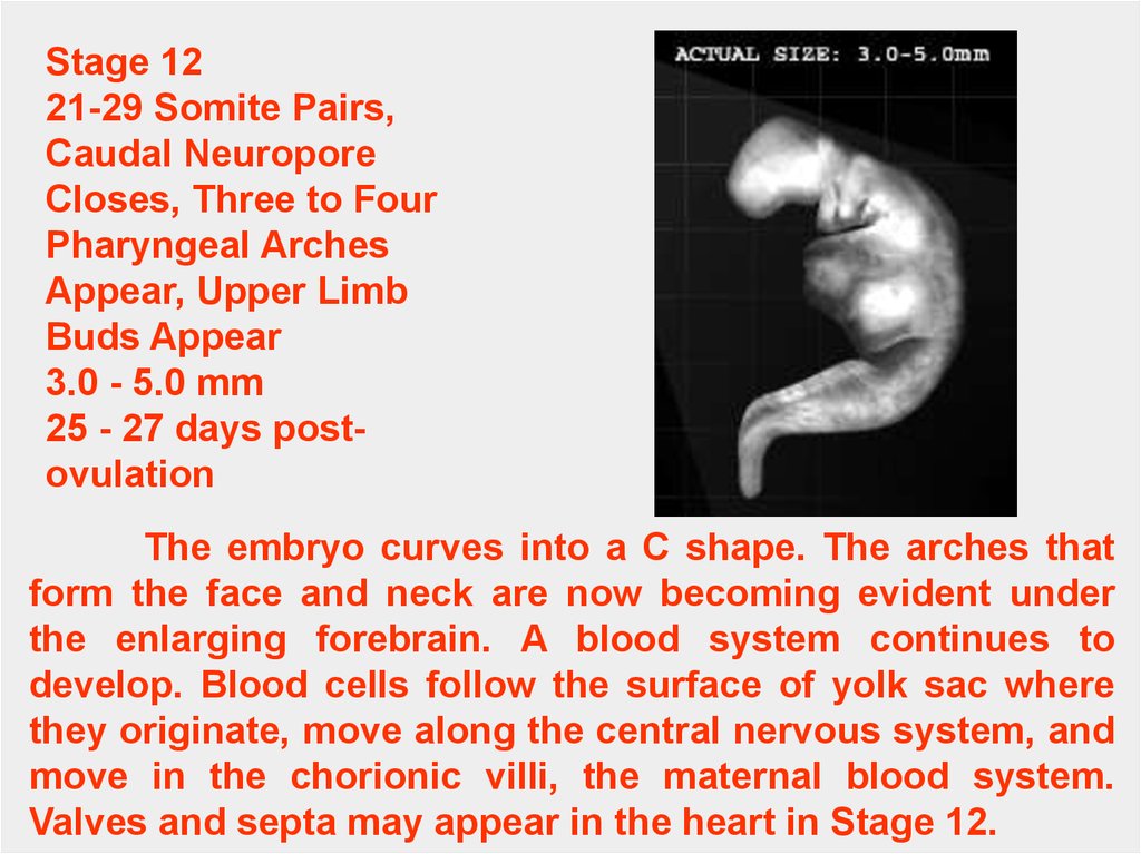
cavities
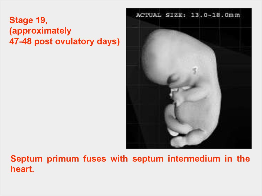
in lateral
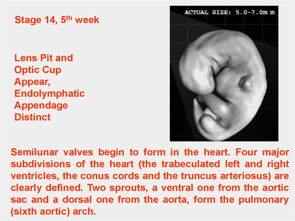
plate
B
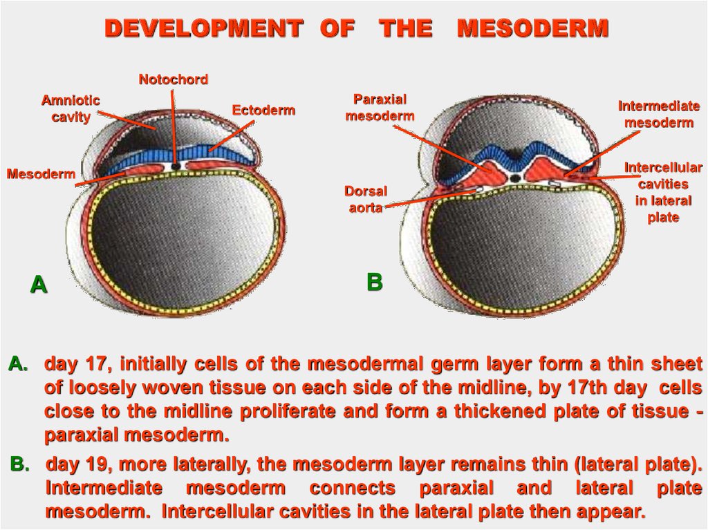
A. day 17, initially cells of the mesodermal germ layer form a thin sheet
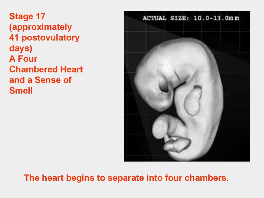
of loosely woven tissue on each side of the midline, by 17th day cells
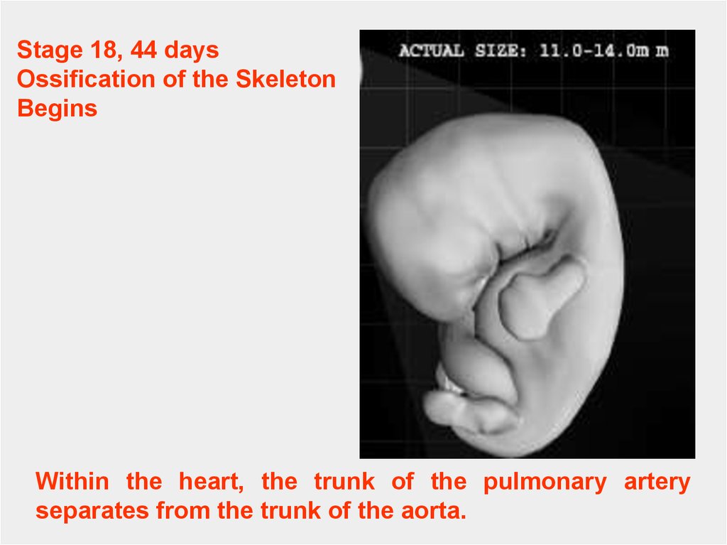
close to the midline proliferate and form a thickened plate of tissue paraxial mesoderm.
B. day 19, more laterally, the mesoderm layer remains thin (lateral plate).
Intermediate mesoderm connects paraxial and lateral plate
mesoderm. Intercellular cavities in the lateral plate then appear.
6.
DEVELOPMENT OF THE MESODERMAmnion
Parietal
mesoderm
layer
Endoderm
Neural groove
Somite
Visceral
mesoderm
layer
Intraembryonic
coelomic
cavity
Intermediate
mesoderm
Endoderm
C
D
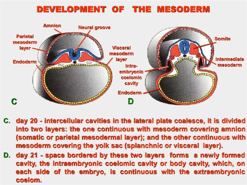
C. day 20 - intercellular cavities in the lateral plate coalesce, it is divided
into two layers: the one continuous with mesoderm covering amnion
(somatic or parietal mesodermal layer); and the other continuous with
mesoderm covering the yolk sac (splanchnic or visceral layer).
D. day 21 - space bordered by these two layers forms a newly formed
cavity, the intraembryonic coelomic cavity or body cavity, which, on
each side of the embryo, is continuous with the extraembryonic
coelom.
7.
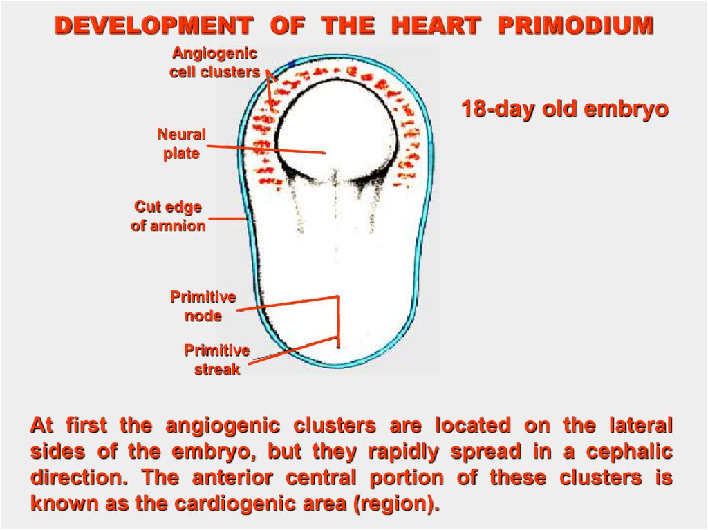
DEVELOPMENT OF THE HEART PRIMODIUMAngiogenic
cell clusters
18-day old embryo
Neural
plate
Cut edge
of amnion
Primitive
node
Primitive
streak
At first the angiogenic clusters are located on the lateral
sides of the embryo, but they rapidly spread in a cephalic
direction. The anterior central portion of these clusters is
known as the cardiogenic area (region).
8.
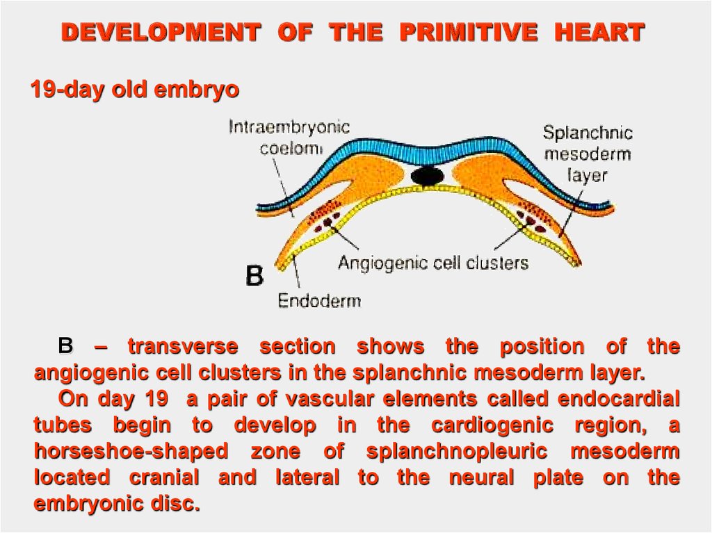
DEVELOPMENT OF THE PRIMITIVE HEART19-day old embryo
B – transverse section shows the position of the
angiogenic cell clusters in the splanchnic mesoderm layer.
On day 19 a pair of vasсular elements called endocardial
tubes begin to develop in the cardiogenic region, a
horseshoe-shaped zone of splanchnopleuric mesoderm
located cranial and lateral to the neural plate on the
embryonic disc.
9.
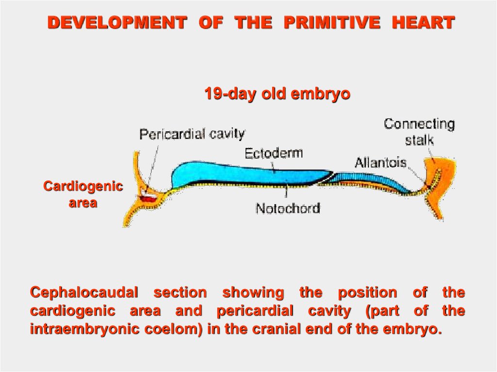
DEVELOPMENT OF THE PRIMITIVE HEART19-day old embryo
Cardiogenic
area
Cephalocaudal section showing the position of the
cardiogenic area and pericardial cavity (part of the
intraembryonic coelom) in the cranial end of the embryo.
10.
DEVELOPMENTOF THE
PRIMITIVE HEART
Hindgut
Foregut
Heart
tube
Pericardial
cavity
21-22 day – cephalocaudal folding, formation of the
fore-, hind-and midgut. The heart primodium is pulled
caudally.
11.
DEVELOPMENT OF THE HEARTBuccopharyngeal
membrane
Cloacal
membrane
Lung bud
Heart tube
Heart
tube
Liver
bud
Midgut
Remnant
of the
buccopharyngeal
membrane
Vitelline duct
Allantois
Yolk sac
D
C
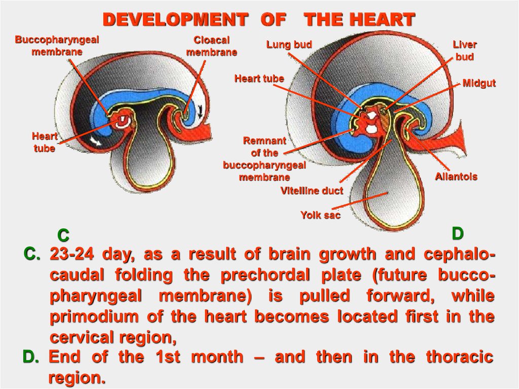
C. 23-24 day, as a result of brain growth and cephalocaudal folding the prechordal plate (future buccopharyngeal membrane) is pulled forward, while
primodium of the heart becomes located first in the
cervical region,
D. End of the 1st month – and then in the thoracic
region.
12.
DEVELOPMENT OF THE SEROUS MEMBRANESAmniotic cavity
Ectoderm
Dorsal
mesentery
Mesonephros
Visceral
mesoderm
layer
Body wall
Parietal
mesoderm
layer
A
B.
Parietal
mesoderm
layer
Intraembryonic
coelomic cavity
Endoderm of
yolk sac
B
Wall of gut
Serous membrane
(peritoneum)
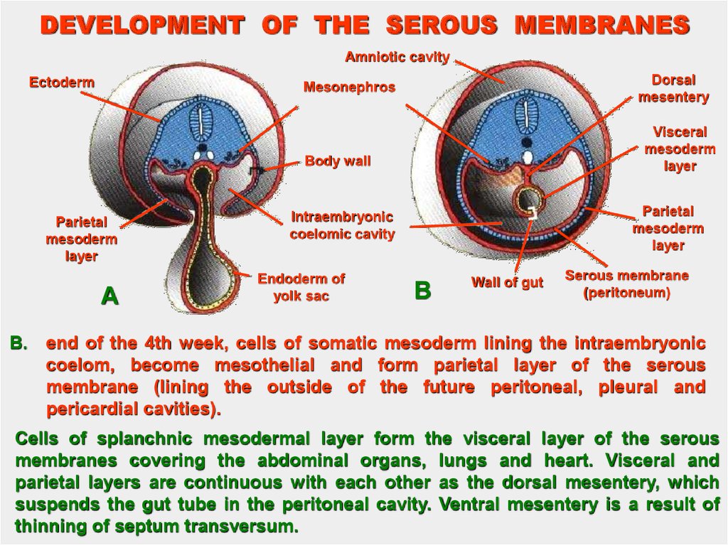
end of the 4th week, cells of somatic mesoderm lining the intraembryonic
coelom, become mesothelial and form parietal layer of the serous
membrane (lining the outside of the future peritoneal, pleural and
pericardial cavities).
Cells of splanchnic mesodermal layer form the visceral layer of the serous
membranes covering the abdominal organs, lungs and heart. Visceral and
parietal layers are continuous with each other as the dorsal mesentery, which
suspends the gut tube in the peritoneal cavity. Ventral mesentery is a result of
thinning of septum transversum.
13.
EARTY DEVELOPMENTOF HEART
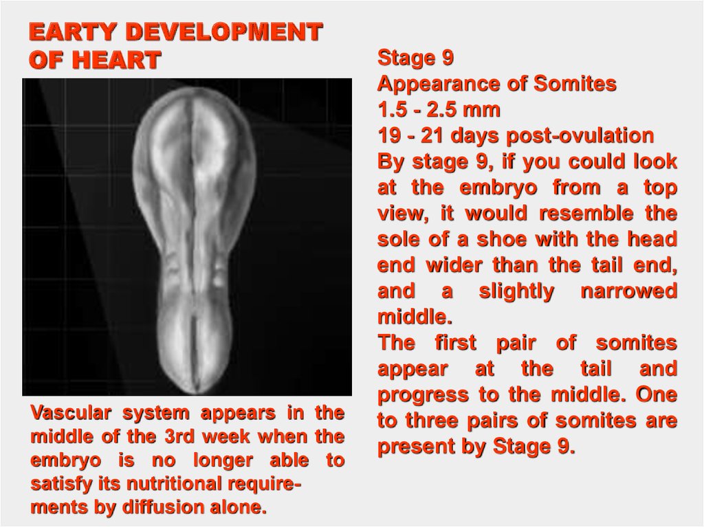
Vascular system appears in the
middle of the 3rd week when the
embryo is no longer able to
satisfy its nutritional requirements by diffusion alone.
Stage 9
Appearance of Somites
1.5 - 2.5 mm
19 - 21 days post-ovulation
By stage 9, if you could look
at the embryo from a top
view, it would resemble the
sole of a shoe with the head
end wider than the tail end,
and a slightly narrowed
middle.
The first pair of somites
appear at the tail and
progress to the middle. One
to three pairs of somites are
present by Stage 9.
14.
WeeksDays
Somites
Length in mm
Cardiac Events
1-2
0-20
1
1,5
No heart or great vessels
3
20
2
1,5
Cardiogenic plate
3
21
5
1,5
Endocardial tubes
4
22
10
2
Fusion of endocardial tubes
4
23
12
2
Single median cardiac tube, first contraction
(ineffective)
4
25
17
2,5
4
26
20
3
Single atrium
5
29
25
4
Bilobed atrium
5
31
26
4
Beginning of circulation
5
31
28
4,8
Septum primum
5
35
7,5
A-V orifice, 3 chamber heart
6
36
8,5
Septum secundum
6
39
10
Complete inferior septum
6
40
10,5
6
42
13
Divided truncus arteriosus
7
49
20
4-chambered heart, Absorption of pulmonary
veins
Cardiogenic loop
Septation of bulbus and ventricle
15.
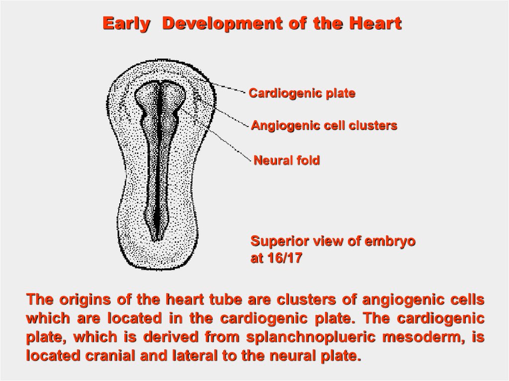
Early Development of the HeartCardiogenic plate
Angiogenic cell clusters
Neural fold
Superior view of embryo
at 16/17
The origins of the heart tube are clusters of angiogenic cells
which are located in the cardiogenic plate. The cardiogenic
plate, which is derived from splanchnoplueric mesoderm, is
located cranial and lateral to the neural plate.
16.
EarlyDevelopment
of the Heart
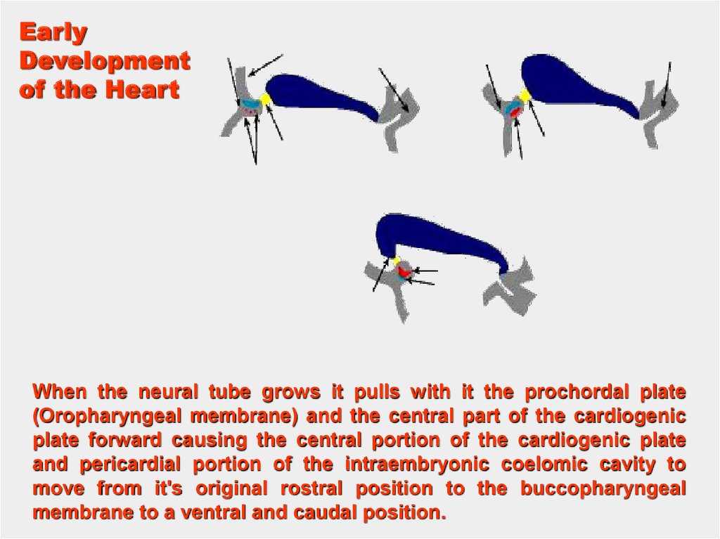
When the neural tube grows it pulls with it the prochordal plate
(Oropharyngeal membrane) and the central part of the cardiogenic
plate forward causing the central portion of the cardiogenic plate
and pericardial portion of the intraembryonic coelomic cavity to
move from it's original rostral position to the buccopharyngeal
membrane to a ventral and caudal position.
17.
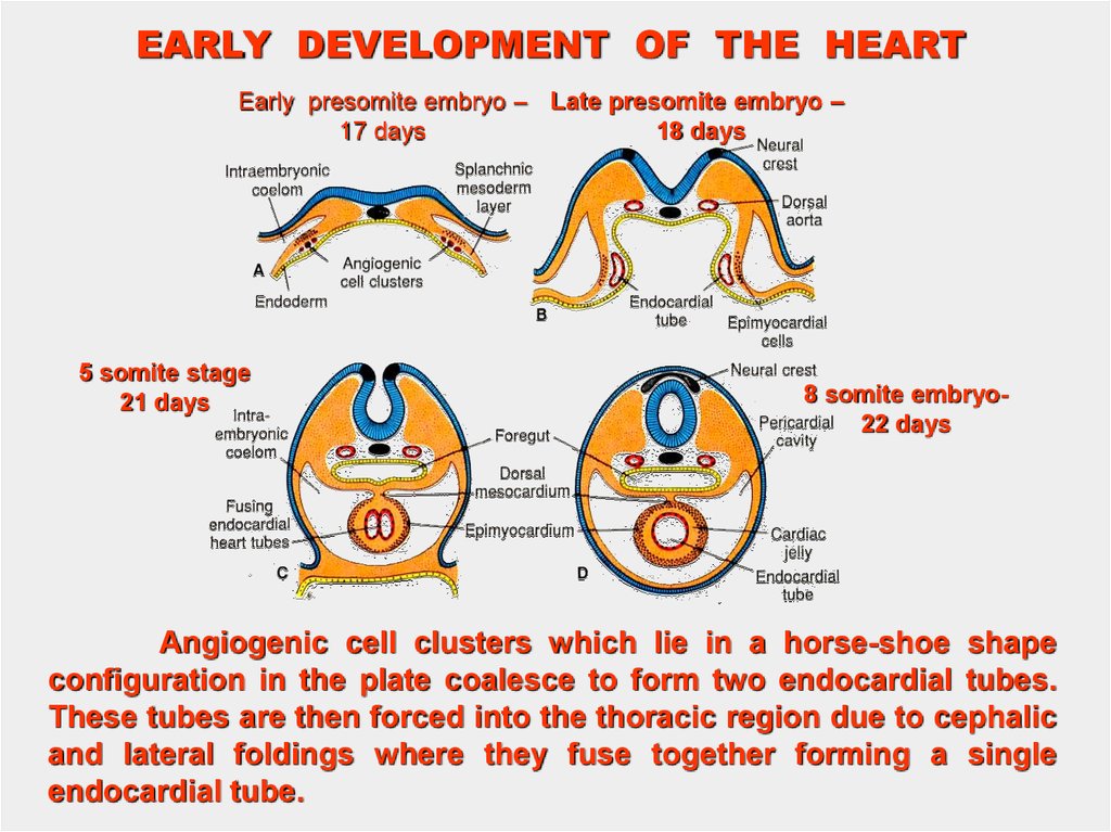
EARLY DEVELOPMENT OF THE HEARTEarly presomite embryo – Late presomite embryo –
18 days
17 days
5 somite stage
21 days
8 somite embryo22 days
Angiogenic cell clusters which lie in a horse-shoe shape
configuration in the plate coalesce to form two endocardial tubes.
These tubes are then forced into the thoracic region due to cephalic
and lateral foldings where they fuse together forming a single
endocardial tube.
18.
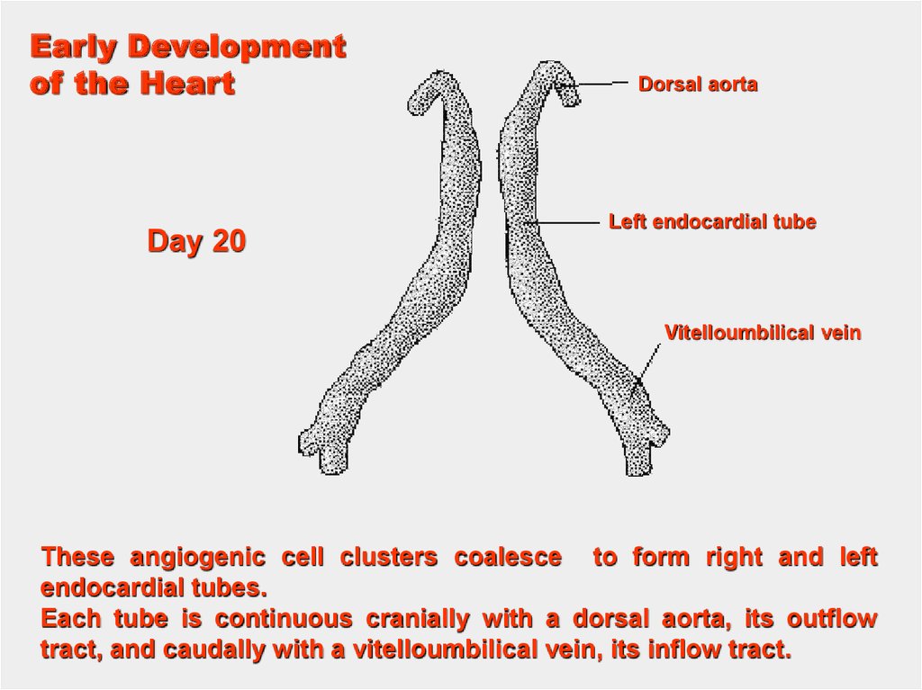
Early Developmentof the Heart
Day 20
Dorsal aorta
Left endocardial tube
Vitelloumbilical vein
These angiogenic cell clusters coalesce to form right and left
endocardial tubes.
Each tube is continuous cranially with a dorsal aorta, its outflow
tract, and caudally with a vitelloumbilical vein, its inflow tract.
19.
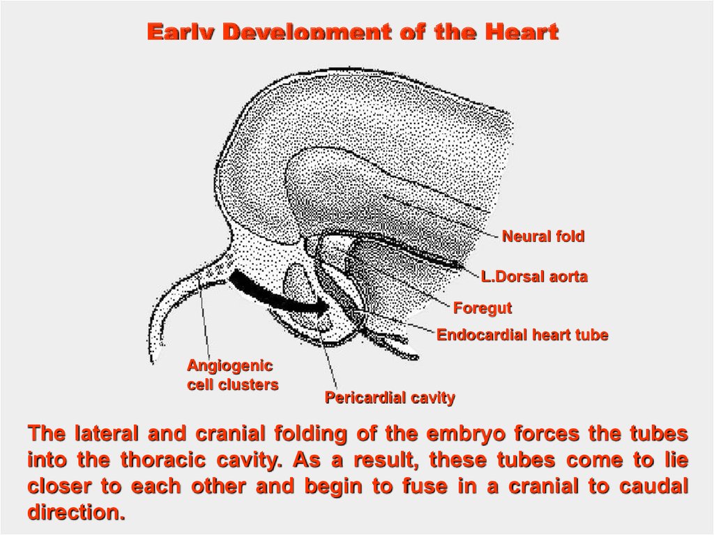
Early Development of the HeartNeural fold
L.Dorsal aorta
Foregut
Endocardial heart tube
Angiogenic
cell clusters
Pericardial cavity
The lateral and cranial folding of the embryo forces the tubes
into the thoracic cavity. As a result, these tubes come to lie
closer to each other and begin to fuse in a cranial to caudal
direction.
20.
Formationof the
Myocardium
and
Epicardium
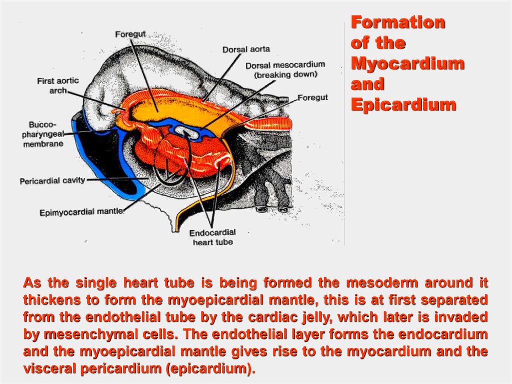
As the single heart tube is being formed the mesoderm around it
thickens to form the myoepicardial mantle, this is at first separated
from the endothelial tube by the cardiac jelly, which later is invaded
by mesenchymal cells. The endothelial layer forms the endocardium
and the myoepicardial mantle gives rise to the myocardium and the
visceral pericardium (epicardium).
21.
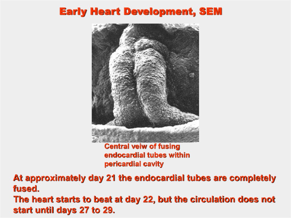
Early Heart Development, SEMCentral veiw of fusing
endocardial tubes within
pericardial cavity
At approximately day 21 the endocardial tubes are completely
fused.
The heart starts to beat at day 22, but the circulation does not
start until days 27 to 29.
22.
23.
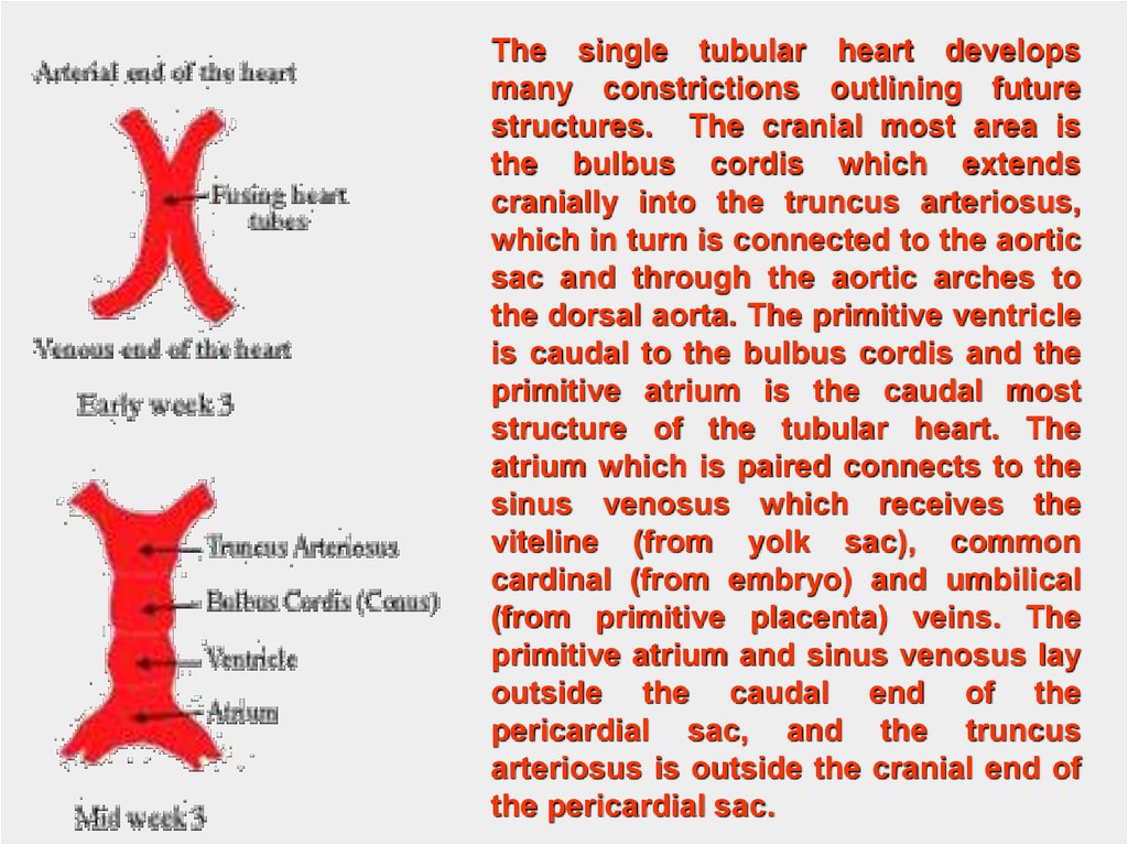
The single tubular heart developsmany constrictions outlining future
structures. The cranial most area is
the bulbus cordis which extends
cranially into the truncus arteriosus,
which in turn is connected to the aortic
sac and through the aortic arches to
the dorsal aorta. The primitive ventricle
is caudal to the bulbus cordis and the
primitive atrium is the caudal most
structure of the tubular heart. The
atrium which is paired connects to the
sinus venosus which receives the
viteline (from yolk sac), common
cardinal (from embryo) and umbilical
(from primitive placenta) veins. The
primitive atrium and sinus venosus lay
outside the caudal end of the
pericardial sac, and the truncus
arteriosus is outside the cranial end of
the pericardial sac.
24.
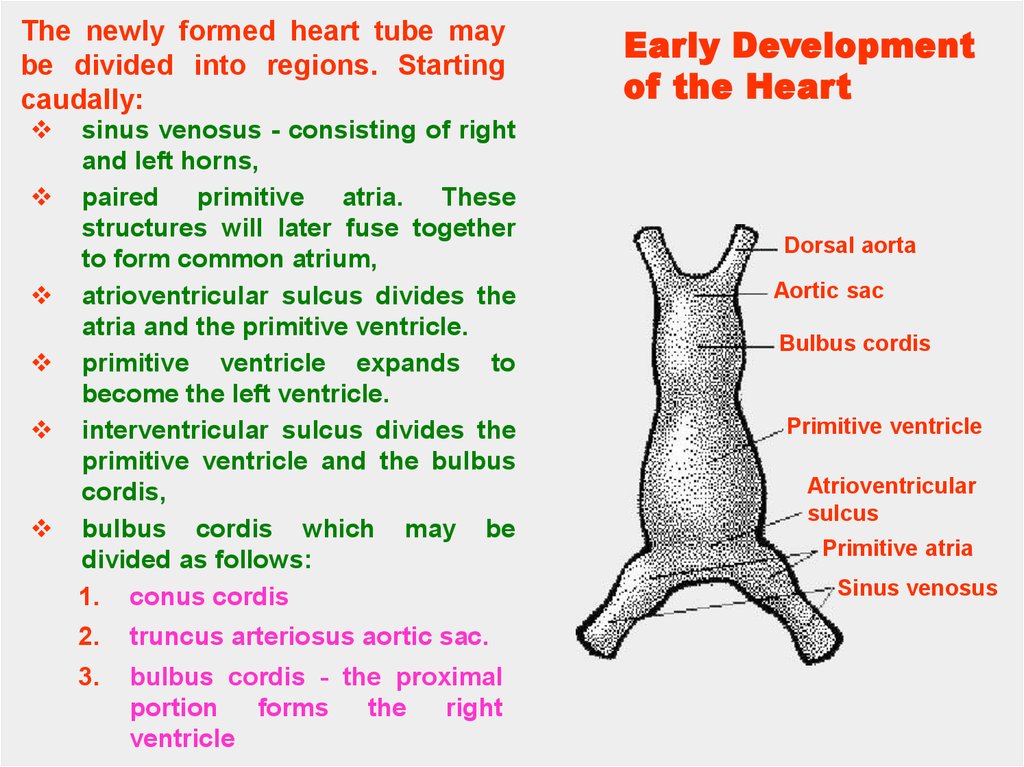
The newly formed heart tube maybe divided into regions. Starting
caudally:
sinus venosus - consisting of right
and left horns,
paired primitive atria. These
structures will later fuse together
to form common atrium,
atrioventricular sulcus divides the
atria and the primitive ventricle.
primitive ventricle expands to
become the left ventricle.
interventricular sulcus divides the
primitive ventricle and the bulbus
cordis,
bulbus cordis which may be
divided as follows:
1. conus cordis
2.
truncus arteriosus aortic sac.
3.
bulbus cordis - the proximal
portion
forms
the
right
ventricle
Early Development
of the Heart
Dorsal aorta
Aortic sac
Bulbus cordis
Primitive ventricle
Atrioventricular
sulcus
Primitive atria
Sinus venosus
25.
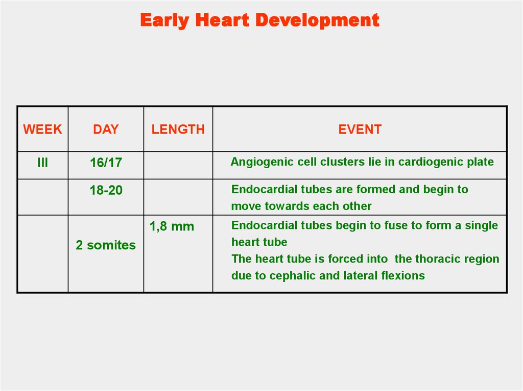
Early Heart DevelopmentWEEK
DAY
LENGTH
III
16/17
Angiogenic cell clusters lie in cardiogenic plate
18-20
Endocardial tubes are formed and begin to
move towards each other
1,8 mm
2 somites
EVENT
Endocardial tubes begin to fuse to form a single
heart tube
The heart tube is forced into the thoracic region
due to cephalic and lateral flexions
26.
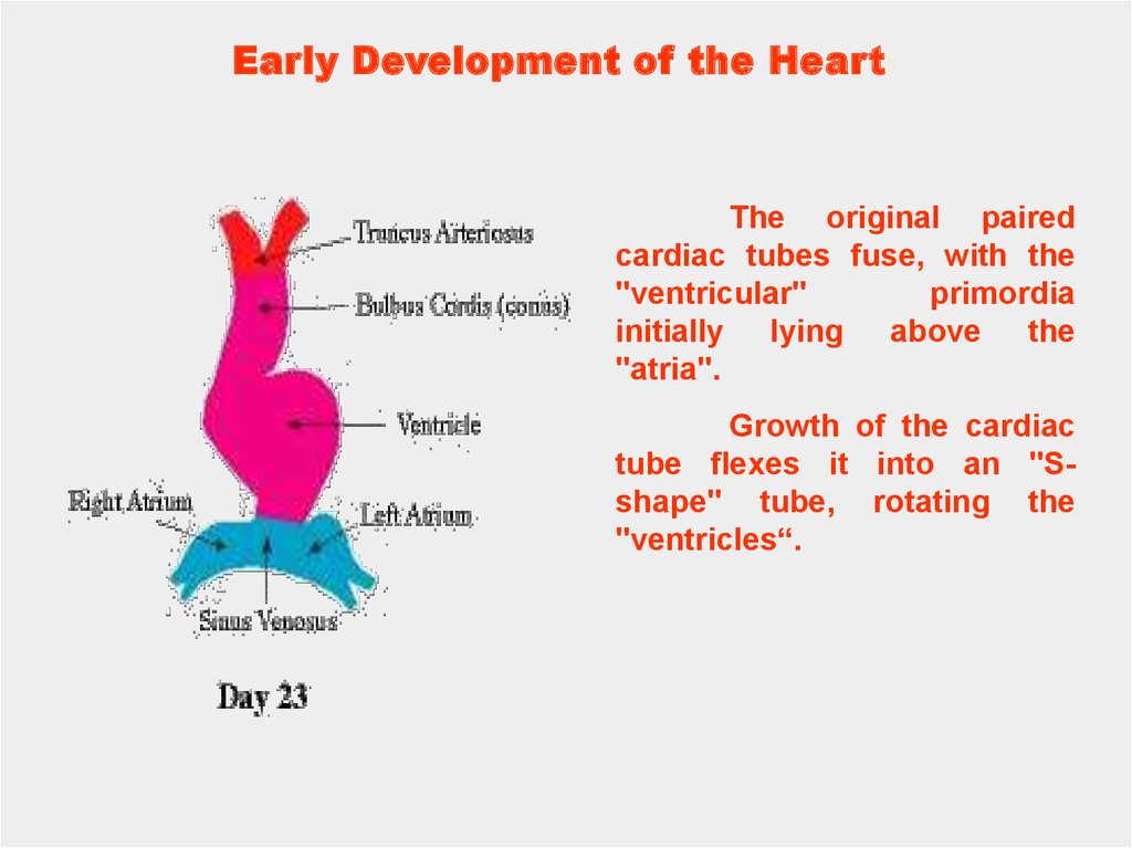
Early Development of the HeartThe original paired
cardiac tubes fuse, with the
"ventricular"
primordia
initially lying above the
"atria".
Growth of the cardiac
tube flexes it into an "Sshape" tube, rotating the
"ventricles“.
27.
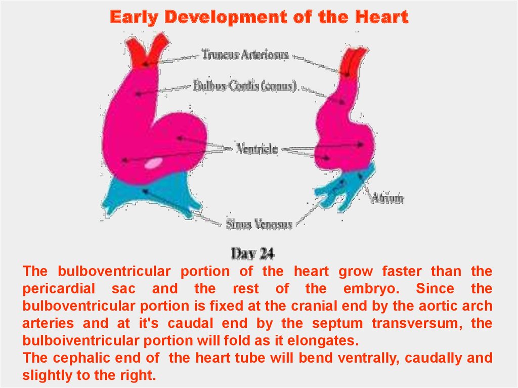
Early Development of the HeartThe bulboventricular portion of the heart grow faster than the
pericardial sac and the rest of the embryo. Since the
bulboventricular portion is fixed at the cranial end by the aortic arch
arteries and at it's caudal end by the septum transversum, the
bulboiventricular portion will fold as it elongates.
The cephalic end of the heart tube will bend ventrally, caudally and
slightly to the right.
28.
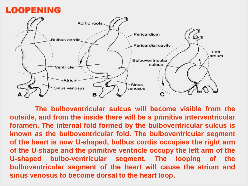
LOOPENINGThe bulboventricular sulcus will become visible from the
outside, and from the inside there will be a primitive interventricular
foramen. The internal fold formed by the bulboventricular sulcus is
known as the bulboventricular fold. The bulboventricular segment
of the heart is now U-shaped, bulbus cordis occupies the right arm
of the U-shape and the primitive ventricle occupy the left arm of the
U-shaped bulbo-ventricular segment. The looping of the
bulboventricular segment of the heart will cause the atrium and
sinus venosus to become dorsal to the heart loop.
29.
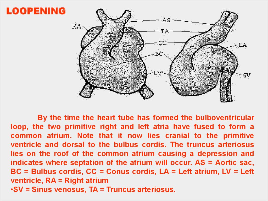
LOOPENINGBy the time the heart tube has formed the bulboventricular
loop, the two primitive right and left atria have fused to form a
common atrium. Note that it now lies cranial to the primitive
ventricle and dorsal to the bulbus cordis. The truncus arteriosus
lies on the roof of the common atrium causing a depression and
indicates where septation of the atrium will occur. AS = Aortic sac,
BC = Bulbus cordis, CC = Conus cordis, LA = Left atrium, LV = Left
ventricle, RA = Right atrium
•SV = Sinus venosus, TA = Truncus arteriosus.
30.
The newly formed heart tube bulges into the pericardial cavityand is attached to the dorsal wall by a fold of tissue, the dorsal
mesoderm. This is a derivative of foregut splanchnoplueric mesoderm.
Eventually this will rupture leaving the heart tube suspended in the
pericardial cavity anchored cranially by the dorsal aortae and caudally
by the vitelloumbilical veins.
As it bulges into the cavity it becomes invested in a layer of
myocardium. A layer of acellular matrix, the cardiac jelly, separates the
myocardium and the endothelial heart tube.
31.
The primitive heart tube can be subdivided intoprimordial heart chambers starting caudally at the inflow
end: the sinus venosus, primitive atria, ventricle, and
bulbus cordis (conus).
32.
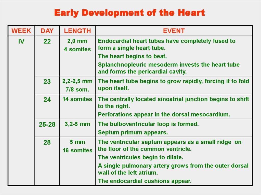
Early Development of the HeartWEEK
DAY
LENGTH
IV
22
2,0 mm
4 somites
23
2,2-2,5 mm The heart tube begins to grow rapidly, forcing it to fold
upon itself.
7/8 som.
24
14 somites
25-28
3,2-5 mm
28
EVENT
Endocardial heart tubes have completely fused to
form a single heart tube.
The heart begins to beat.
Splanchnopleuric mesoderm invests the heart tube
and forms the pericardial cavity.
The centrally located sinoatrial junction begins to shift
to the right.
Perforations appear in the dorsal mesocardium.
The bulboventricular loop is formed.
Septum primum appears.
5 mm
The ventricular septum appears as a small ridge on
16 somites the floor of the common ventricle.
The ventricules begin to dilate.
A single pulmonary artery grows from the outer dorsal
wall of the left atrium.
The endocardial cushions appear.
33.
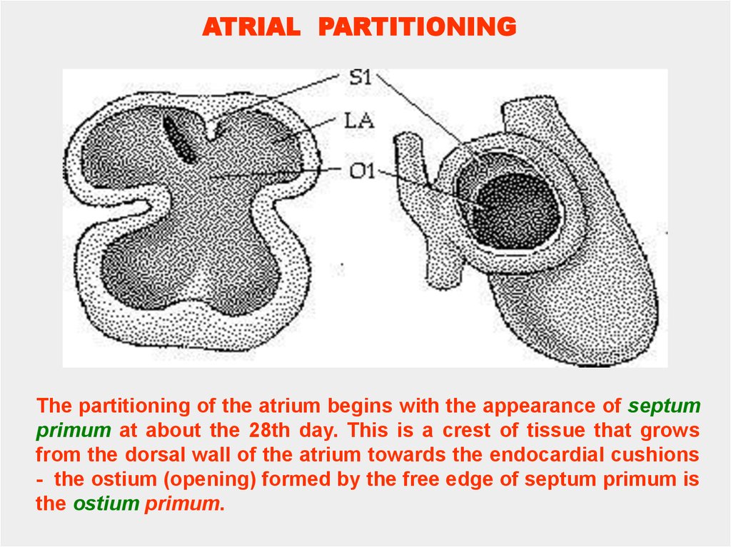
ATRIAL PARTITIONINGThe partitioning of the atrium begins with the appearance of septum
primum at about the 28th day. This is a crest of tissue that grows
from the dorsal wall of the atrium towards the endocardial cushions
- the ostium (opening) formed by the free edge of septum primum is
the ostium primum.
34.
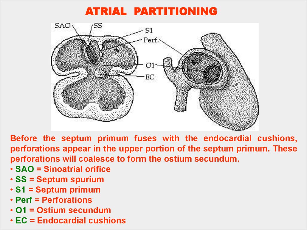
ATRIAL PARTITIONINGBefore the septum primum fuses with the endocardial cushions,
perforations appear in the upper portion of the septum primum. These
perforations will coalesce to form the ostium secundum.
• SAO = Sinoatrial orifice
• SS = Septum spurium
• S1 = Septum primum
• Perf = Perforations
• O1 = Ostium secundum
• EC = Endocardial cushions
35.
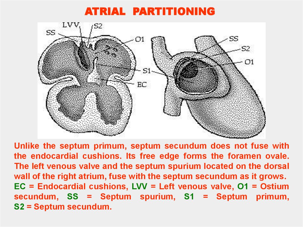
ATRIAL PARTITIONINGUnlike the septum primum, septum secundum does not fuse with
the endocardial cushions. Its free edge forms the foramen ovale.
The left venous valve and the septum spurium located on the dorsal
wall of the right atrium, fuse with the septum secundum as it grows.
EC = Endocardial cushions, LVV = Left venous valve, O1 = Ostium
secundum, SS = Septum spurium, S1 = Septum primum,
S2 = Septum secundum.
36.
ATRIALPARTITIONING
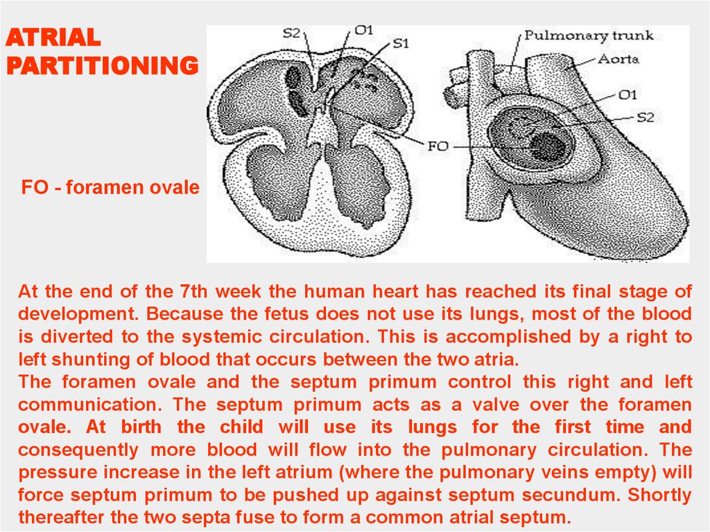
FO - foramen ovale
At the end of the 7th week the human heart has reached its final stage of
development. Because the fetus does not use its lungs, most of the blood
is diverted to the systemic circulation. This is accomplished by a right to
left shunting of blood that occurs between the two atria.
The foramen ovale and the septum primum control this right and left
communication. The septum primum acts as a valve over the foramen
ovale. At birth the child will use its lungs for the first time and
consequently more blood will flow into the pulmonary circulation. The
pressure increase in the left atrium (where the pulmonary veins empty) will
force septum primum to be pushed up against septum secundum. Shortly
thereafter the two septa fuse to form a common atrial septum.
37.
DEVELOPMENT OF THE HEARTWEEK
DAY
LENGTH
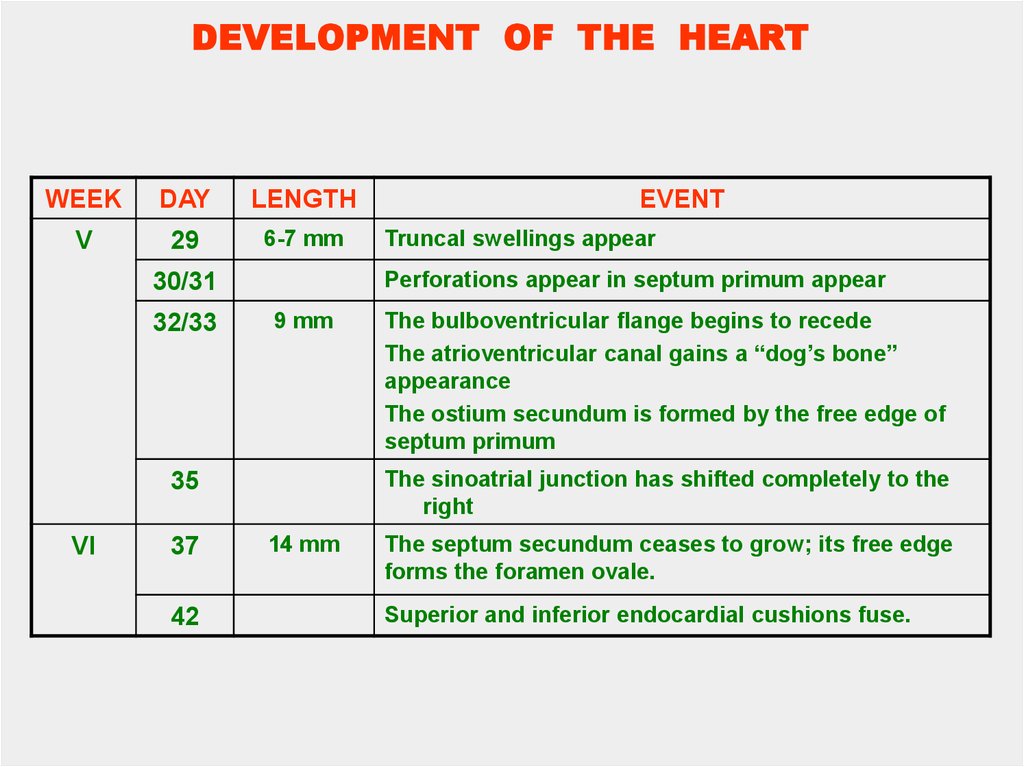
V
29
6-7 mm
9 mm
37
42
The bulboventricular flange begins to recede
The atrioventricular canal gains a “dog’s bone”
appearance
The ostium secundum is formed by the free edge of
septum primum
The sinoatrial junction has shifted completely to the
right
35
VI
Truncal swellings appear
Perforations appear in septum primum appear
30/31
32/33
EVENT
14 mm
The septum secundum ceases to grow; its free edge
forms the foramen ovale.
Superior and inferior endocardial cushions fuse.
38.
Early Development of the HeartWEEK
DAY
VII
46
end of
7th week
VIII
early
In wk
LENGTH
EVENT
The ventricular septum ceases to grow.
The coronary sinus is formed.
The interventricular canal is completely obliterated.
The outflow tracts (the aorta and the pulmonary
trunk) are completely separated.
39.
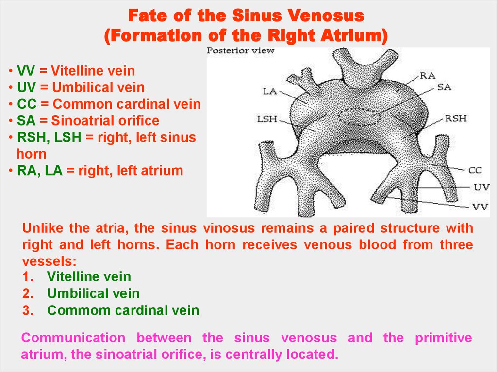
Fate of the Sinus Venosus(Formation of the Right Atrium)
• VV = Vitelline vein
• UV = Umbilical vein
• CC = Common cardinal vein
• SA = Sinoatrial orifice
• RSH, LSH = right, left sinus
horn
• RA, LA = right, left atrium
Unlike the atria, the sinus vinosus remains a paired structure with
right and left horns. Each horn receives venous blood from three
vessels:
1. Vitelline vein
2. Umbilical vein
3. Commom cardinal vein
Communication between the sinus venosus and the primitive
atrium, the sinoatrial orifice, is centrally located.
40.
DEVELOPMENTOF THE ATRIA
• SVC = Superior vena cava
• IVC = Inferior vena cava
• SA = Sinoatrial junction
• CS/OV = Coronary sinus/ oblique
vein of left ventricle
• LA, RA = Left, right atrium
• LV, RV = Left, right ventricle
Gradually the sinoatrial oriface shifts to the right, due to the shunting of
blood to the right, until the sinus venosus communicates with only the
right atrium. The fate of each structure is as follows:
the right sinus horn becomes enlarged
the right anterior cardinal vein becomes the superior vena cava
the right vitelline vein becomes the inferior vena cava
the right umbilical vein is obliterated
Conversely, the left vein counterparts are obliterated and the left
sinus horn diminishes in size and forms the coronary sinus and the
oblique vein of the left ventricle.
41.
SEPTATION• LVV, RVV = left, right venous
valve
• SS = Septum spurium
• SA = Sinoatrial oriface
• OCS = orifice of the coronary
sinus
Internally, the sinoatrial orifice is flanked by two valves,
the right and left venous valves. Superiorly these two
valves meet to form the septum spurium. Note that the left
horn opens up underneath the orifice of the right horn
(sinoatrial oriface). This is the orifice of the coronary
sinus.
42.
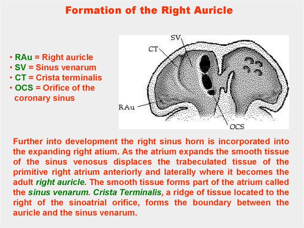
Formation of the Right Auricle• RAu = Right auricle
• SV = Sinus venarum
• CT = Crista terminalis
• OCS = Orifice of the
coronary sinus
Further into development the right sinus horn is incorporated into
the expanding right atium. As the atrium expands the smooth tissue
of the sinus venosus displaces the trabeculated tissue of the
primitive right atrium anteriorly and laterally where it becomes the
adult right auricle. The smooth tissue forms part of the atrium called
the sinus venarum. Crista Terminalis, a ridge of tissue located to the
right of the sinoatrial orifice, forms the boundary between the
auricle and the sinus venarum.
43.
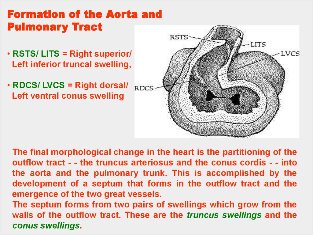
Formation of the Aorta andPulmonary Tract
• RSTS/ LITS = Right superior/
Left inferior truncal swelling,
• RDCS/ LVCS = Right dorsal/
Left ventral conus swelling
The final morphological change in the heart is the partitioning of the
outflow tract - - the truncus arteriosus and the conus cordis - - into
the aorta and the pulmonary trunk. This is accomplished by the
development of a septum that forms in the outflow tract and the
emergence of the two great vessels.
The septum forms from two pairs of swellings which grow from the
walls of the outflow tract. These are the truncus swellings and the
conus swellings.
44.
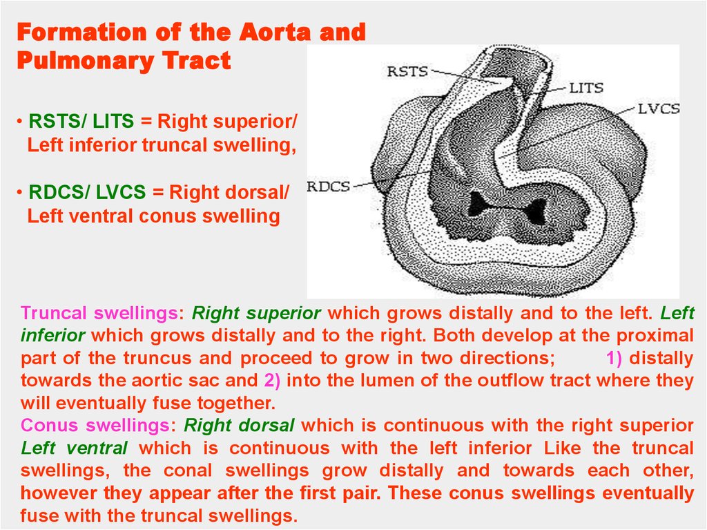
Formation of the Aorta andPulmonary Tract
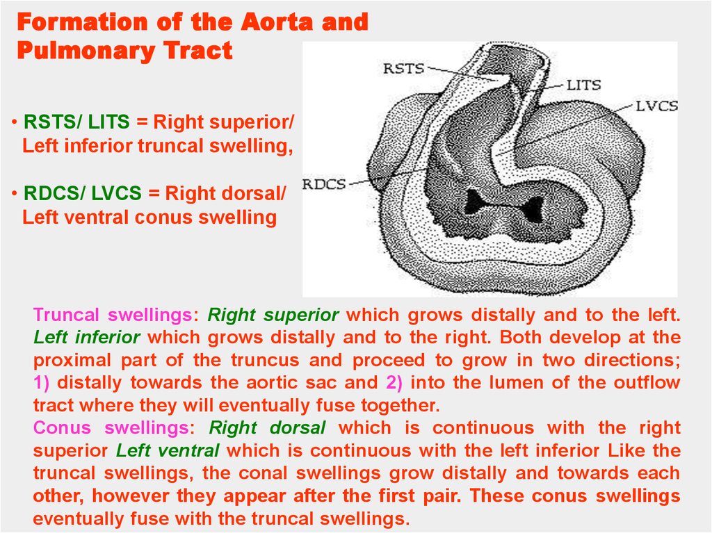
• RSTS/ LITS = Right superior/
Left inferior truncal swelling,
• RDCS/ LVCS = Right dorsal/
Left ventral conus swelling
Truncal swellings: Right superior which grows distally and to the left. Left
inferior which grows distally and to the right. Both develop at the proximal
part of the truncus and proceed to grow in two directions;
1) distally
towards the aortic sac and 2) into the lumen of the outflow tract where they
will eventually fuse together.
Conus swellings: Right dorsal which is continuous with the right superior
Left ventral which is continuous with the left inferior Like the truncal
swellings, the conal swellings grow distally and towards each other,
however they appear after the first pair. These conus swellings eventually
fuse with the truncal swellings.
45.
Pulmonary Veins (Formation of the Left Atrium)• LA = Left atrium
• OPV = orifice of
pulmonary vein
• PV = pulmonary vein
Development of the left atrium occurs concurrently with that of the
right atrium. During the early part of the fourth week an outgrowth of
the pulmonary veins appear from the left atrium. This "sprout" will
bifurcate until there are four veins. These vessels will then grow
towards the lung buds.
46.
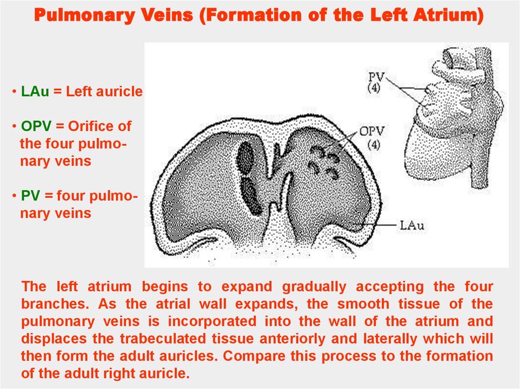
Pulmonary Veins (Formation of the Left Atrium)• LAu = Left auricle
• OPV = Orifice of
the four pulmonary veins
• PV = four pulmonary veins
The left atrium begins to expand gradually accepting the four
branches. As the atrial wall expands, the smooth tissue of the
pulmonary veins is incorporated into the wall of the atrium and
displaces the trabeculated tissue anteriorly and laterally which will
then form the adult auricles. Compare this process to the formation
of the adult right auricle.
47.
AtrioventricularCanals
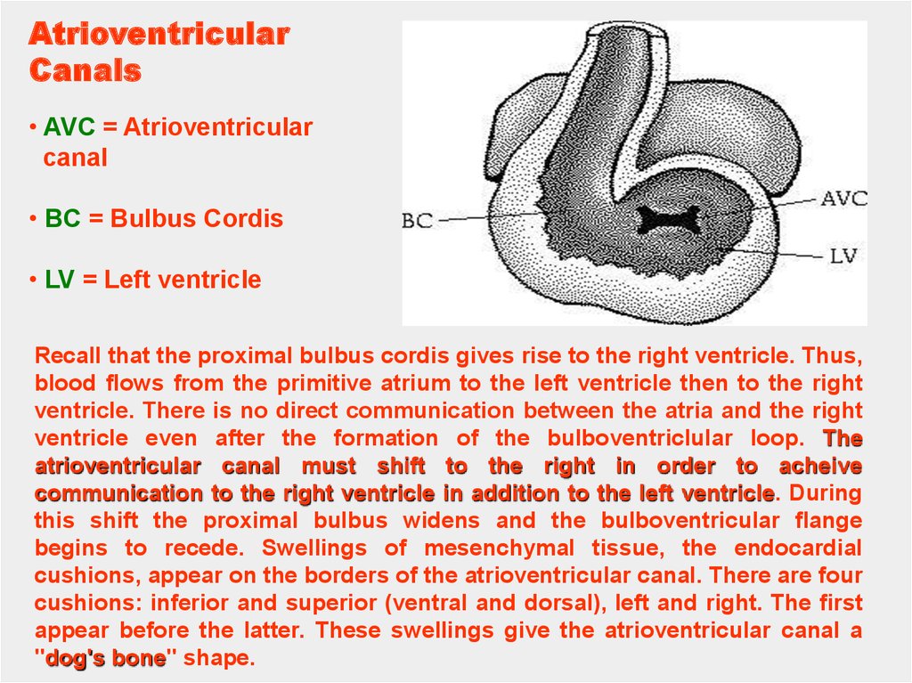
• AVC = Atrioventricular
canal
• BC = Bulbus Cordis
• LV = Left ventricle
Recall that the proximal bulbus cordis gives rise to the right ventricle. Thus,
blood flows from the primitive atrium to the left ventricle then to the right
ventricle. There is no direct communication between the atria and the right
ventricle even after the formation of the bulboventriclular loop. The
atrioventricular canal must shift to the right in order to acheive
communication to the right ventricle in addition to the left ventricle. During
this shift the proximal bulbus widens and the bulboventricular flange
begins to recede. Swellings of mesenchymal tissue, the endocardial
cushions, appear on the borders of the atrioventricular canal. There are four
cushions: inferior and superior (ventral and dorsal), left and right. The first
appear before the latter. These swellings give the atrioventricular canal a
"dog's bone" shape.
48.
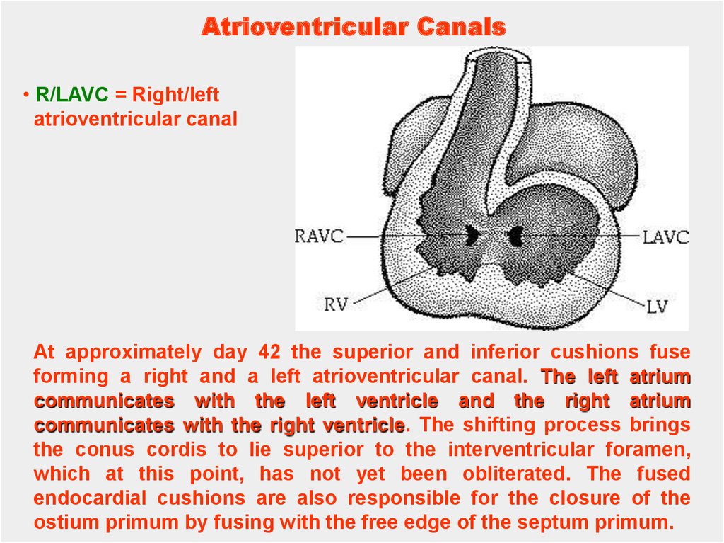
Atrioventricular Canals• R/LAVC = Right/left
atrioventricular canal
At approximately day 42 the superior and inferior cushions fuse
forming a right and a left atrioventricular canal. The left atrium
communicates with the left ventricle and the right atrium
communicates with the right ventricle. The shifting process brings
the conus cordis to lie superior to the interventricular foramen,
which at this point, has not yet been obliterated. The fused
endocardial cushions are also responsible for the closure of the
ostium primum by fusing with the free edge of the septum primum.
49.
Formation of the Aorta andPulmonary Tract
• RSTS/ LITS = Right superior/
Left inferior truncal swelling,
• RDCS/ LVCS = Right dorsal/
Left ventral conus swelling
The final morphological change in the heart is the partitioning of the
outflow tract - - the truncus arteriosus and the conus cordis - - into
the aorta and the pulmonary trunk. This is accomplished by the
development of a septum that forms in the outflow tract and the
emergence of the two great vessels.
The septum forms from two pairs of swellings which grow from the
walls of the outflow tract. These are the truncus swellings and the
conus swellings.
50.
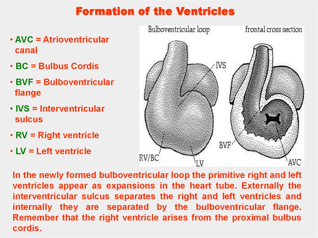
Formation of the Ventricles• AVC = Atrioventricular
canal
• BC = Bulbus Cordis
• BVF = Bulboventricular
flange
• IVS = Interventricular
sulcus
• RV = Right ventricle
• LV = Left ventricle
In the newly formed bulboventricular loop the primitive right and left
ventricles appear as expansions in the heart tube. Externally the
interventricular sulcus separates the right and left ventricles and
internally they are separated by the bulboventricular flange.
Remember that the right ventricle arises from the proximal bulbus
cordis.
51.
Formation of theVentricles
• BC = Bulbus cordis
• IVS = Interventricular
septum
• MC = Myocardium
• RV = Right ventricle
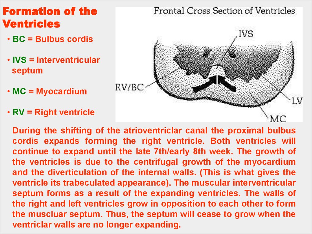
During the shifting of the atrioventriclar canal the proximal bulbus
cordis expands forming the right ventricle. Both ventricles will
continue to expand until the late 7th/early 8th week. The growth of
the ventricles is due to the centrifugal growth of the myocardium
and the diverticulation of the internal walls. (This is what gives the
ventricle its trabeculated appearance). The muscular interventricular
septum forms as a result of the expanding ventricles. The walls of
the right and left ventricles grow in opposition to each other to form
the muscluar septum. Thus, the septum will cease to grow when the
ventriclar walls are no longer expanding.
52.
Atrial Septal DefectIn a heart with an Atrial Septal Defect (ASD) there is
communication between the right and left atria which
causes a left to right shunting of blood due to the lower
pressure in the pulmonary circulatory system.
Consequently there is a mixing of oxygenated (systemic)
and deoxygenated (pulmonary) blood.
There are two types of ASD:
Primum type involves the endocardial cushions.
Secundum type involves septum primum or septum
secundum.
53.
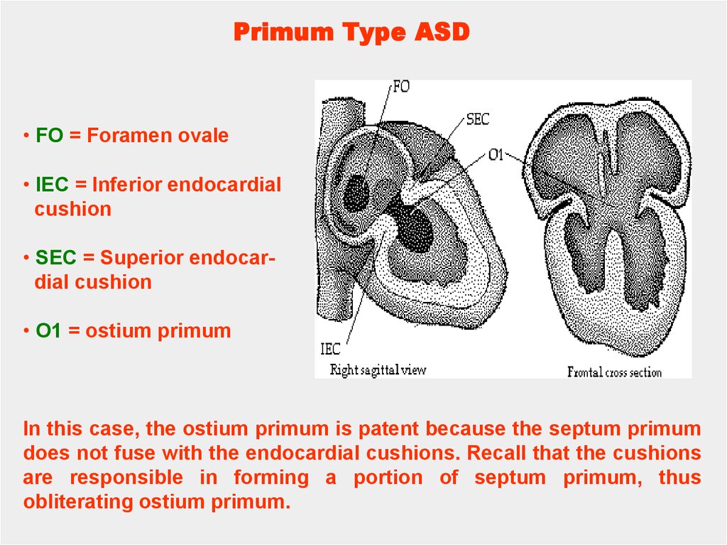
Primum Type ASD• FO = Foramen ovale
• IEC = Inferior endocardial
cushion
• SEC = Superior endocardial cushion
• O1 = ostium primum
In this case, the ostium primum is patent because the septum primum
does not fuse with the endocardial cushions. Recall that the cushions
are responsible in forming a portion of septum primum, thus
obliterating ostium primum.
54.
Formation of the Aorta andPulmonary Tract
• RSTS/ LITS = Right superior/
Left inferior truncal swelling,
• RDCS/ LVCS = Right dorsal/
Left ventral conus swelling
Truncal swellings: Right superior which grows distally and to the left.
Left inferior which grows distally and to the right. Both develop at the
proximal part of the truncus and proceed to grow in two directions;
1) distally towards the aortic sac and 2) into the lumen of the outflow
tract where they will eventually fuse together.
Conus swellings: Right dorsal which is continuous with the right
superior Left ventral which is continuous with the left inferior Like the
truncal swellings, the conal swellings grow distally and towards each
other, however they appear after the first pair. These conus swellings
eventually fuse with the truncal swellings.
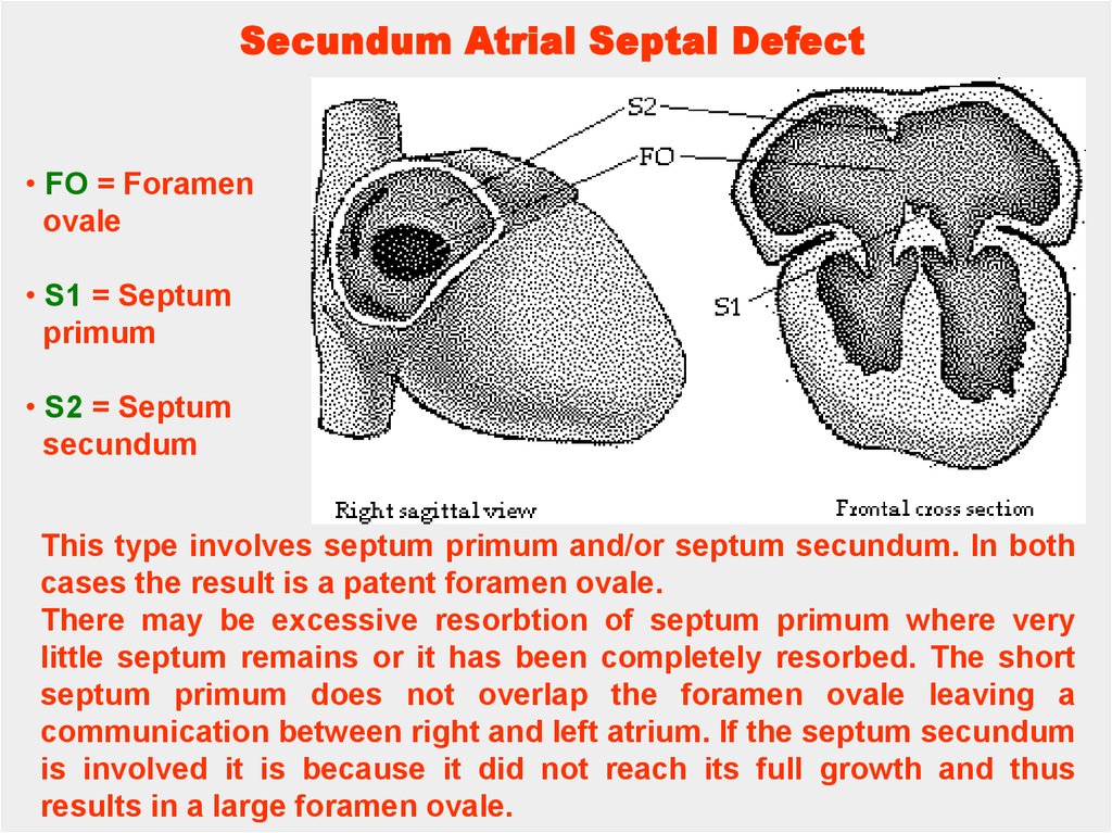
55.
Secundum Atrial Septal Defect• FO = Foramen
ovale
• S1 = Septum
primum
• S2 = Septum
secundum
This type involves septum primum and/or septum secundum. In both
cases the result is a patent foramen ovale.
There may be excessive resorbtion of septum primum where very
little septum remains or it has been completely resorbed. The short
septum primum does not overlap the foramen ovale leaving a
communication between right and left atrium. If the septum secundum
is involved it is because it did not reach its full growth and thus
results in a large foramen ovale.
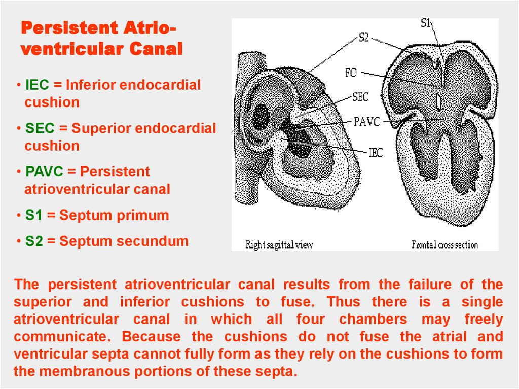
56.
Persistent Atrioventricular Canal• IEC = Inferior endocardial
cushion
• SEC = Superior endocardial
cushion
• PAVC = Persistent
atrioventricular canal
• S1 = Septum primum
• S2 = Septum secundum
The persistent atrioventricular canal results from the failure of the
superior and inferior cushions to fuse. Thus there is a single
atrioventricular canal in which all four chambers may freely
communicate. Because the cushions do not fuse the atrial and
ventricular septa cannot fully form as they rely on the cushions to form
the membranous portions of these septa.
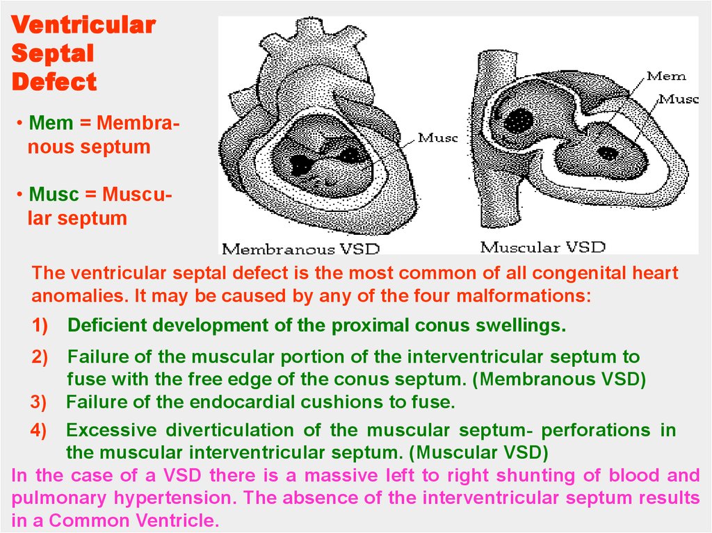
57.
VentricularSeptal
Defect
• Mem = Membranous septum
• Musc = Muscular septum
The ventricular septal defect is the most common of all congenital heart
anomalies. It may be caused by any of the four malformations:
1)
2)
Deficient development of the proximal conus swellings.
Failure of the muscular portion of the interventricular septum to
fuse with the free edge of the conus septum. (Membranous VSD)
3) Failure of the endocardial cushions to fuse.
4) Excessive diverticulation of the muscular septum- perforations in
the muscular interventricular septum. (Muscular VSD)
In the case of a VSD there is a massive left to right shunting of blood and
pulmonary hypertension. The absence of the interventricular septum results
in a Common Ventricle.
58.
Transposition ofthe Great
Vessels
• AO = Aorta
• PT = Pulmonary
trunk
• PDA = Persistent
Dunctus Arteriosus
• RV/LV = right and
left ventricles
Transposition is a condition in which the aorta arises from the
right ventricle and the pulmonary trunk from the left. This anomally is
due to the failure of the truncoconal swellings to grow in the normal
spiral direction. There is also a ventricular septal defect and a patent
ductus arteriosus. However, these secondary defects make life
possible as they provide a way for oxygenated blood to reach the
entire body.
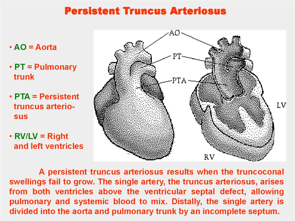
59.
Persistent Truncus Arteriosus• AO = Aorta
• PT = Pulmonary
trunk
• PTA = Persistent
truncus arteriosus
• RV/LV = Right
and left ventricles
A persistent truncus arteriosus results when the truncoconal
swellings fail to grow. The single artery, the truncus arteriosus, arises
from both ventricles above the ventricular septal defect, allowing
pulmonary and systemic blood to mix. Distally, the single artery is
divided into the aorta and pulmonary trunk by an incomplete septum.
60.
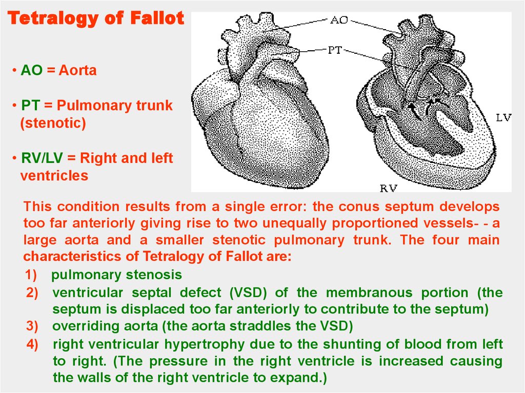
Tetralogy of Fallot• AO = Aorta
• PT = Pulmonary trunk
(stenotic)
• RV/LV = Right and left
ventricles
This condition results from a single error: the conus septum develops
too far anteriorly giving rise to two unequally proportioned vessels- - a
large aorta and a smaller stenotic pulmonary trunk. The four main
characteristics of Tetralogy of Fallot are:
1) pulmonary stenosis
2) ventricular septal defect (VSD) of the membranous portion (the
septum is displaced too far anteriorly to contribute to the septum)
3) overriding aorta (the aorta straddles the VSD)
4) right ventricular hypertrophy due to the shunting of blood from left
to right. (The pressure in the right ventricle is increased causing
the walls of the right ventricle to expand.)
61.
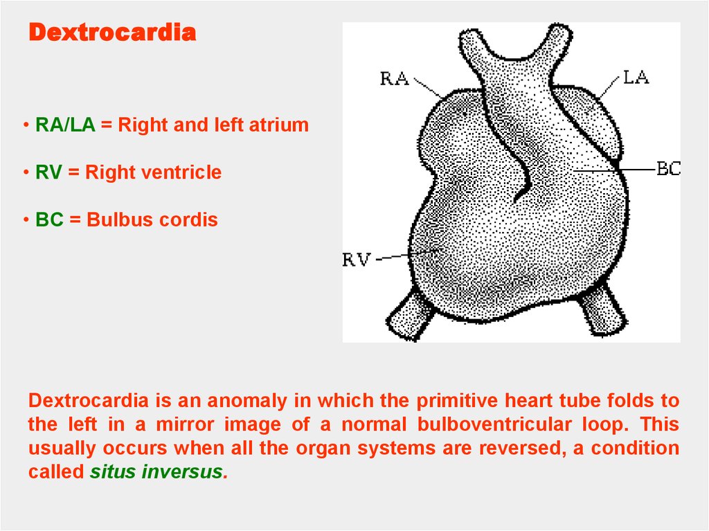
Dextrocardia• RA/LA = Right and left atrium
• RV = Right ventricle
• BC = Bulbus cordis
Dextrocardia is an anomaly in which the primitive heart tube folds to
the left in a mirror image of a normal bulboventricular loop. This
usually occurs when all the organ systems are reversed, a condition
called situs inversus.
62.
Time-Line Schedule of Heart DevelopmentStage 11
13-20 Somite Pairs, Rostral
Neuropore Closes, Optic Vesicle
Appears,
Two Pharyngeal Arches Appear
2.5 - 3.0 mm
23 - 25 days post-ovulation
The embryo is shaped in a modified S curve. The embryo has a bulblike tail and a connecting stalk to the developing placenta.
A primitive S-shaped tubal heart is beating and peristalsis, the
rhythmic flow propelling fluids throughout the body, begins.
However, this is not true circulation because blood vessel
development is still incomplete.
63.
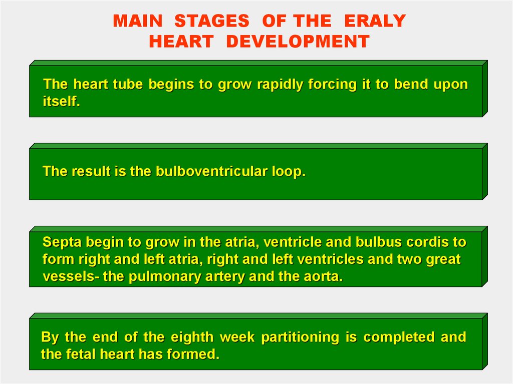
MAIN STAGES OF THE ERALYHEART DEVELOPMENT
The heart tube begins to grow rapidly forcing it to bend upon
itself.
The result is the bulboventricular loop.
Septa begin to grow in the atria, ventricle and bulbus cordis to
form right and left atria, right and left ventricles and two great
vessels- the pulmonary artery and the aorta.
By the end of the eighth week partitioning is completed and
the fetal heart has formed.
64.
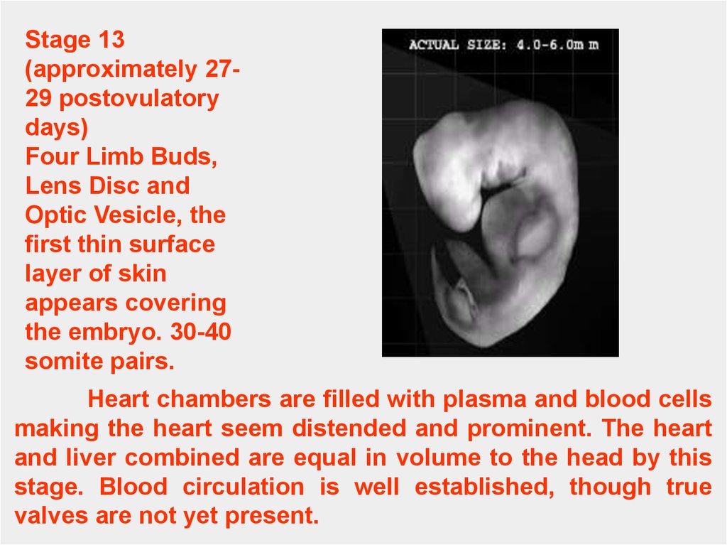
Stage 13(approximately 2729 postovulatory
days)
Four Limb Buds,
Lens Disc and
Optic Vesicle, the
first thin surface
layer of skin
appears covering
the embryo. 30-40
somite pairs.
Heart chambers are filled with plasma and blood cells
making the heart seem distended and prominent. The heart
and liver combined are equal in volume to the head by this
stage. Blood circulation is well established, though true
valves are not yet present.
65.
Stage 1221-29 Somite Pairs,
Caudal Neuropore
Closes, Three to Four
Pharyngeal Arches
Appear, Upper Limb
Buds Appear
3.0 - 5.0 mm
25 - 27 days postovulation
The embryo curves into a C shape. The arches that
form the face and neck are now becoming evident under
the enlarging forebrain. A blood system continues to
develop. Blood cells follow the surface of yolk sac where
they originate, move along the central nervous system, and
move in the chorionic villi, the maternal blood system.
Valves and septa may appear in the heart in Stage 12.
66.
Stage 19,(approximately
47-48 post ovulatory days)
Septum primum fuses with septum intermedium in the
heart.
67.
Stage 14, 5th weekLens Pit and
Optic Cup
Appear,
Endolymphatic
Appendage
Distinct
Semilunar valves begin to form in the heart. Four major
subdivisions of the heart (the trabeculated left and right
ventricles, the conus cords and the truncus arteriosus) are
clearly defined. Two sprouts, a ventral one from the aortic
sac and a dorsal one from the aorta, form the pulmonary
(sixth aortic) arch.
68.
Stage 15(6 to 8 weeks post
fertilization)
Lens Vesicle,
Nasal Pit, Hand
Plate; Trunk
Widens, Future
Cerebral
Hemispheres
Distinct
Blood flow through the arioventricular canal is divided into
left and right streams, which continue through the outflow
tract and aortic sac. The left ventricle is larger than the right
and has a thicker wall.
69.
Stage 16(6th weeks post
fertilization)
Primary cardiac tube separates into aortic and pulmonary
channels and the ventricular pouches deepen and
enlarge, forming a common wall with their myocardial
shells.
70.
Stage 17(approximately
41 postovulatory
days)
A Four
Chambered Heart
and a Sense of
Smell
The heart begins to separate into four chambers.
71.
Stage 18, 44 daysOssification of the Skeleton
Begins
Within the heart, the trunk of the pulmonary artery
separates from the trunk of the aorta.
72.
CONGENITAL MALFORMATIONS OF THE HEART ANDGREAT VESSELS.
1. They are common.
2. The overall incidence is 0.7% of live births and 2.7% of
stillbirths.
- Atrial Septal Defects (ASD) – are among the most common
(6.4/10,000births).
- One of the most significant defects is ostium secundum
defect. This anomaly is characterized by a large opening
between the left and the right atria and is caused either by
excessive cell death or resoption of the septum primum. Or
by inadequate development of the septum secundum.
Depending on the size of the opening considerable
intercardiac shunting may occur from left to the right.
73.
Ventricular Septal Defects involve the membranousportion of the septum. The occur in 12/10,000.
Depending on the size of the opening, blood carried by the
pulmonary artery may be 1.5 times more abundant than that
carried by aorta. The defect may be not restricted to the
membranous part and involve the muscular part of the septum.
Tetralogy of Fallot is the most frequently occured
abnormality of the conotruncal region. The defect is due to an
unequal division of the conus, resulting from anterior
displacement of the conotruncal septum. Displacement of the
septum produces four CV-alterations:
-a narrow right ventricular outflow region,i.e. Pulmonary
infundibular stenosis,
-a large defect of the interventricular septum,
-an overriding aorta that arrises directly above the septal
defect,
-hypertrophy of the right ventricular wall due to the hight
pressure on the right side. The rate is 9.6/10,000.