


 medicine
Similar presentations:


Hypertrophic cardiomyopathy
1. HYPERTROPHIC CARDIOMYOPATHY
AKIMBEKOVA DINARAGM-13 48-02
ISKAKOVA E.E.
2. Plan:
ClassificationDefinition of disease
Etiology
Morphology:
- macro image;
- micro image.
Complications
Conclusion
Reference
3.
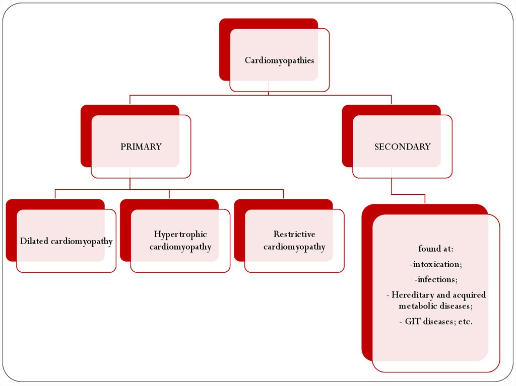
CardiomyopathiesPRIMARY
Dilated cardiomyopathy
Hypertrophic
cardiomyopathy
SECONDARY
Restrictive
cardiomyopathy
found at:
-intoxication;
-infections;
- Hereditary and acquired
metabolic diseases;
- GIT diseases; etc.
4.
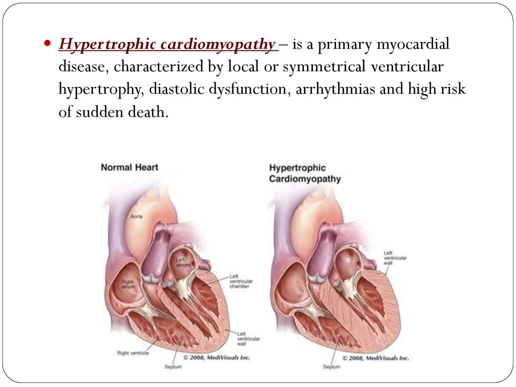
Hypertrophic cardiomyopathy – is a primary myocardialdisease, characterized by local or symmetrical ventricular
hypertrophy, diastolic dysfunction, arrhythmias and high risk
of sudden death.
5. It is characterized by myocardial hypertrophy, abnormal diastolic filling, and in about one third of cases, intermittent
ventricular outflowobstruction.
6. Etiology
Hypertrophiccardiomyopathy
Obstructive
Nonobstructive
Inherited
by abnormal genes (gene
mutations) that cause the
heart muscle to grow
abnormally thick
have a form of the disease in which the wall
(septum) between the two bottom chambers of
the heart (ventricles) becomes enlarged and
impedes blood flow out of the heart.
significant blocking of blood flow. However, the
heart's main pumping chamber (left ventricle) may
become stiff, reducing the amount of blood the
ventricle can hold and the amount pumped out to
the body with each heartbeat.
7. Macro image
The ventricular cavity loses itsusual round-to-ovoid shape and
may be compressed into a
‘banana-like’ configuration by
bulging of the ventricular septum
into the lumen.
Often present are endocardial
thickening or mural plaque
formation in the left ventricular
outflow tract and thickening of
the anterior mitral leaflet.
8.
Extensive myocyte hypertrophy with transverse myocyte diameters frequently greaterthan 40 μm (n: ~ 15μm)
Haphazard disarray of bundles of myocytes, individual myocytes, and contractile elements
in sarcomeres within cells
Interstitial and replacement fibrosis
9. Literature:
V.Kumar, A.K. Abbas, S.N. Fauso. Pathologic Basis of Disease,7th edition, 2008 – 1525 p.
V.V.Serov, V.S.Paukov. Pathological anatomy, 2010 – 800 p.
R.A.Cooke, B.Stewart. Colour Atlas of Anatomical
Pathology, 3rd edition, 2004 – 300 p.
10.
Thank you forattention!