


 medicine
Similar presentations:


Pediatric cardiomyopathy and anesthesia
1. Pediatric Cardiomyopathy and Anesthesia
Alexander Zlotnik MD, PhDProfessor and Chairman,
Soroka University Medical Center,
Ben Gurion University of the Negev
Beer Sheva,
Israel
2.
3.
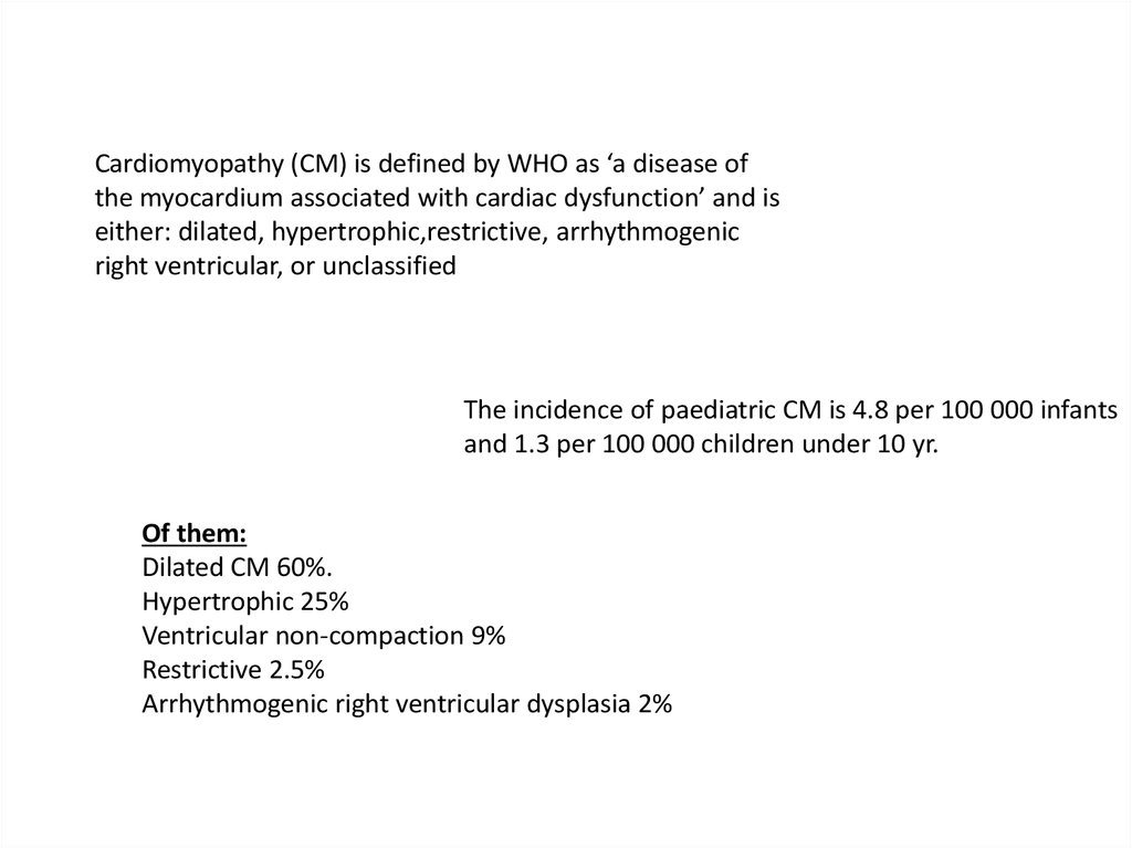
Cardiomyopathy (CM) is defined by WHO as ‘a disease ofthe myocardium associated with cardiac dysfunction’ and is
either: dilated, hypertrophic,restrictive, arrhythmogenic
right ventricular, or unclassified
The incidence of paediatric CM is 4.8 per 100 000 infants
and 1.3 per 100 000 children under 10 yr.
Of them:
Dilated CM 60%.
Hypertrophic 25%
Ventricular non-compaction 9%
Restrictive 2.5%
Arrhythmogenic right ventricular dysplasia 2%
4.
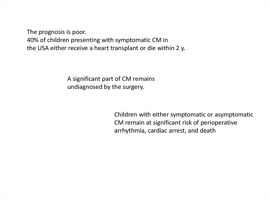
The prognosis is poor.40% of children presenting with symptomatic CM in
the USA either receive a heart transplant or die within 2 y.
A significant part of CM remains
undiagnosed by the surgery.
Children with either symptomatic or asymptomatic
CM remain at significant risk of perioperative
arrhythmia, cardiac arrest, and death
5.
Dilated cardiomyopathy (DCM)DCM, also called congestive CM, is characterized by dilatation
and impaired contractility of one or both ventricles.
Annual incidence of DCM is 0.58 per 100 000 children
14% mortality rate in
the 2 years after diagnosis
Ethyology:
congenital
Infection
Inflammation
metabolic or endocrine disease
malnutrition.
longstanding SVT
Idiopathic (66%)
7% of children who sustain burn injuries
>70% BSA may develop a reversible DCM.
This often presents 100 days after the injury.
Inflammatory mediators?
6.
Pathopfysiology of DCMBiventricular dilatation
Systolic and diastolic myocardial dysfunction
Decreased EF
Decreased CO
Atrial filling pressure and LVEDP are elevated
Associated mitral and tricuspid valve regurgitation.
The dilated myocardium is potentially arrhythmogenic
7.
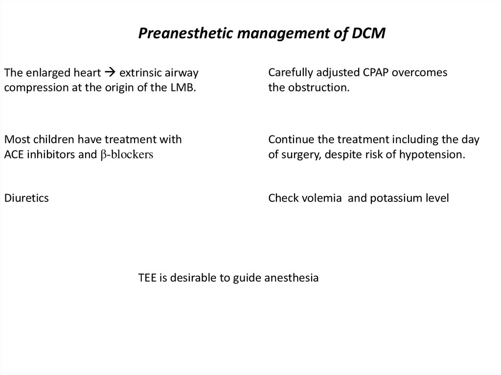
Preanesthetic management of DCMThe enlarged heart extrinsic airway
compression at the origin of the LMB.
Carefully adjusted CPAP overcomes
the obstruction.
Most children have treatment with
ACE inhibitors and β-blockers
Continue the treatment including the day
of surgery, despite risk of hypotension.
Diuretics
Check volemia and potassium level
TEE is desirable to guide anesthesia
8.
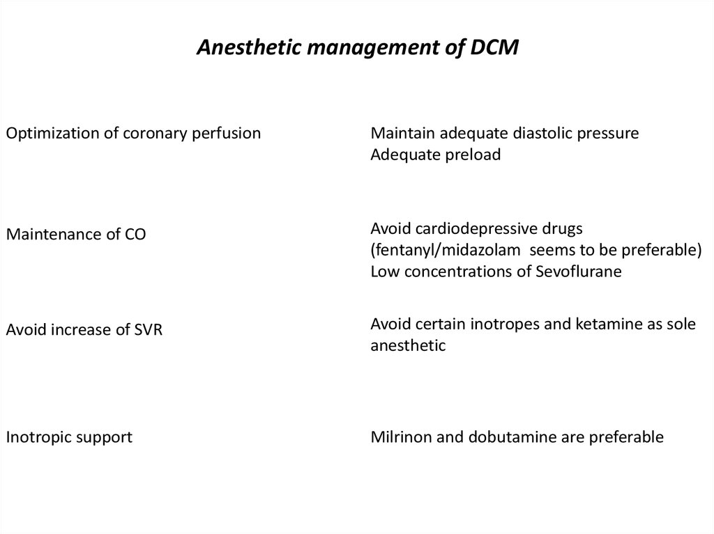
Anesthetic management of DCMOptimization of coronary perfusion
Maintain adequate diastolic pressure
Adequate preload
Maintenance of CO
Avoid cardiodepressive drugs
(fentanyl/midazolam seems to be preferable)
Low concentrations of Sevoflurane
Avoid increase of SVR
Avoid certain inotropes and ketamine as sole
anesthetic
Inotropic support
Milrinon and dobutamine are preferable
9.
Hypertrophic cardiomyopathy HCMMore common in adults, the incidence is low in
children (5/1,000,000).
As patients can be asymptomatic, the diagnosis is often PM
A focal area of hypertrophy may also incorporate and surround
a coronary vessel, so-called myocardial bridging significant
coronary hypoperfusion risk of sudden death.
10. Pathophysiology of HCM
Asymmetric hypertrophy of septum & dynamic obstruction to LV outflow due tomitral valve systolic anterior motion and ventricular septal contact;
11.
12. Factors affecting hemodynamics in patients with HCM
ImprovingDeteriorating
Good preload/filling of LV, hypervolemia
Poor preload/ filling of LV, hypovolemia
Low contractility
High contractility
Bradycardia
Tachycardia, stress
High SVR
Low SVR
Low transaortic pressure gradient
High transaortic pressure gradient
Laying
Standing, Valsalva man.
13.
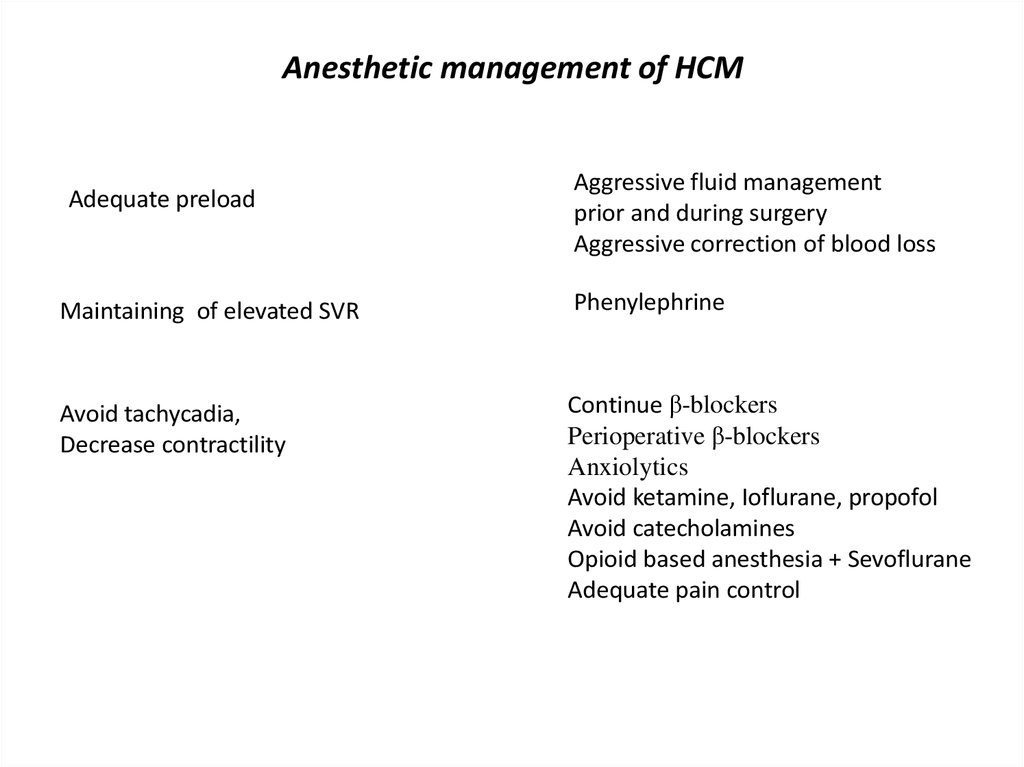
Anesthetic management of HCMAdequate preload
Aggressive fluid management
prior and during surgery
Aggressive correction of blood loss
Maintaining of elevated SVR
Phenylephrine
Avoid tachycadia,
Decrease contractility
Continue β-blockers
Perioperative β-blockers
Anxiolytics
Avoid ketamine, Ioflurane, propofol
Avoid catecholamines
Opioid based anesthesia + Sevoflurane
Adequate pain control
14.
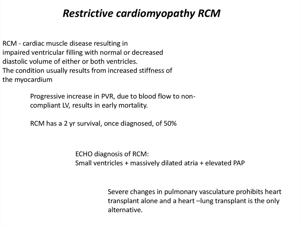
Restrictive cardiomyopathy RCMRCM - cardiac muscle disease resulting in
impaired ventricular filling with normal or decreased
diastolic volume of either or both ventricles.
The condition usually results from increased stiffness of
the myocardium
Progressive increase in PVR, due to blood flow to noncompliant LV, results in early mortality.
RCM has a 2 yr survival, once diagnosed, of 50%
ECHO diagnosis of RCM:
Small ventricles + massively dilated atria + elevated PAP
Severe changes in pulmonary vasculature prohibits heart
transplant alone and a heart –lung transplant is the only
alternative.
15.
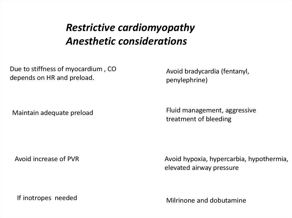
Restrictive cardiomyopathyAnesthetic considerations
Due to stiffness of myocardium , CO
depends on HR and preload.
Maintain adequate preload
Avoid increase of PVR
If inotropes needed
Avoid bradycardia (fentanyl,
penylephrine)
Fluid management, aggressive
treatment of bleeding
Avoid hypoxia, hypercarbia, hypothermia,
elevated airway pressure
Milrinone and dobutamine
16.
Arrhythmogenic right ventricular dysplasia/CMCharacterized by the gradual replacement of myocytes by adipose and
fibrous tissue, it usually presents between the ages of 10–50 yr
ARVD/CM and long QT syndrome are the most common primary
arrhythmic causes of SCD.
The inheritance of this disorder is autosomal-dominant
Pathologically, the free wall of the RV is replaced by fibro-fatty
infiltration locuses for arrhythmias.
17.
Arrhythmogenic right ventricular dysplasia/CMSymptoms include palpitations, syncope, atypical chest pain,
or dyspnea, but SCD may be the initial manifestation.
Of 50 autopsies performed for perioperative
death, ARVD/CM was detected in 18 (36%).
Four of the patients died on induction, 9
during surgery, and 5 within 2 h after
surgery.
50% of patients have an abnormal ECG:
complete or incomplete RBBB,
QRS prolongation without RBBB,
epsilon wave immediately after the QRS in V1–V2,
T-wave inversion in V1–V3
Diagnosis:
1. ECHO: regional or global RV hypokinesis with or
without dilatation
2. Angiography: RV wall anomalies in the absence of
other structural heart defects
3. Histologically after an endomyocardial biopsy
18.
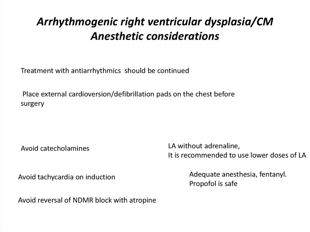
Arrhythmogenic right ventricular dysplasia/CMAnesthetic considerations
Treatment with antiarrhythmics should be continued
Place external cardioversion/defibrillation pads on the chest before
surgery
Avoid catecholamines
Avoid tachycardia on induction
Avoid reversal of NDMR block with atropine
LA without adrenaline,
It is recommended to use lower doses of LA
Adequate anesthesia, fentanyl.
Propofol is safe