


 medicine
Similar presentations:


Aortic Insufficiency
1. Aortic Insufficiency
2. Classification -1
Abnormalitiesof the Leaflets
• Rheumatic, Bicuspid, Degenerative
• Endocarditis
Dilation
of the Aortic Annulus
• Aortic Aneurysm / Dissection
• Inflammatory (Syphyllis, Giant Cell Arteritis.
Coll Vasc Dis-Ankylosis Spondylitis, Reiters)
• Inheritable (Marfans, Osteogensis Imperfecta)
3. Classification -2
4. Chronic AI - Pathophysiology
increased LV EDVaddition of new sarcomeres in series/ elongation of
myocytes and myocardial fibers (Eccentric Hypertrophy)
enlarged chamber/ increased wall stress is stimulus for
concentric hypertrophy
dilatation and hypertrophy with resultant recruitment of
preload reserve allow compensation and maintenance of
LV systolic function
may be asymptomatic for decades until decompensated
state develops, wall thickening unable to keep pace with
hemodynamic load, increased interstitial fibrosis and
decreased compliance symptoms of CHF ensue
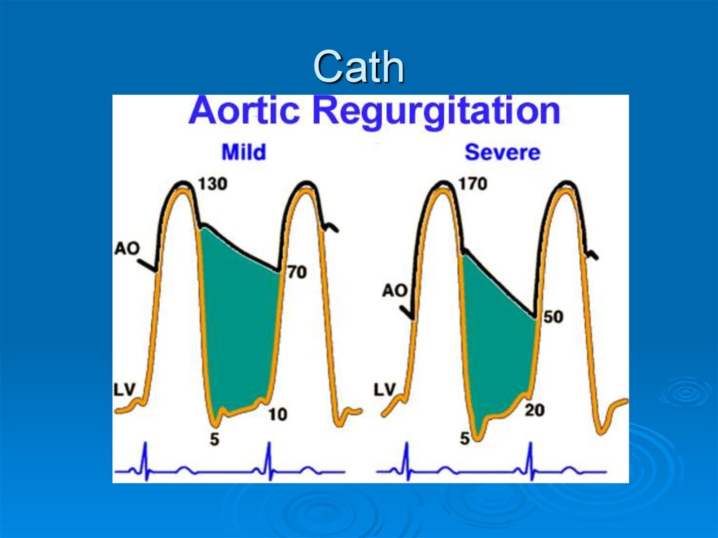
5. Pressure Volume Relationships in Chronic AI
CO at rest may approach 25 L/min in severe AI with little increase in EDPvery large EDV (Cor Bovinum)
Braunwald 6th ed
6. History
DOE, Orthopnea, PNDAngina pectoris
usually after 4th / 5th decade and significant
cardiomegaly and LV dysfx
develops later, nocturnal symptoms prominent; often
with diaphoresis due to HR slowing with arterial DBP
falling to low levels
Palpitations / Head pounding
especially in supine position, pounding of heart
against chest wall
tachycardia from stress/exertion may precipitate and
cause extreme discomfort for pt
7. Physical Findings
Diastolic murmurAustin Flint murmur
high frequency, sitting up, leaning forward
duration > intensity correlates with severity
mild AR – early diastole, hi pitched blowing
severe AR – holodiastolic, rough
musical (“cooing dove”) – eversion/perforation of Ao cusp
Primary valve dz – heard best LSB 3-4 intercostal
Ao Root dz – heard best RSB
mid-late diastolic apical rumble – severe AR
Wide Pulse Pressure
Systolic flow murmur (/thrill)
8. Peripheral Signs of Severe Aortic Regurgitation
CXR9. CXR
ECHO2D/ M-Mode
Doppler
AV/ Ao Root anatomic abnormalities
LV dimension / sphericity
AMVL – fluttering, reverse doming
increased EPSS
Color Flow Mapping
Continuous Wave
Flow reversal in desc Ao (100% sens 97% spec for
severe AI)
Limitations – What is severe AI?
10. ECHO
AMVL flutteringColor Flow – top mild, bottom moderate
11.
Continuous Wave DopplerChronic AI
Acute AI
12.
Cath13. Cath
Medical ManagementVasodilators
Uses
goal is to reduce SBP, improve forward SV, reduce regurgitant
volume
severe AR + symptoms of LV dysfxn
short term hemodynamic improvement in pt with symptomatic
AR before AVR
prolong compensated phase of asymptomatic patients
No indication for asymptomatic pt with mild AI and normal LV
fxn
Studied in AI
Nifedipine, Hydralizine, ACEI, Nipride, Prazosin
Children/ severe AR – ACEI reversed LV dilatation/wall stress
avoid (-) inotrope in LV dysfx
14. Medical Management
Timing of SurgeryGoal is to intervene before irreversible LV
systolic dysfx ensues
initially reversible, mainly due to afterload
excess – full recovery in LV size/fx possible
with progressive chamber dilatation,
decreased myocardial contractility >>
afterload excess as cause of LV dysfx.
associated with worse recovery of LV fx and
increased mortality
15. Timing of Surgery
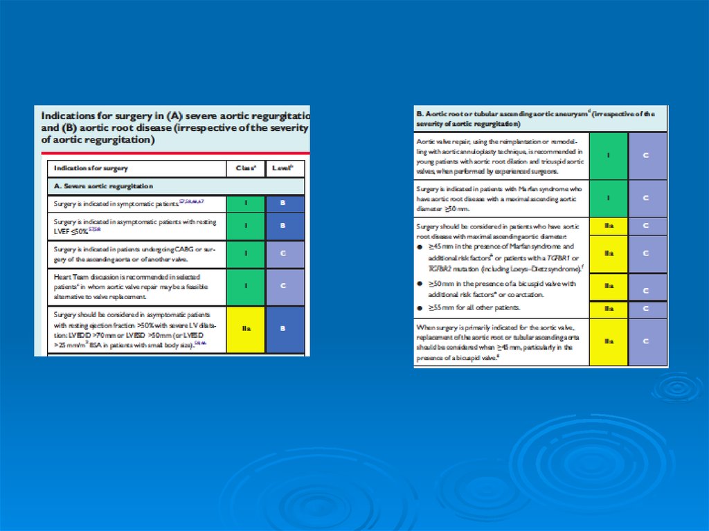
Surgical TherapyIndications for AVR (Severe AR)1
Predictors of Postoperative Prognosis
1
Symptoms (NYHA III-IV) regardless of LV fxn
Symptoms (NYHA II) with evidence of progressing
LV dysfx ( LV ESD ~ 55, LV EF <50-55%)
Angina (CHA Class II or higher) w or w/o CAD
mild-mod LV dysfx (EF 25-49%) regardless of
symptoms
mod-sev AR and undergoing CABG or other valvular
surgery
LV systolic function
LV End Systolic Size ( LV ESD)
Bonow, et al. Circulation 1998;98:1949-84
16. Surgical Therapy
17.
18.
19.
Aortic Valve Replacement20.
Surgical OptionsAo Root disease
annuloplasty or other valve sparing surgery
possible if pure Ao Root dz
Primary AV disease
valve replacement
21. Surgical Options
AV sparing conduitFigure 46-42 Repair of the aortic valve in patient with severe AR. Conduit tailoring in the
supravalvular position. The conduit is cut to replace three (left), two (middle), or one (right)
individual sinuses. The aortic aneurysm is replaced and the valve is spared.
(From David TE, Feindel CM, Bos J: Repair of the aortic valve in patients with aortic
insufficiency and aortic root aneurysm. J Thorac Cardiovasc Surg 109:345, 1995.)
Braunwauld 6th ed
22. AV sparing conduit
Rx of Acute AITreat
cause of acute AI
Dissection/Trauma
Endocarditis
Prosthesis malfunction
Urgent AVR + aortoplasty in most cases