


 electronics
Similar presentations:


 - Technical Highlights")


Ultrasonic B-Scanner UD-8000. Inspire by Digital Tomey Corporation
1.
Ultrasonic B-ScannerUD-8000
Inspire by Digital
Tomey
Corporation
2.
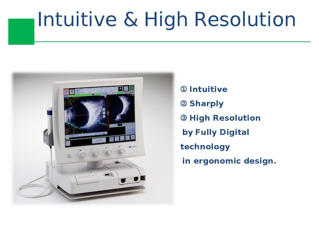
Intuitive & High Resolution① Intuitive
② Sharply
③ High Resolution
by Fully Digital
technology
in ergonomic design.
3.
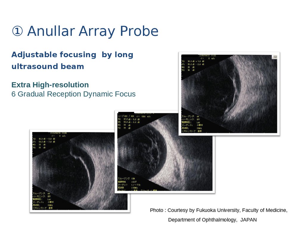
① Anullar Array ProbeAdjustable focusing by long
ultrasound beam
Extra High-resolution
6 Gradual Reception Dynamic Focus
Photo : Courtesy by Fukuoka University, Faculty of Medicine,
Department of Ophthalmology, JAPAN
4.
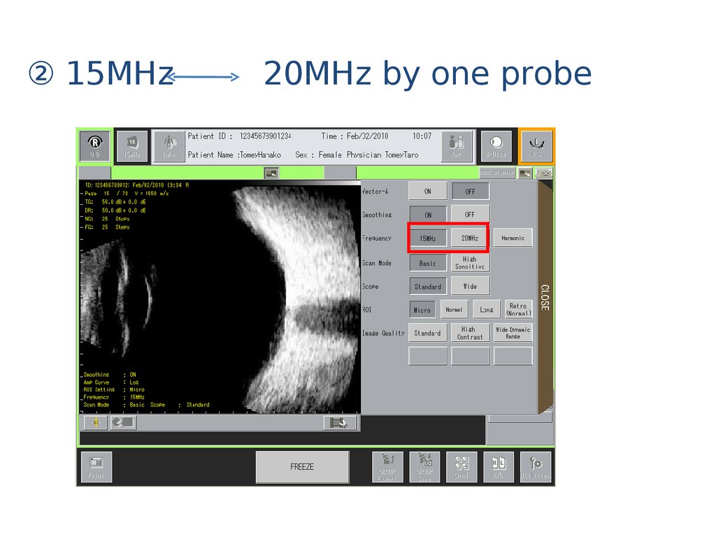
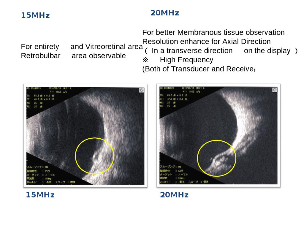
② 15MHz20MHz by one probe
5.
15MHzFor entirety
Retrobulbar
15MHz
20MHz
For better Membranous tissue observation
Resolution enhance for Axial Direction
and Vitreoretinal area
In a transverse direction on the display
area observable
※ High Frequency
(Both of Transducer and Receive)
20MHz
6.
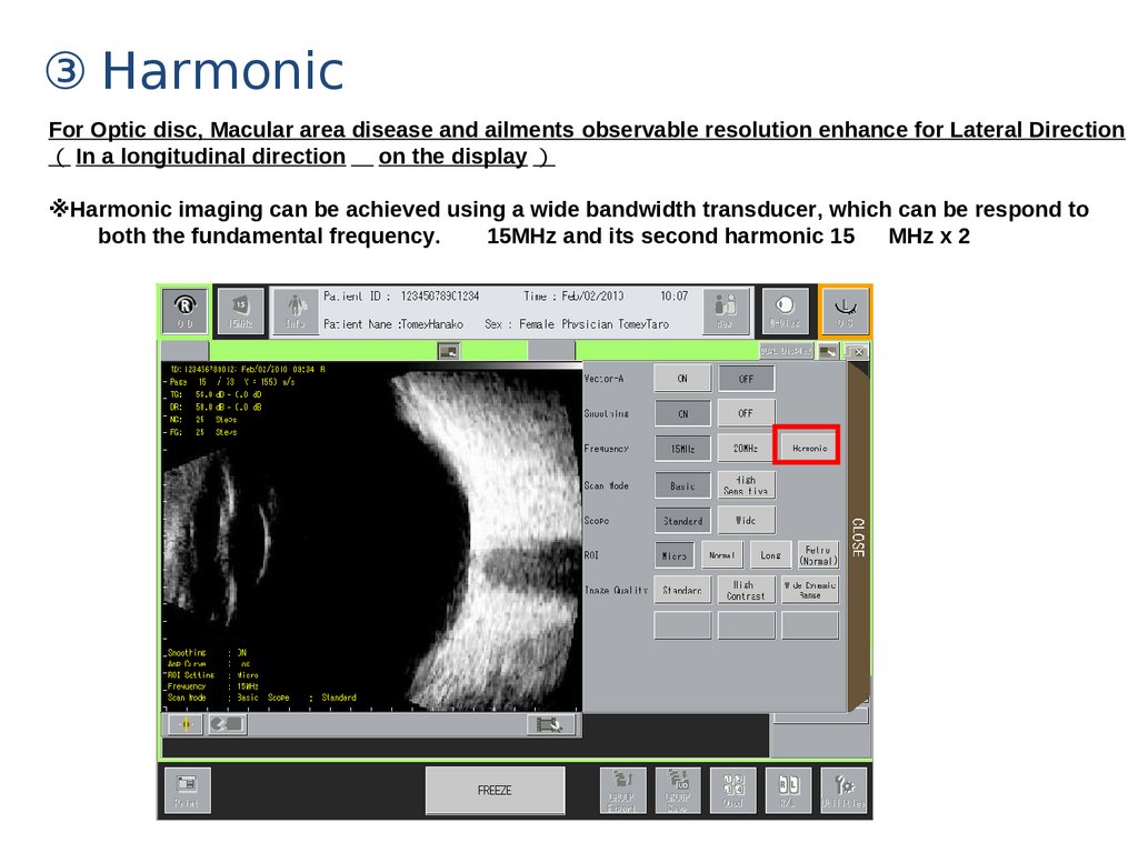
③ HarmonicFor Optic disc, Macular area disease and ailments observable resolution enhance for Lateral Direction
In a longitudinal direction on the display
※Harmonic imaging can be achieved using a wide bandwidth transducer, which can be respond to
both the fundamental frequency.
15MHz and its second harmonic 15
MHz x 2
7.
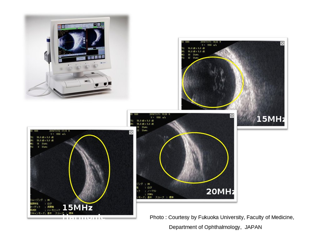
15MHz20MHz
15MHz
Harmonic
Photo : Courtesy by Fukuoka University, Faculty of Medicine,
Department of Ophthalmology, JAPAN
8.
④ Indicate of probe direction9.
⑤ New designed probe10.
Function setting① Scan mode
① Standard mode
② High Sensitive
Mode
※ Deduct noise, improve
SNR
Frame rate 】
① Standard mode 22
Frames/sec
② High sensitive mode
11
Frames/sec
11.
② Scope Displayed depthStandard
Eye
ball
or
Wide
Orbit
Display Range 】
Standard
42
mm
Wide
mm
54
12.
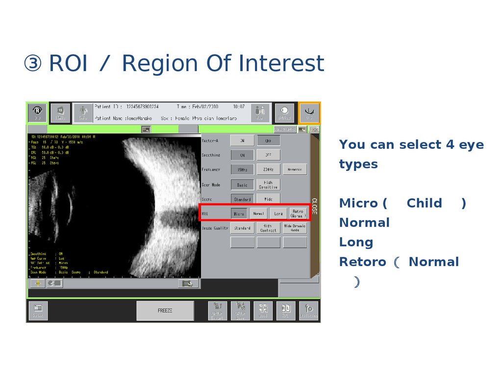
③ ROI Region Of InterestYou can select 4 eye
types
Micro (
Child
Normal
Long
Retoro Normal
)
13.
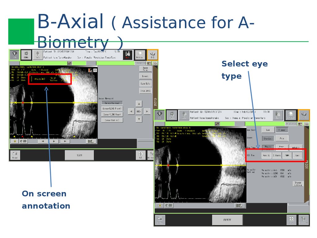
B-Axial ( Assistance for ABiometrySelect eye
type
On screen
annotation
14.
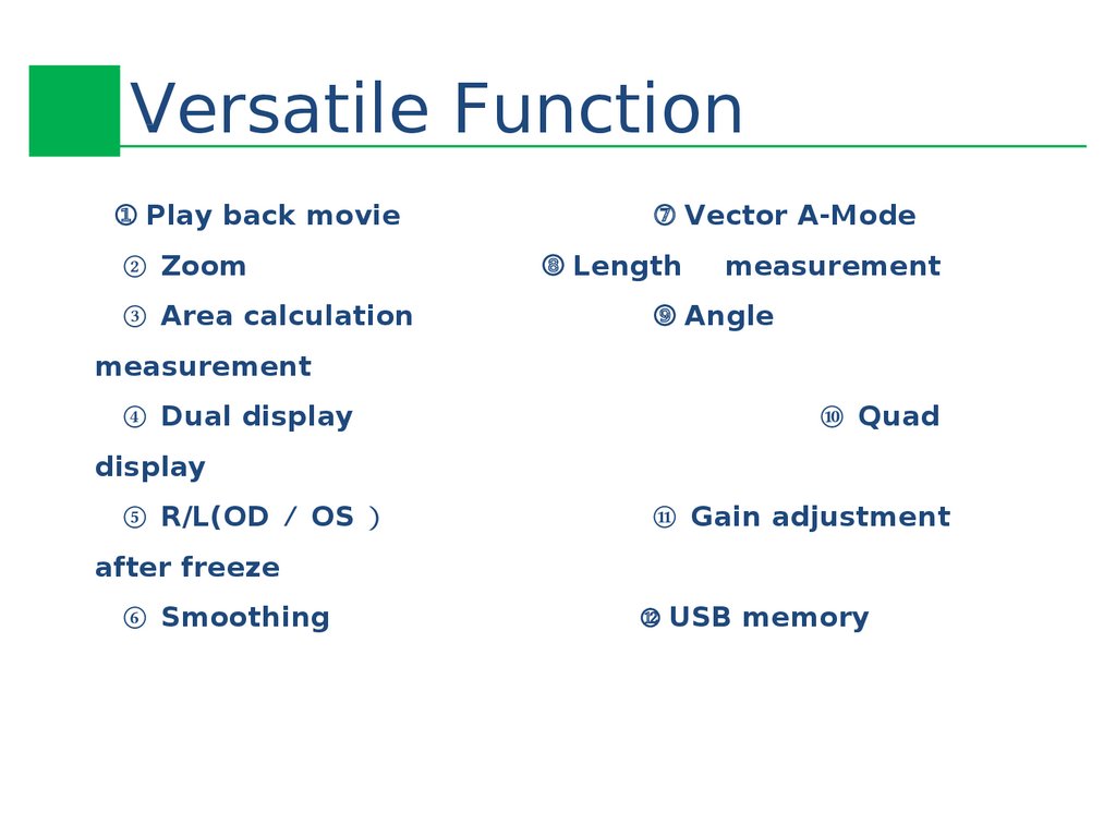
Versatile Function① Play back movie
② Zoom
③ Area calculation
⑦ Vector A-Mode
⑧ Length
measurement
⑨ Angle
measurement
④ Dual display
⑩ Quad
display
⑤ R/L(OD OS
⑪ Gain adjustment
after freeze
⑥ Smoothing
⑫ USB memory
15.
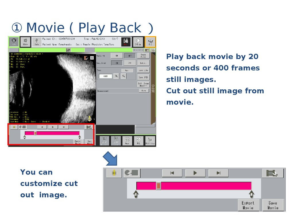
① Movie ( Play BackPlay back movie by 20
seconds or 400 frames
still images.
Cut out still image from
movie.
You can
customize cut
out image.
16.
② Vector A-ModeTemporary ABiometry
by Compare with
pathology
wave form and
retina wave form.
17.
③ Zoom5 steps Zoom
100%, 125%, 150%,
200%, 300%
Intuitive navigation monitor
18.
④ Length calculationUp
to 3 spots on
same time
calculation
19.
⑤ Area calculationUp
to 2 spots on
same time
calculation
20.
⑥ Angle calculationUp
to 2 spots on
same time
calculation
21.
⑦ Dual/Quad Display22.
⑧ R(OD L OS Display23.
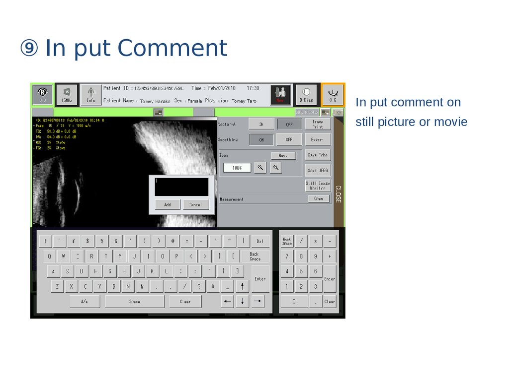
⑨ In put CommentIn put comment on
still picture or movie
24.
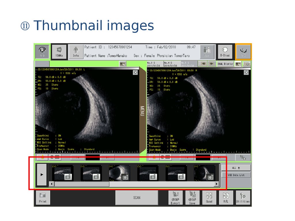
⑩ Print out by still image part only25.
⑪ Thumbnail images26.
⑫ Group ExportGroup Save
27.
Save Out put①Still image
Save Echo or JPEG
format
( Re-Display, Edit
possible )
② Movie
Save in USB memory by
Echo format and out put.
28.
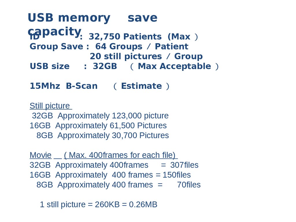
USB memory savecapacity
ID
: 32,750 Patients
(Max
Group Save : 64 Groups Patient
20 still pictures Group
USB size
: 32GB Max Acceptable
15Mhz B-Scan
Estimate
Still picture
32GB Approximately 123,000 picture
16GB Approximately 61,500 Pictures
8GB Approximately 30,700 Pictures
Movie ( Max. 400frames for each file)
32GB Approximately 400frames
= 307files
16GB Approximately 400 frames = 150files
8GB Approximately 400 frames =
70files
1 still picture = 260KB = 0.26MB
29.
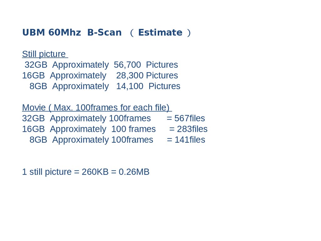
UBM 60Mhz B-Scan EstimateStill picture
32GB Approximately 56,700 Pictures
16GB Approximately 28,300 Pictures
8GB Approximately 14,100 Pictures
Movie ( Max. 100frames for each file)
32GB Approximately 100frames
= 567files
16GB Approximately 100 frames
= 283files
8GB Approximately 100frames
= 141files
1 still picture = 260KB = 0.26MB
30.
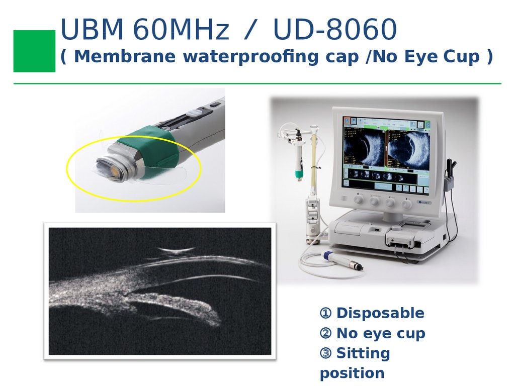
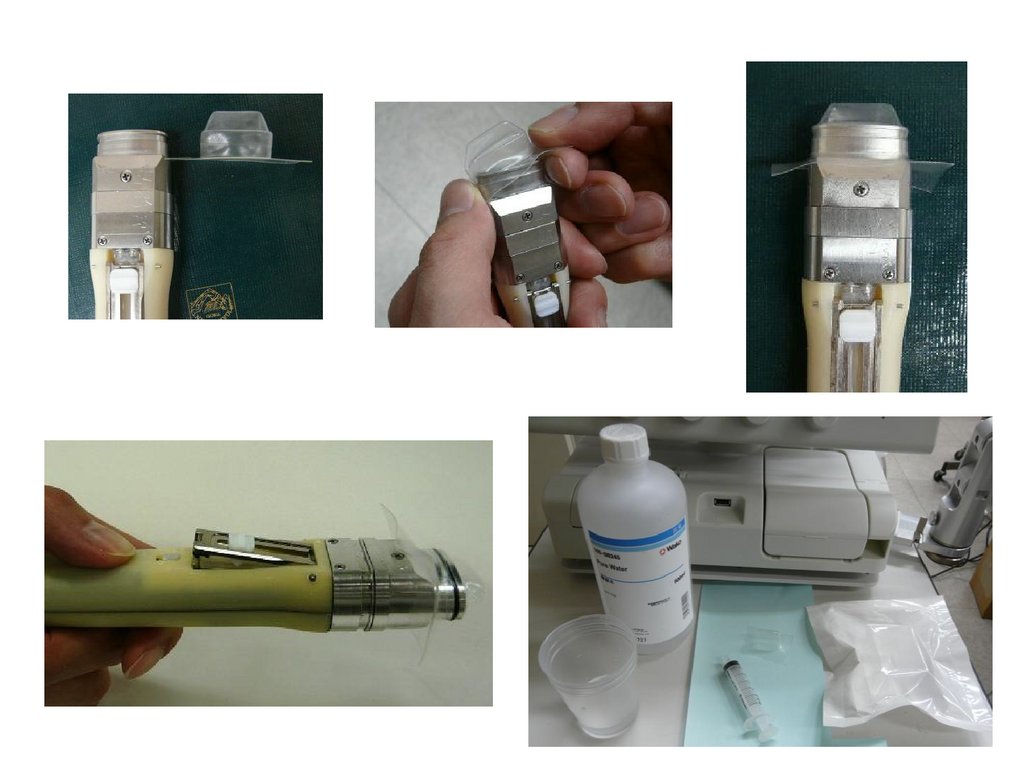
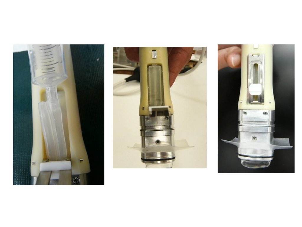
UBM 60MHz UD-8060( Membrane waterproofng cap /No Eye Cup )
① Disposable
② No eye cup
③ Sitting
position
31.
32.
33.
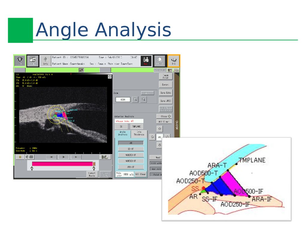
Angle Analysis34.
SS:
Sclera: The angle corner side of the line segment that
constitutes the trabeculum plain
TMPLANE
:
The cornea side of the line segment that constitutes the
trabeculum plain
SS-IF
:
The intersection of the iris anterior surface and the line that
crosses SS and is vertical to the line that crosses SS and
TMPLANE
AOD250-T
:
The measurement point on the trabeculum side of AOD250
AOD250-IF
AOD500-T
:
:
The measurement point on the iris side of AOD250
The measurement point on the trabeculum side of AOD500
AOD500-IF
ARA-T
:
:
The measurement point on the iris side of AOD500
A point, on the trabeculum (on the corneal surface), that is
750 um away from the sclera.
ARA-IF
:
The intersection of the iris anterior surface and the line that
crosses ARA-T and is vertical to the line that crosses SS and
ARA-T
AR
:
Angle point
AOD250
:
Distance between AOD250-T and AOD250-IF
AOD500
:
Distance between AOD500-T and AOD500-IF
AOD700
:
Distance between AOD700-T and AOD700-IF
ARA500
:
The area of the angle area defined by the line that crosses
AOD500-T and AOD500IF
ARA750
:
TISA500
:
TISA700
:
TIA500
:
The area of the angle area defined by the line that crosses
AOD700-T and AOD700-IF
The area of the angle area defined by the line that crosses
SS and SS-IF and the line that crosses AOD500-T and
AOD500IF
The area of the angle area defined by the line that crosses
SS and SS-IF and the line that crosses AOD700-T and
AOD700-IF
The angle between the line AB to AOD500-T and the line
AB to AOD500-IF.
Angle Analysis
Parameter 】
35.
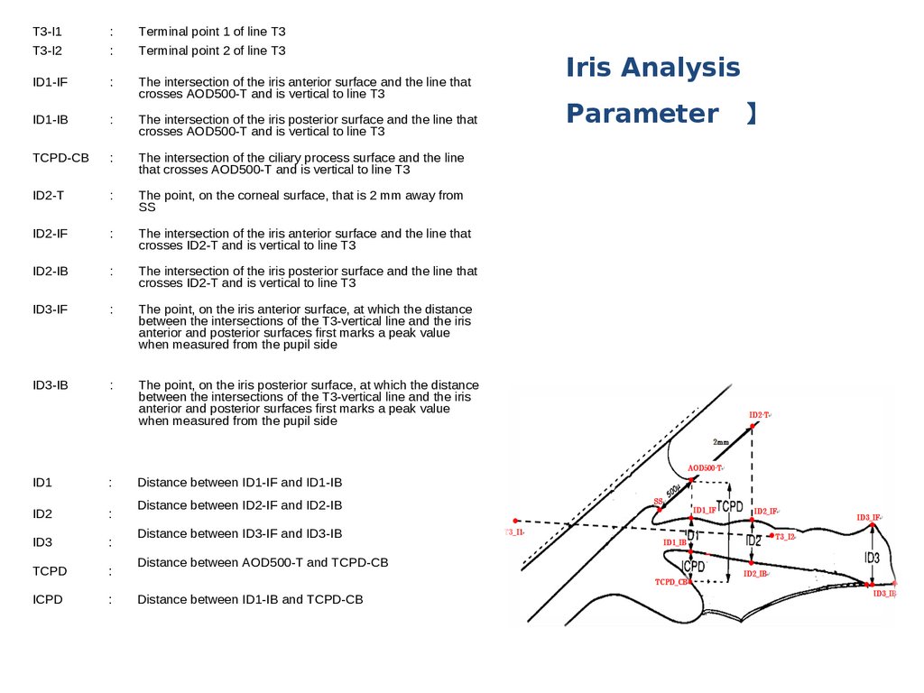
Iris Analysis36.
T3-I1T3-I2
:
:
Terminal point 1 of line T3
Terminal point 2 of line T3
ID1-IF
:
The intersection of the iris anterior surface and the line that
crosses AOD500-T and is vertical to line T3
ID1-IB
:
The intersection of the iris posterior surface and the line that
crosses AOD500-T and is vertical to line T3
TCPD-CB
:
The intersection of the ciliary process surface and the line
that crosses AOD500-T and is vertical to line T3
ID2-T
:
The point, on the corneal surface, that is 2 mm away from
SS
ID2-IF
:
The intersection of the iris anterior surface and the line that
crosses ID2-T and is vertical to line T3
ID2-IB
:
The intersection of the iris posterior surface and the line that
crosses ID2-T and is vertical to line T3
ID3-IF
:
The point, on the iris anterior surface, at which the distance
between the intersections of the T3-vertical line and the iris
anterior and posterior surfaces first marks a peak value
when measured from the pupil side
ID3-IB
:
The point, on the iris posterior surface, at which the distance
between the intersections of the T3-vertical line and the iris
anterior and posterior surfaces first marks a peak value
when measured from the pupil side
ID1
:
Distance between ID1-IF and ID1-IB
ID2
:
ID3
:
TCPD
:
ICPD
:
Distance between ID2-IF and ID2-IB
Distance between ID3-IF and ID3-IB
Distance between AOD500-T and TCPD-CB
Distance between ID1-IB and TCPD-CB
Iris Analysis
Parameter 】
37.
AL-4000y
( Biometry Pachymetr
① A-Biometry
② IOL Power
calculation
③ Pachymetry
USB or Bluetooth communication
38.
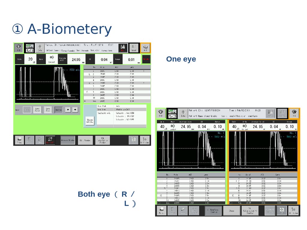
① A-BiometeryOne eye
Both eye R
L
39.
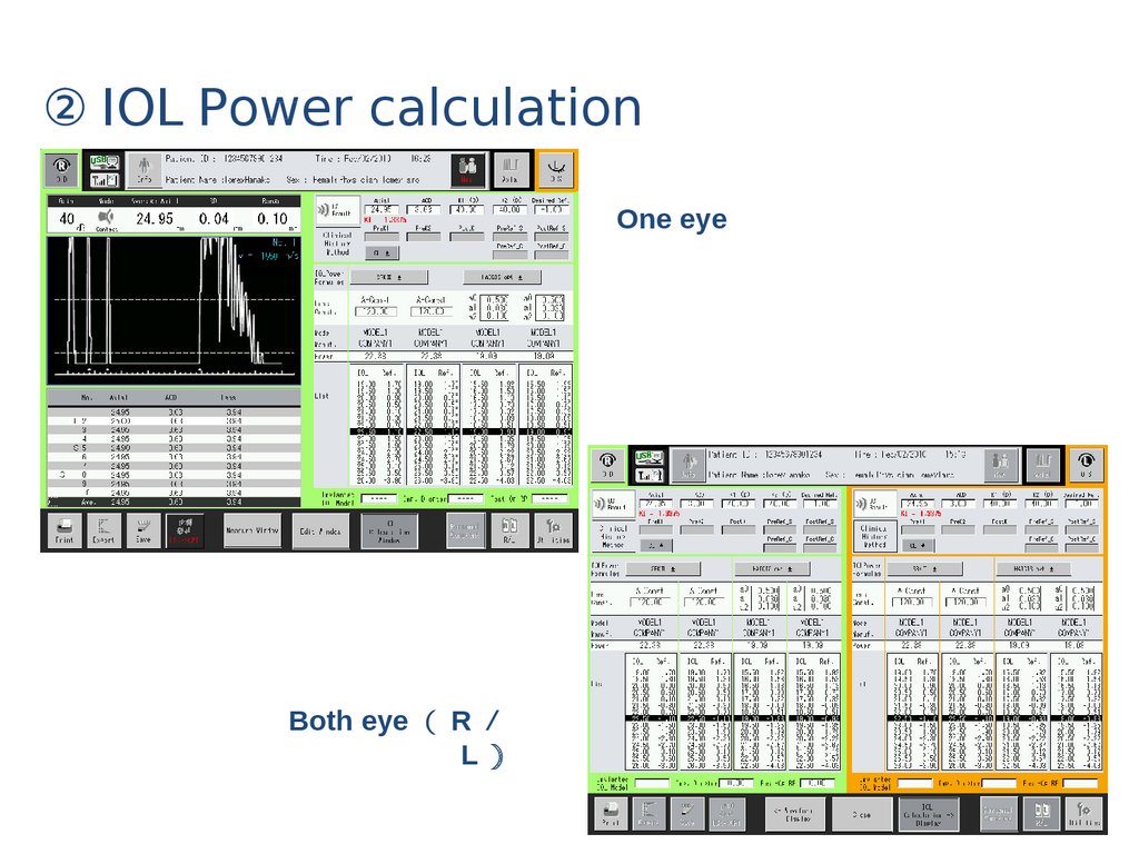
② IOL Power calculationOne eye
Both eye R
L
40.
③ PachymetryOne eye
Both eye R
L
41.
<15MHz B Probe>Focus
Dynamic Focus
Frame rate
Basic mode
High Sensitive mode
Maximum number of pages in a movie
Image display range
Standard
Wide
Color scale
Scan type
Transducer type
Transducer frequency
<60MHz UBM Probe >
Frame rate
Basic mode
High Sensitive mode
Maximum number of pages in a movie
Image display range
Color scale
Scan type
Transducer type
Transducer frequency
Dimensions and weights
Dimension
Weight
Display
TFT LCD
Power source
Input Voltage
Frequency
Power Consumption
UD-8000
Specifcation 】
22 frame / sec
11 frame / sec
400 pages x 2
42mm / 52° (at ultrasound velocity=1550 m/sec)
54mm / 52°(at ultrasound velocity=1550 m/sec)
256 scale level
Sector scanning
Annular array
15 MHz
10 frame / sec
7 frame / sec
100 pages x 2
9 mm(W) x 7mm(D)
256 scale level
Linear scanning
Single
60 MHz
(at ultrasound velocity=1550 m/sec)
398(W)×359(D)×456(H) mm
15.0kg
15 inches, color touch screen
100-120V / 220-240 VAC
50/60Hz
125/125 VA
42.
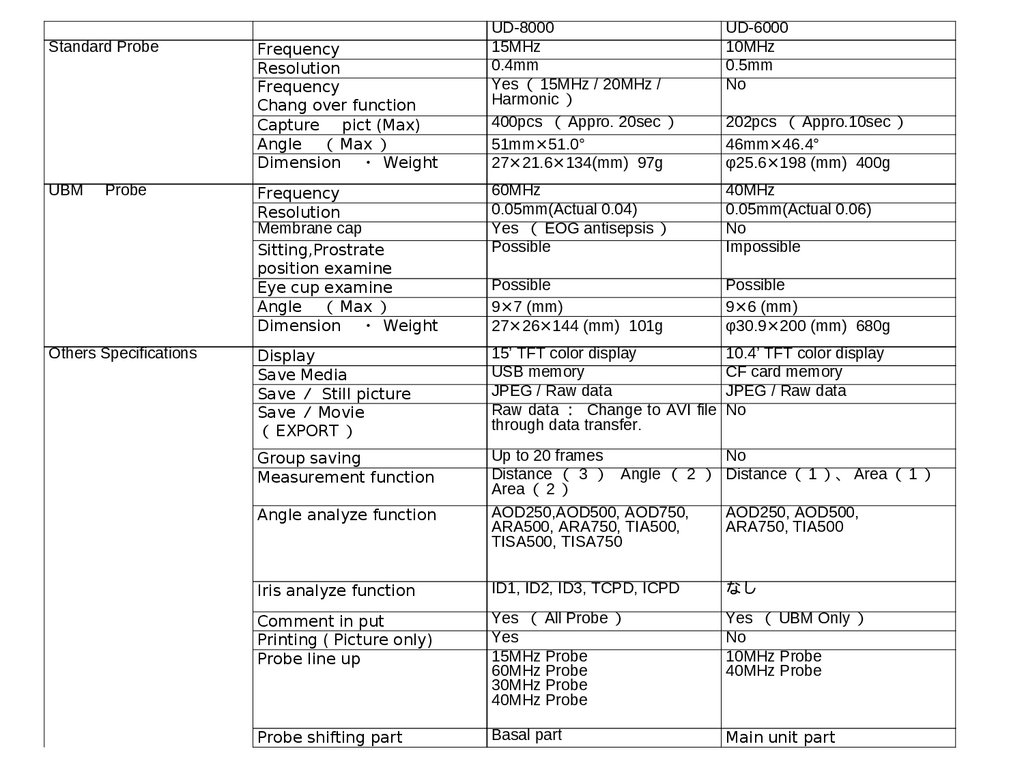
Standard ProbeUBM
Probe
Others Specifications
UD-8000
15MHz
0.4mm
Yes 15MHz / 20MHz /
Harmonic
400pcs Appro. 20sec
51mm×51.0°
27×21.6×134(mm) 97g
UD-6000
10MHz
0.5mm
No
Frequency
Resolution
Membrane cap
Sitting,Prostrate
position examine
Eye cup examine
Angle Max
Dimension ・ Weight
60MHz
0.05mm(Actual 0.04)
Yes EOG antisepsis
Possible
40MHz
0.05mm(Actual 0.06)
No
Impossible
Possible
9×7 (mm)
27×26×144 (mm) 101g
Possible
9×6 (mm)
φ30.9×200 (mm) 680g
Display
Save Media
Save Still picture
Save Movie
EXPORT
15’ TFT color display
USB memory
JPEG / Raw data
Raw data Change to AVI file
through data transfer.
10.4’ TFT color display
CF card memory
JPEG / Raw data
No
Group saving
Measurement function
Up to 20 frames
No
Distance 3 Angle 2 Distance 1 、 Area 1
Area 2
AOD250,AOD500, AOD750,
AOD250, AOD500,
ARA500, ARA750, TIA500,
ARA750, TIA500
TISA500, TISA750
Frequency
Resolution
Frequency
Chang over function
Capture pict (Max)
Angle Max
Dimension ・ Weight
Angle analyze function
202pcs Appro.10sec
46mm×46.4°
φ25.6×198 (mm) 400g
Iris analyze function
ID1, ID2, ID3, TCPD, ICPD
なし
Comment in put
Printing ( Picture only)
Probe line up
Yes All Probe
Yes
15MHz Probe
60MHz Probe
30MHz Probe
40MHz Probe
Yes UBM Only
No
10MHz Probe
40MHz Probe
Probe shifting part
Basal part
Main unit part
43.
Thankyou !