


 medicine
 electronics
Similar presentations:


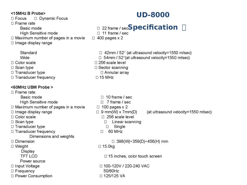
Ultrasonic B-Scanner UD-8000
1.
Ultrasonic B-ScannerUD-8000
Inspire by Digital
Tomey
Corporation
2.
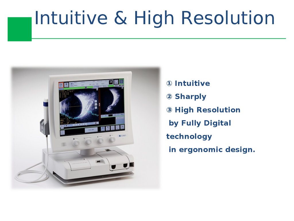
Intuitive & High Resolution① Intuitive
② Sharply
③ High Resolution
by Fully Digital
technology
in ergonomic design.
3.
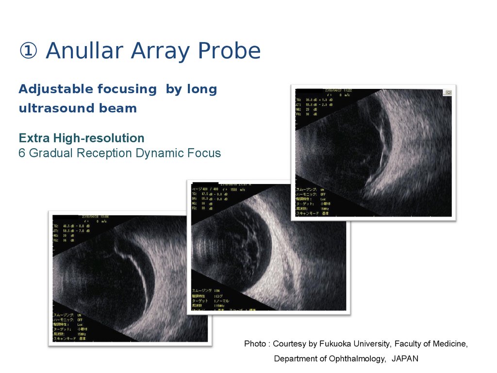
① Anullar Array ProbeAdjustable focusing by long
ultrasound beam
Extra High-resolution
6 Gradual Reception Dynamic Focus
Photo : Courtesy by Fukuoka University, Faculty of Medicine,
Department of Ophthalmology, JAPAN
4.
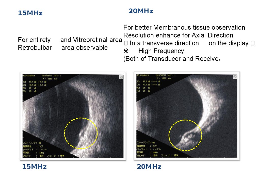
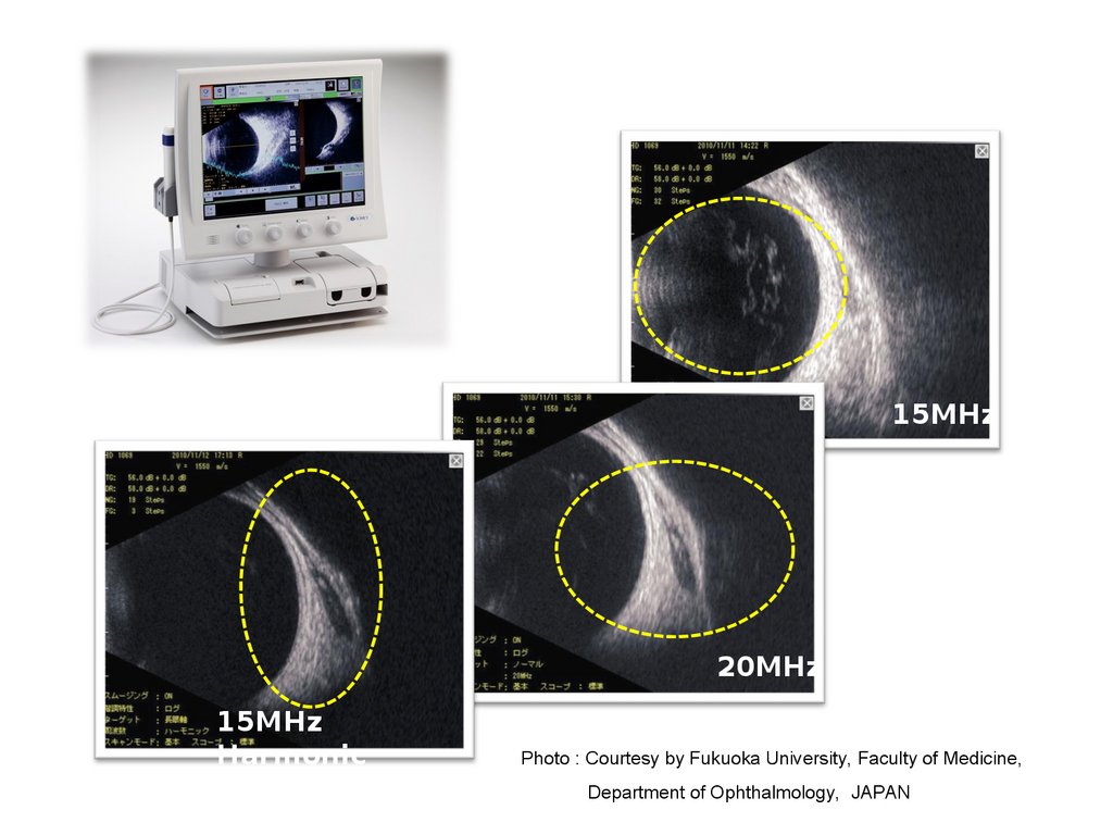
② 15MHz20MHz by one probe
5.
15MHzFor entirety
Retrobulbar
15MHz
20MHz
For better Membranous tissue observation
Resolution enhance for Axial Direction
and Vitreoretinal area
In a transverse direction on the display
area observable
※ High Frequency
(Both of Transducer and Receive)
20MHz
6.
③ HarmonicFor Optic disc, Macular area disease and ailments observable resolution enhance for Lateral Direction
In a longitudinal direction on the display
※Harmonic imaging can be achieved using a wide bandwidth transducer, which can be respond to
both the fundamental frequency.
15MHz and its second harmonic 15 MHz x 2
7.
15MHz20MHz
15MHz
Harmonic
Photo : Courtesy by Fukuoka University, Faculty of Medicine,
Department of Ophthalmology, JAPAN
8.
④ Indicate of probe direction9.
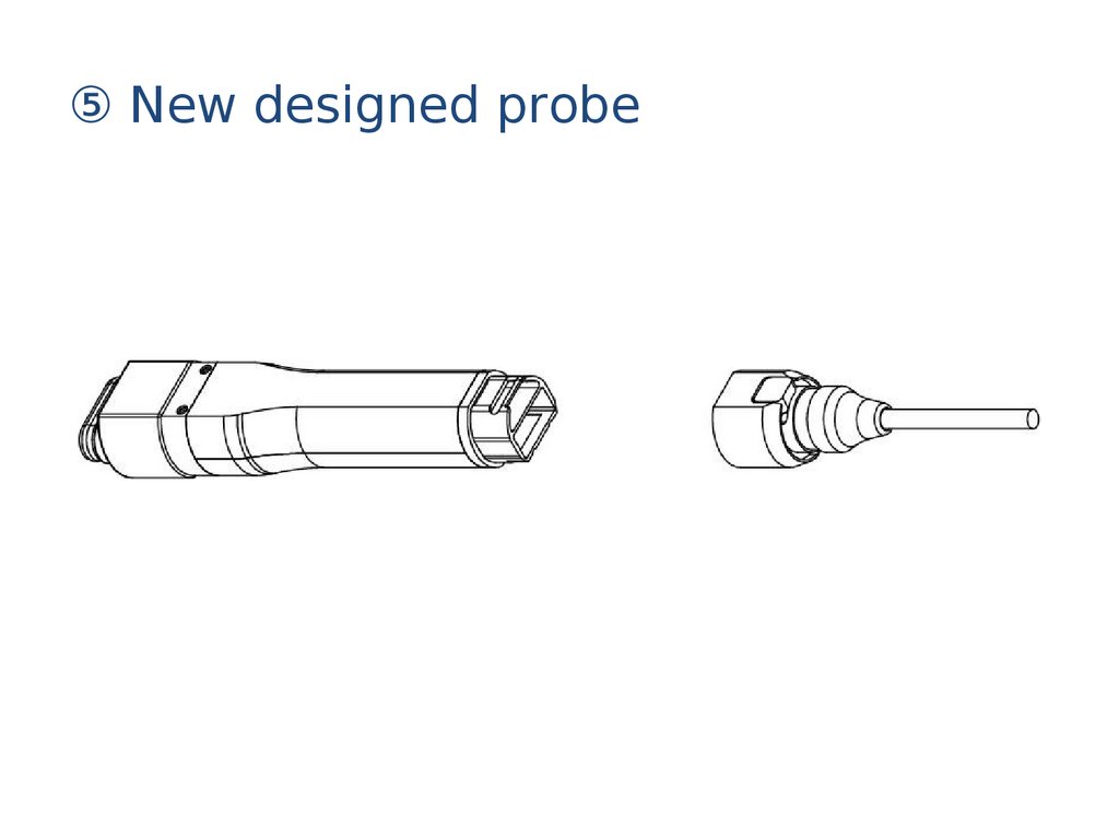
⑤ New designed probe10.
Function setting① Scan mode
① Standard mode
② High Sensitive
Mode
※ Deduct noise, improve
SNR
Frame rate 】
① Standard mode 22
Frames/sec
② High sensitive mode
11
Frames/sec
11.
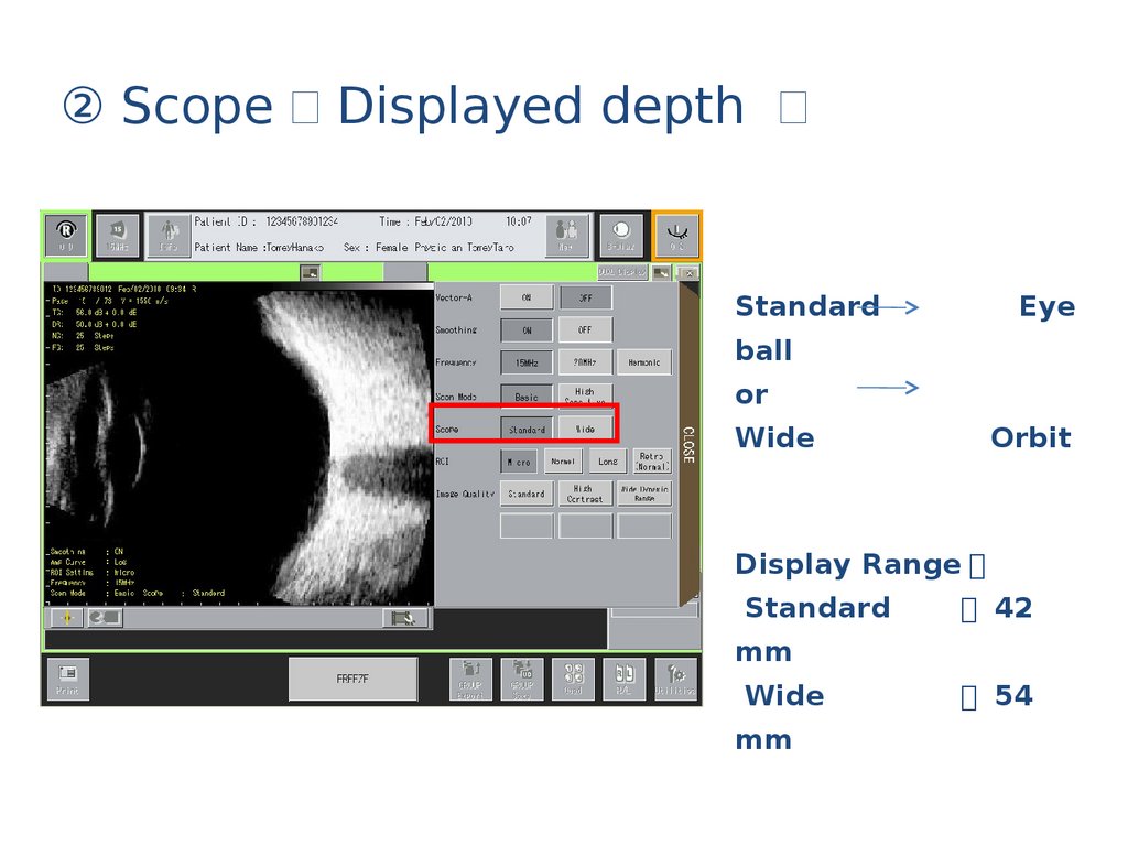
② Scope Displayed depthStandard
Eye
ball
or
Wide
Orbit
Display Range 】
Standard
42
mm
Wide
mm
54
12.
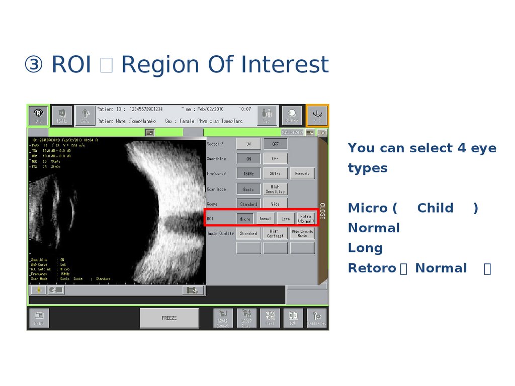
③ ROI Region Of InterestYou can select 4 eye
types
Micro (
Child
)
Normal
Long
Retoro Normal
13.
B-Axial ( Assistance for ABiometrySelect eye
type
On screen
annotation
14.
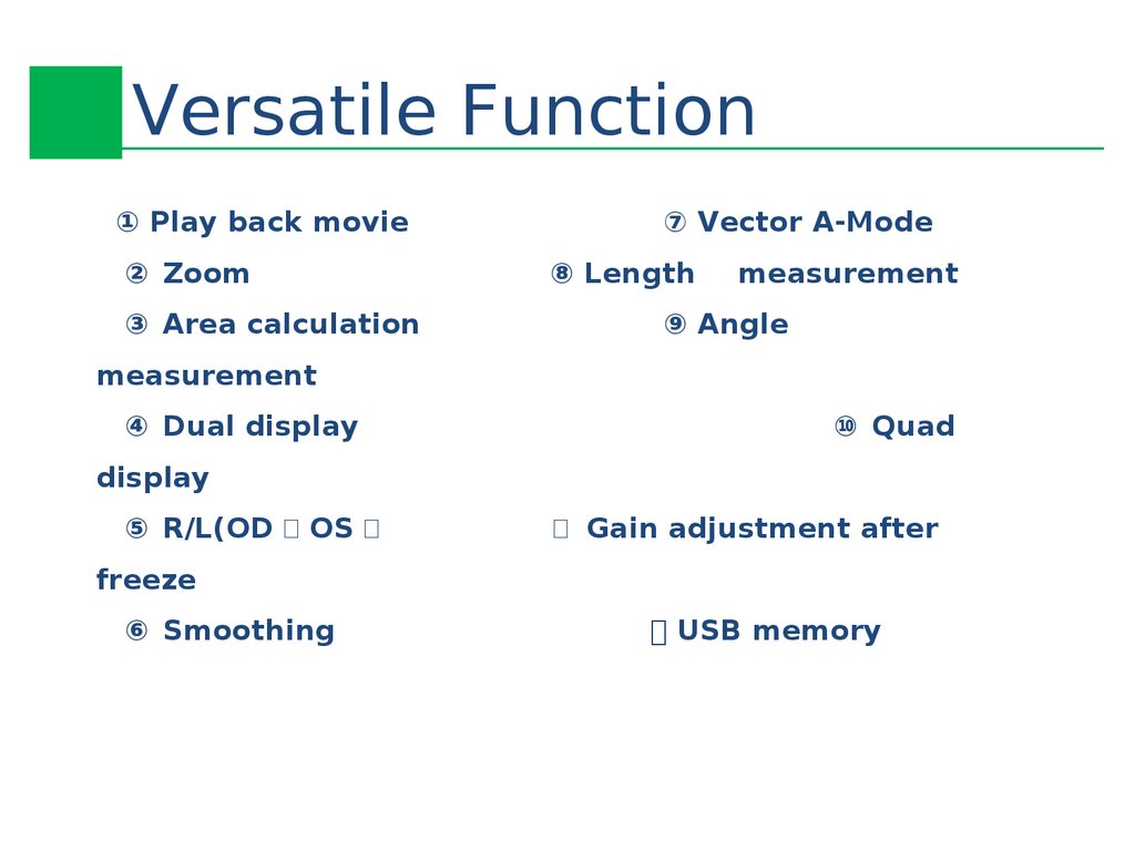
Versatile Function① Play back movie
② Zoom
③ Area calculation
⑦ Vector A-Mode
⑧ Length
measurement
⑨ Angle
measurement
④ Dual display
⑩ Quad
display
⑤ R/L(OD OS
⑪ Gain adjustment after
freeze
⑥ Smoothing
⑫ USB memory
15.
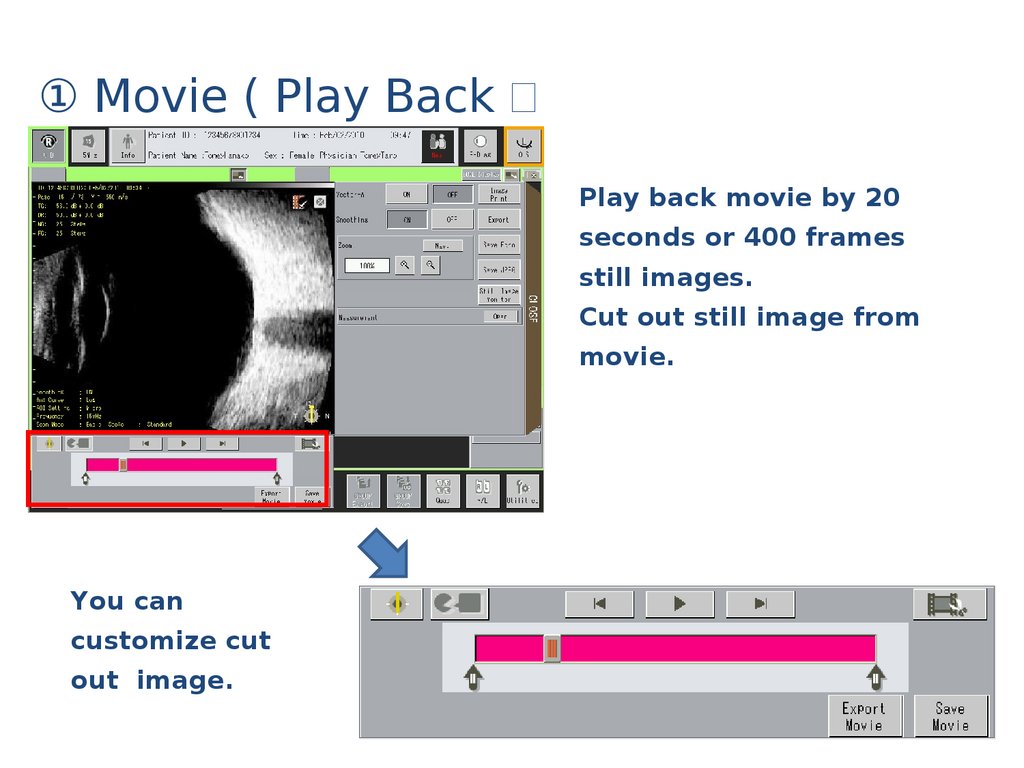
① Movie ( Play BackPlay back movie by 20
seconds or 400 frames
still images.
Cut out still image from
movie.
You can
customize cut
out image.
16.
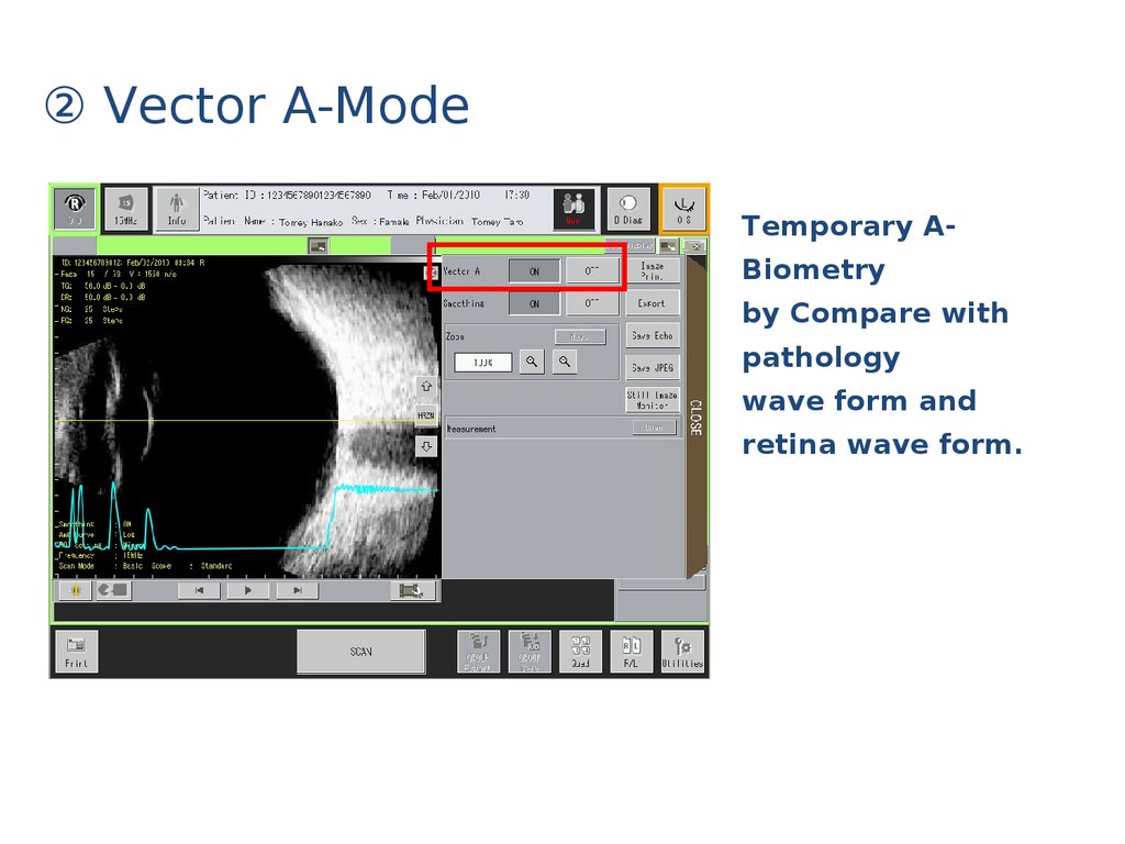
② Vector A-ModeTemporary ABiometry
by Compare with
pathology
wave form and
retina wave form.
17.
③ Zoom5 steps Zoom
100%, 125%, 150%,
200%, 300%
Intuitive navigation monitor
18.
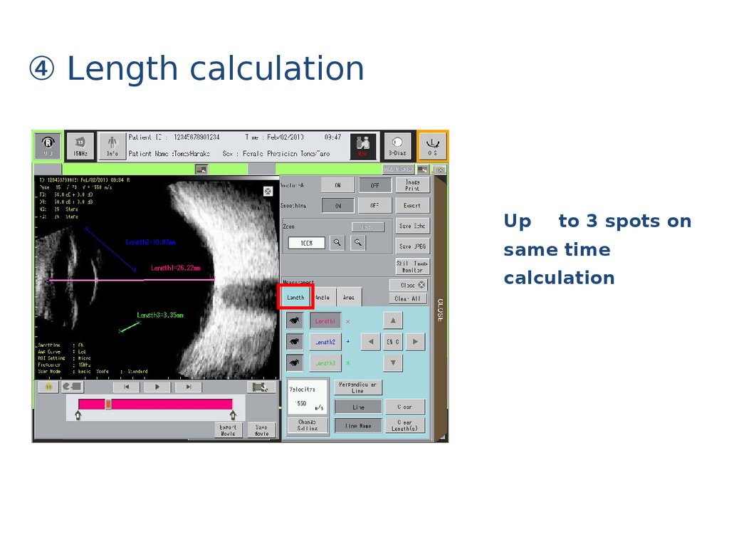
④ Length calculationUp
to 3 spots on
same time
calculation
19.
⑤ Area calculationUp
to 2 spots on
same time
calculation
20.
⑥ Angle calculationUp
to 2 spots on
same time
calculation
21.
⑦ Dual/Quad Display22.
⑧ R(OD L OS Display23.
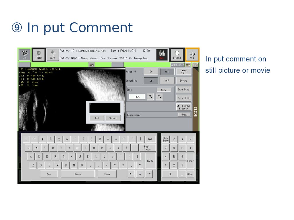
⑨ In put CommentIn put comment on
still picture or movie
24.
⑩ Print out by still image part only25.
⑪ Thumbnail images26.
⑫ Group ExportGroup Save
27.
Save Out put①Still image
Save Echo or JPEG
format
( Re-Display, Edit
possible )
② Movie
Save in USB memory by
Echo format and out put.
28.
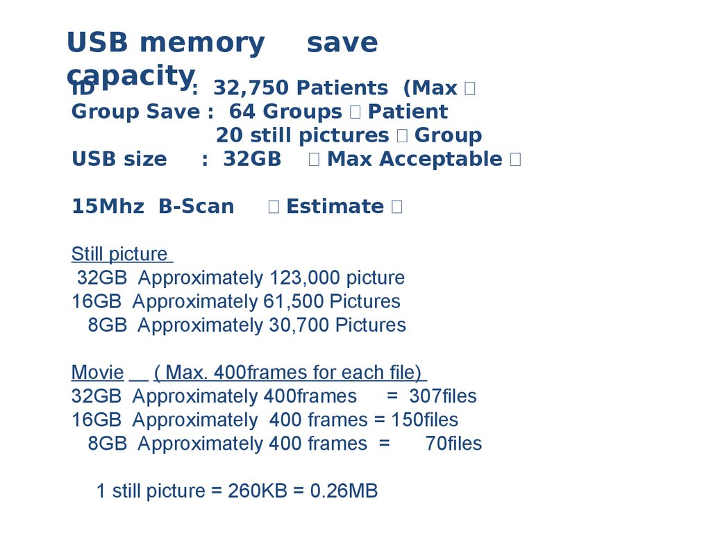
USB memory savecapacity
ID
: 32,750 Patients
(Max
Group Save : 64 Groups Patient
20 still pictures Group
USB size
: 32GB Max Acceptable
15Mhz B-Scan
Estimate
Still picture
32GB Approximately 123,000 picture
16GB Approximately 61,500 Pictures
8GB Approximately 30,700 Pictures
Movie ( Max. 400frames for each file)
32GB Approximately 400frames = 307files
16GB Approximately 400 frames = 150files
8GB Approximately 400 frames =
70files
1 still picture = 260KB = 0.26MB
29.
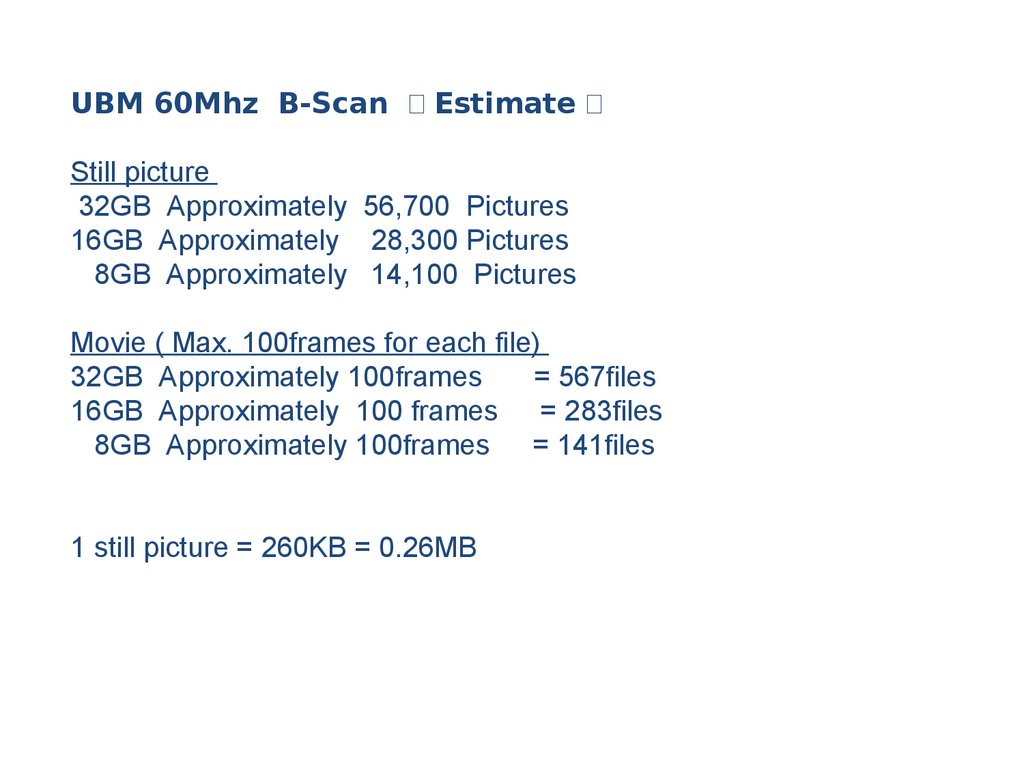
UBM 60Mhz B-Scan EstimateStill picture
32GB Approximately 56,700 Pictures
16GB Approximately 28,300 Pictures
8GB Approximately 14,100 Pictures
Movie ( Max. 100frames for each file)
32GB Approximately 100frames
= 567files
16GB Approximately 100 frames = 283files
8GB Approximately 100frames = 141files
1 still picture = 260KB = 0.26MB
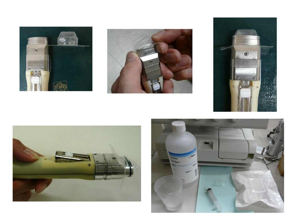
30.
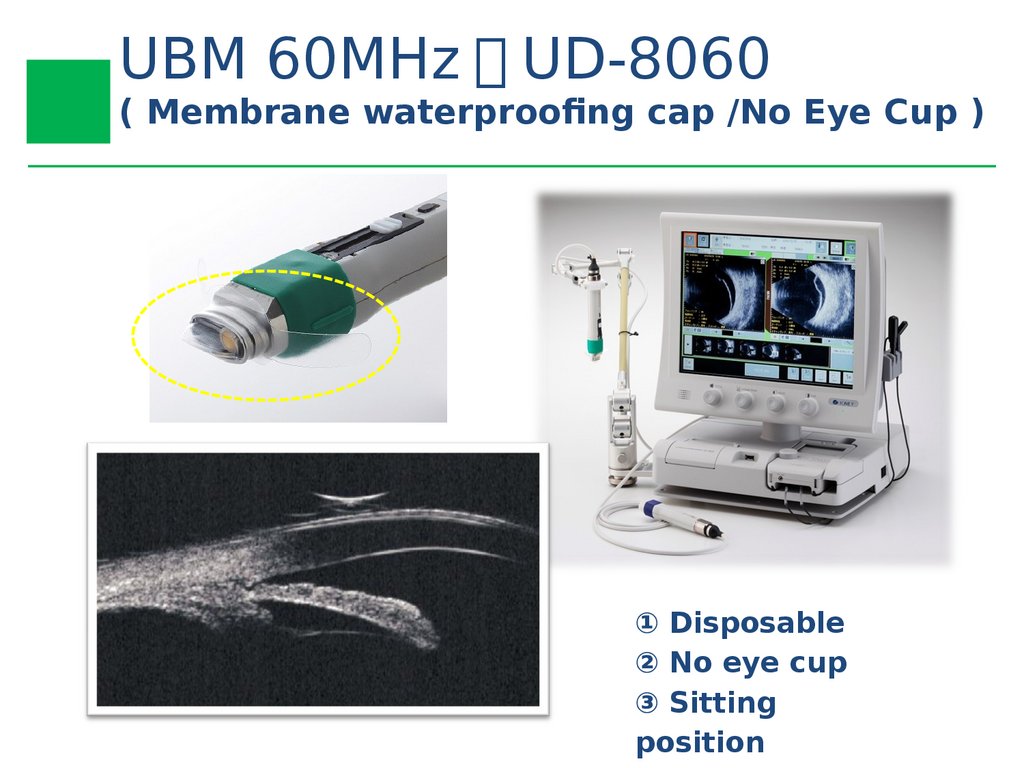
UBM 60MHz UD-8060( Membrane waterproofing cap /No Eye Cup )
① Disposable
② No eye cup
③ Sitting
position
31.
32.
33.
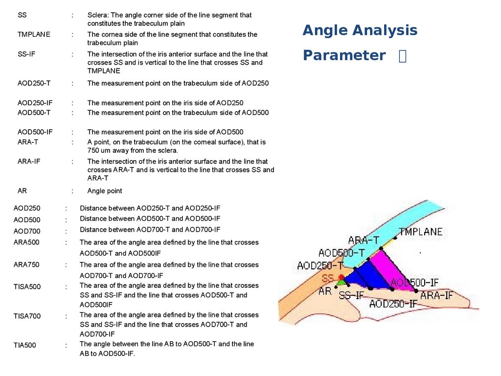
Angle Analysis34.
SS:
Sclera: The angle corner side of the line segment that
constitutes the trabeculum plain
TMPLANE
:
The cornea side of the line segment that constitutes the
trabeculum plain
SS-IF
:
The intersection of the iris anterior surface and the line that
crosses SS and is vertical to the line that crosses SS and
TMPLANE
AOD250-T
:
The measurement point on the trabeculum side of AOD250
AOD250-IF
AOD500-T
:
:
The measurement point on the iris side of AOD250
The measurement point on the trabeculum side of AOD500
AOD500-IF
ARA-T
:
:
The measurement point on the iris side of AOD500
A point, on the trabeculum (on the corneal surface), that is
750 um away from the sclera.
ARA-IF
:
The intersection of the iris anterior surface and the line that
crosses ARA-T and is vertical to the line that crosses SS and
ARA-T
AR
:
Angle point
AOD250
:
AOD500
:
Distance between AOD250-T and AOD250-IF
Distance between AOD500-T and AOD500-IF
AOD700
:
Distance between AOD700-T and AOD700-IF
ARA500
:
The area of the angle area defined by the line that crosses
AOD500-T and AOD500IF
ARA750
:
TISA500
:
TISA700
:
TIA500
:
The area of the angle area defined by the line that crosses
AOD700-T and AOD700-IF
The area of the angle area defined by the line that crosses
SS and SS-IF and the line that crosses AOD500-T and
AOD500IF
The area of the angle area defined by the line that crosses
SS and SS-IF and the line that crosses AOD700-T and
AOD700-IF
The angle between the line AB to AOD500-T and the line
AB to AOD500-IF.
Angle Analysis
Parameter 】
35.
Iris Analysis36.
T3-I1T3-I2
:
:
Terminal point 1 of line T3
Terminal point 2 of line T3
ID1-IF
:
The intersection of the iris anterior surface and the line that
crosses AOD500-T and is vertical to line T3
ID1-IB
:
The intersection of the iris posterior surface and the line that
crosses AOD500-T and is vertical to line T3
TCPD-CB
:
The intersection of the ciliary process surface and the line
that crosses AOD500-T and is vertical to line T3
ID2-T
:
The point, on the corneal surface, that is 2 mm away from
SS
ID2-IF
:
The intersection of the iris anterior surface and the line that
crosses ID2-T and is vertical to line T3
ID2-IB
:
The intersection of the iris posterior surface and the line that
crosses ID2-T and is vertical to line T3
ID3-IF
:
The point, on the iris anterior surface, at which the distance
between the intersections of the T3-vertical line and the iris
anterior and posterior surfaces first marks a peak value
when measured from the pupil side
ID3-IB
:
The point, on the iris posterior surface, at which the distance
between the intersections of the T3-vertical line and the iris
anterior and posterior surfaces first marks a peak value
when measured from the pupil side
ID1
:
Distance between ID1-IF and ID1-IB
ID2
:
Distance between ID2-IF and ID2-IB
ID3
:
Distance between ID3-IF and ID3-IB
TCPD
:
Distance between AOD500-T and TCPD-CB
ICPD
:
Distance between ID1-IB and TCPD-CB
Iris Analysis
Parameter 】
37.
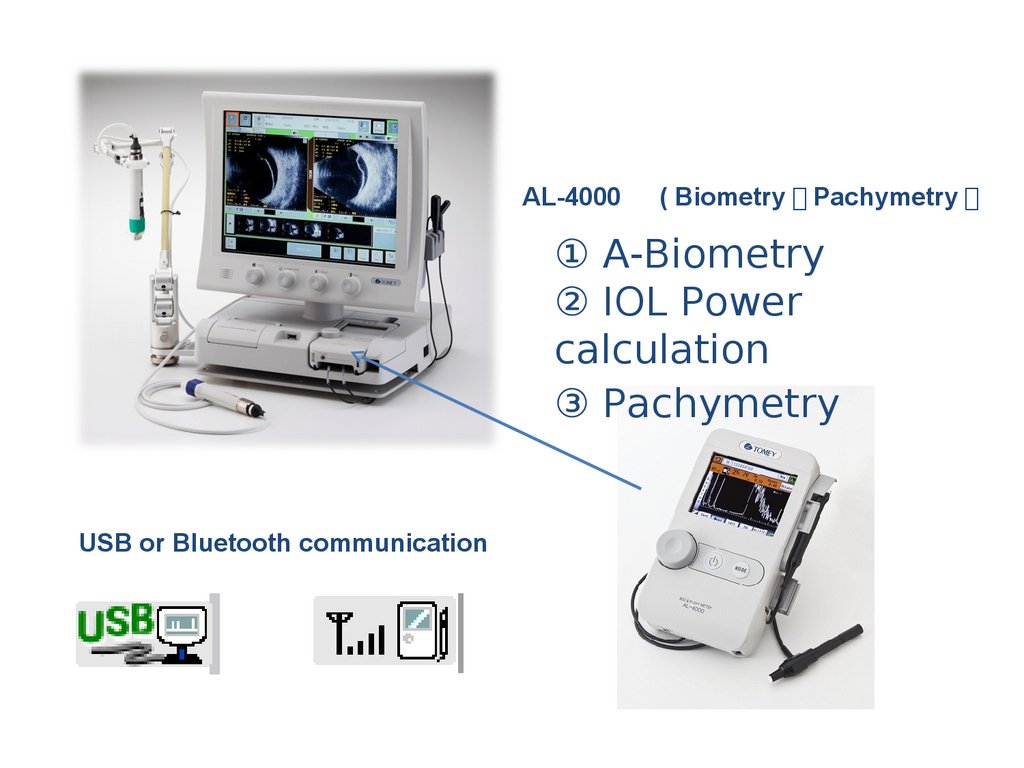
AL-4000( Biometry Pachymetry
① A-Biometry
② IOL Power
calculation
③ Pachymetry
USB or Bluetooth communication
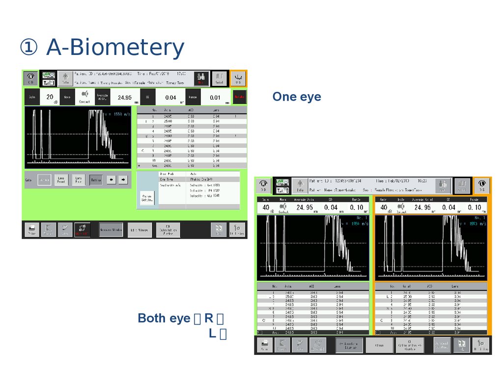
38.
① A-BiometeryOne eye
Both eye R
L
39.
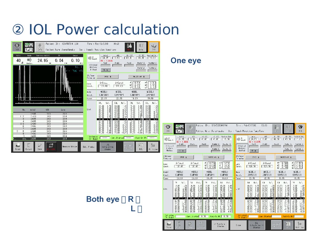
② IOL Power calculationOne eye
Both eye R
L
40.
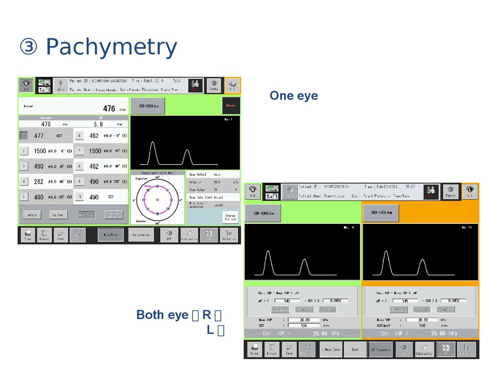
③ PachymetryOne eye
Both eye R
L