


 medicine
Similar presentations:


")


Отек и выпот
1.
2.
Лекція 3РОЗЛАДИ
ЦИРКУЛЯЦІЇ
3.
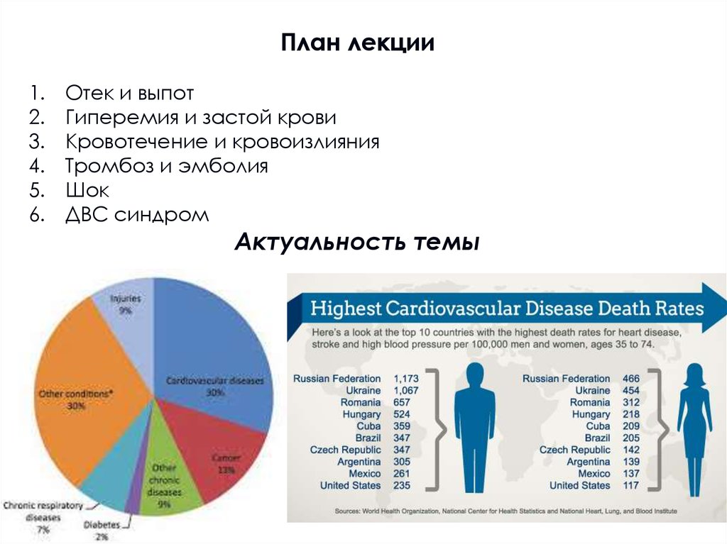
План лекции1.
2.
3.
4.
5.
6.
Отек и выпот
Гиперемия и застой крови
Кровотечение и кровоизлияния
Тромбоз и эмболия
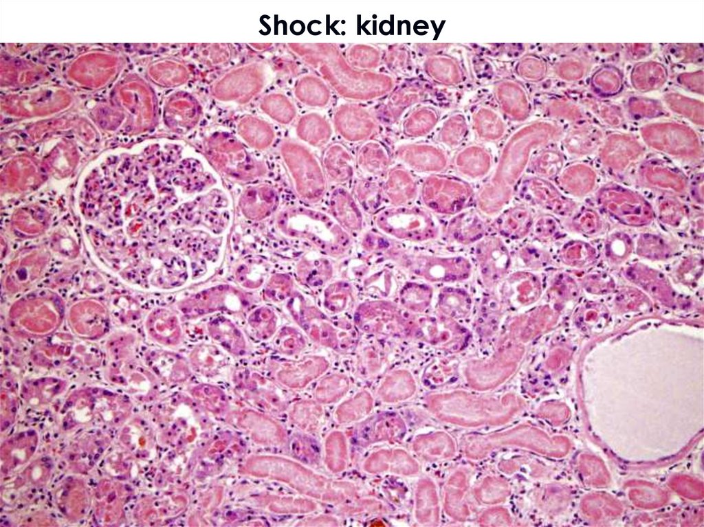
Шок
ДВС синдром
Актуальность темы
4.
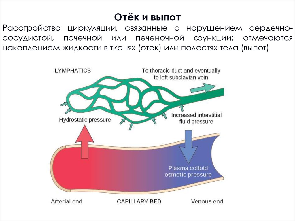
Отёк и выпотРасстройства циркуляции, связанные с нарушением сердечнососудистой, почечной или печеночной функции; отмечаются
накоплением жидкости в тканях (отек) или полостях тела (выпот)
5.
Отек и выпот6.
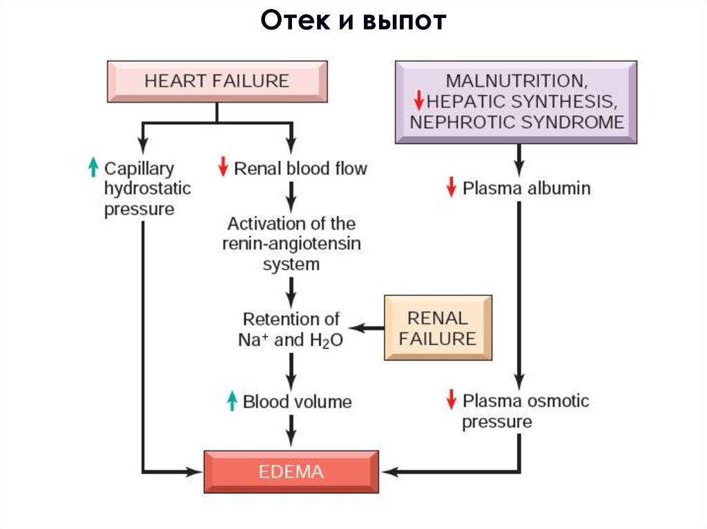
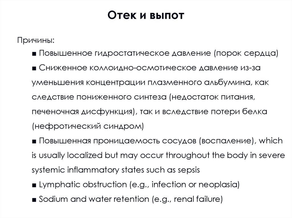
Отек и выпотПричины:
■ Повышенное гидростатическое давление (порок сердца)
■ Сниженное коллоидно-осмотическое давление из-за
уменьшения концентрации плазменного альбумина, как
следствие пониженного синтеза (недостаток питания,
печеночная дисфункция), так и вследствие потери белка
(нефротический синдром)
■ Повышенная проницаемость сосудов (воспаление), which
is usually localized but may occur throughout the body in severe
systemic inflammatory states such as sepsis
■ Lymphatic obstruction (e.g., infection or neoplasia)
■ Sodium and water retention (e.g., renal failure)
7.
Гидродинамический отек8.
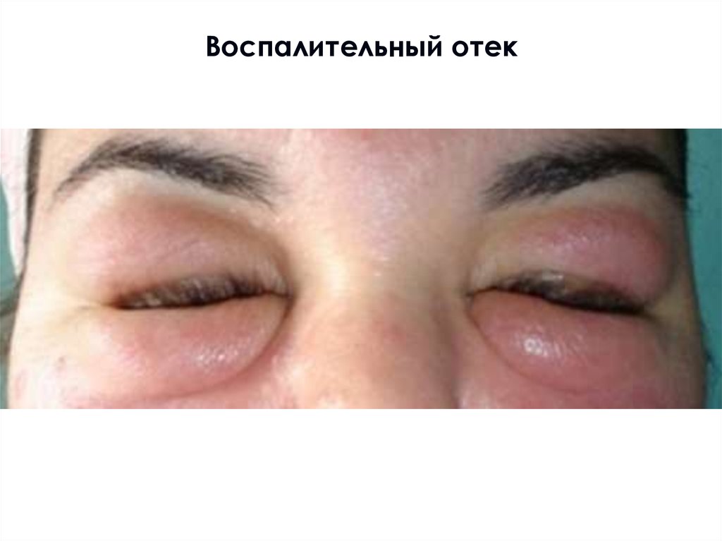
Воспалительный отек9.
Лимфогенный отек: слоновость10.
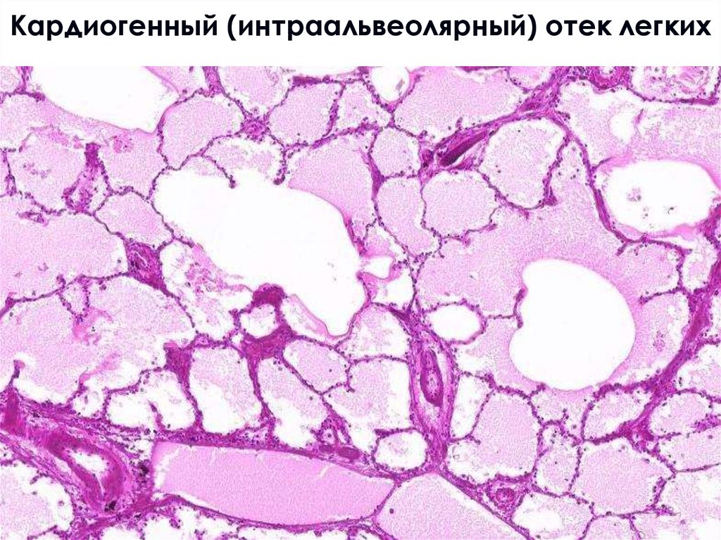
Кардиогенный отек легких11.
Кардиогенный (интраальвеолярный) отек легких12.
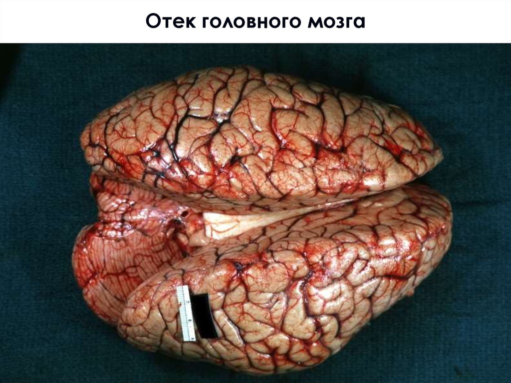
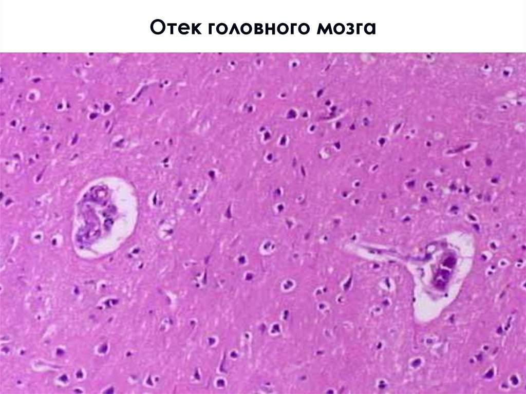
Отек головного мозга13.
Отек мозга: дислокация мозжечка14.
Отек головного мозга15.
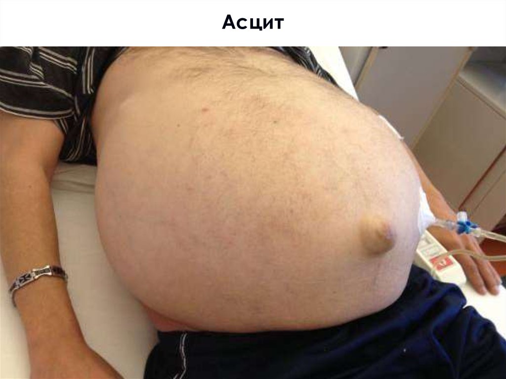
Асцит16.
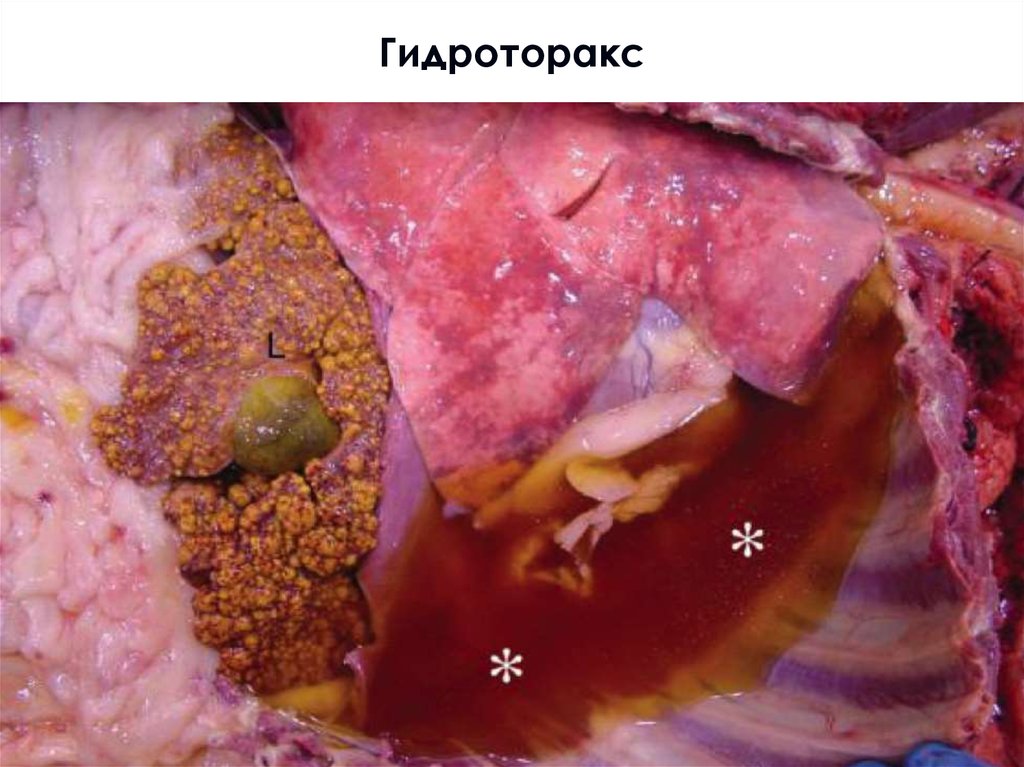
Гидроторакс17.
Гидроперикард18.
Транссудат (уд.плотность <1,012):Небогатая белком жидкость (<0,25 г/л), полупрозрачная,
соломенно-желтая; за исключением выпотов, вызванных
блокировкой лимфатических сосудов (хилёзный выпот) –
молочного цвета, благодаря липидам, поглощенным из
кишечника.
Экссудат (уд. плотность >1,02) :
Богатый белками выпот (>0,29 г/л), часто мутный из-за
присутствия лейкоцитов (серозный, фибринозный, гнойный)
19.
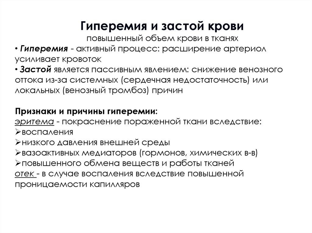
Гиперемия и застой кровиповышенный объем крови в тканях
• Гиперемия - активный процесс: расширение артериол
усиливает кровоток
• Застой является пассивным явлением: снижение венозного
оттока из-за системных (сердечная недостаточность) или
локальных (венозный тромбоз) причин
Признаки и причины гиперемии:
эритема - покраснение пораженной ткани вследствие:
воспаления
низкого давления внешней среды
вазоактивных медиаторов (гормонов, химических в-в)
повышенного обмена веществ и работы тканей
отек - в случае воспаления вследствие повышенной
проницаемости капилляров
20.
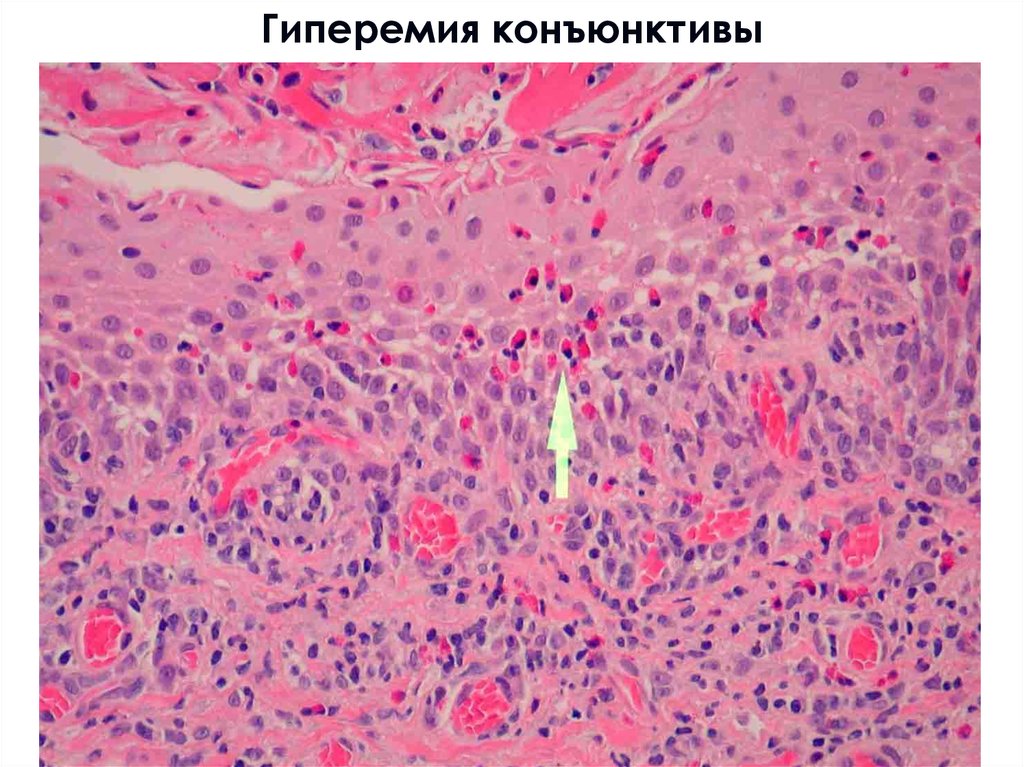
Гиперемия конъюнктивы21.
Гиперемия конъюнктивы22.
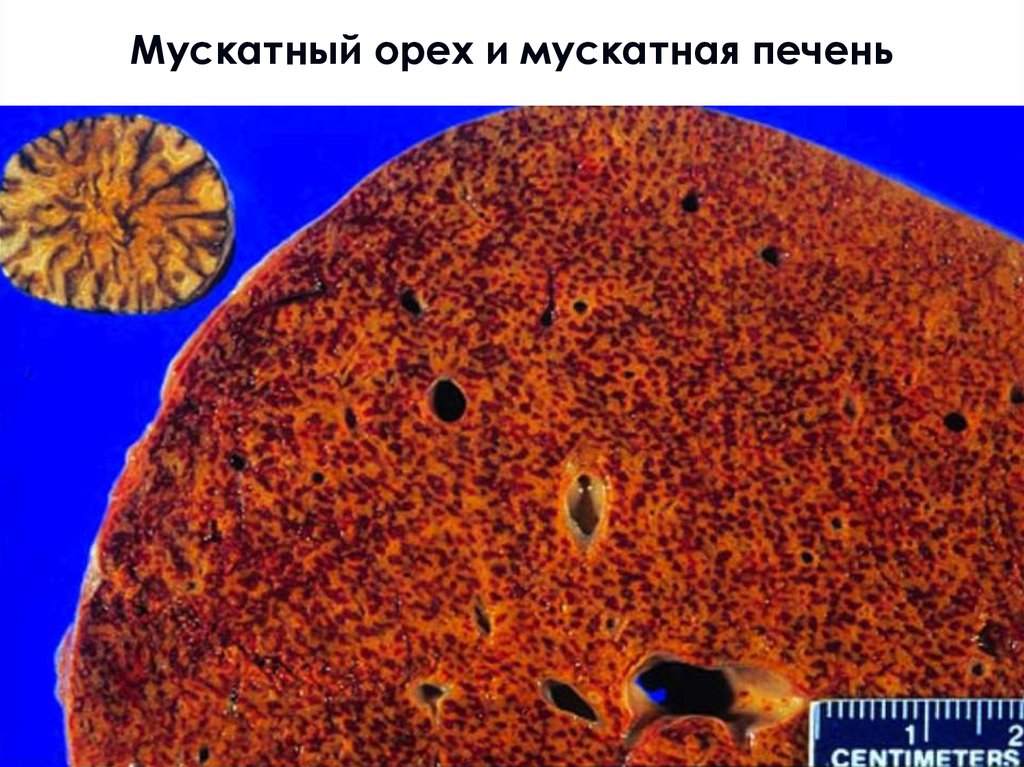
Мускатный орех и мускатная печень23.
Мускатная печень24.
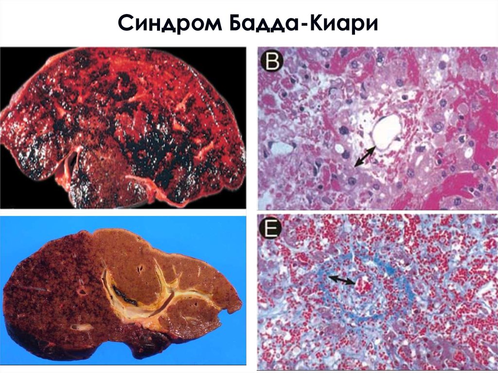
Синдром Бадда-Киари25.
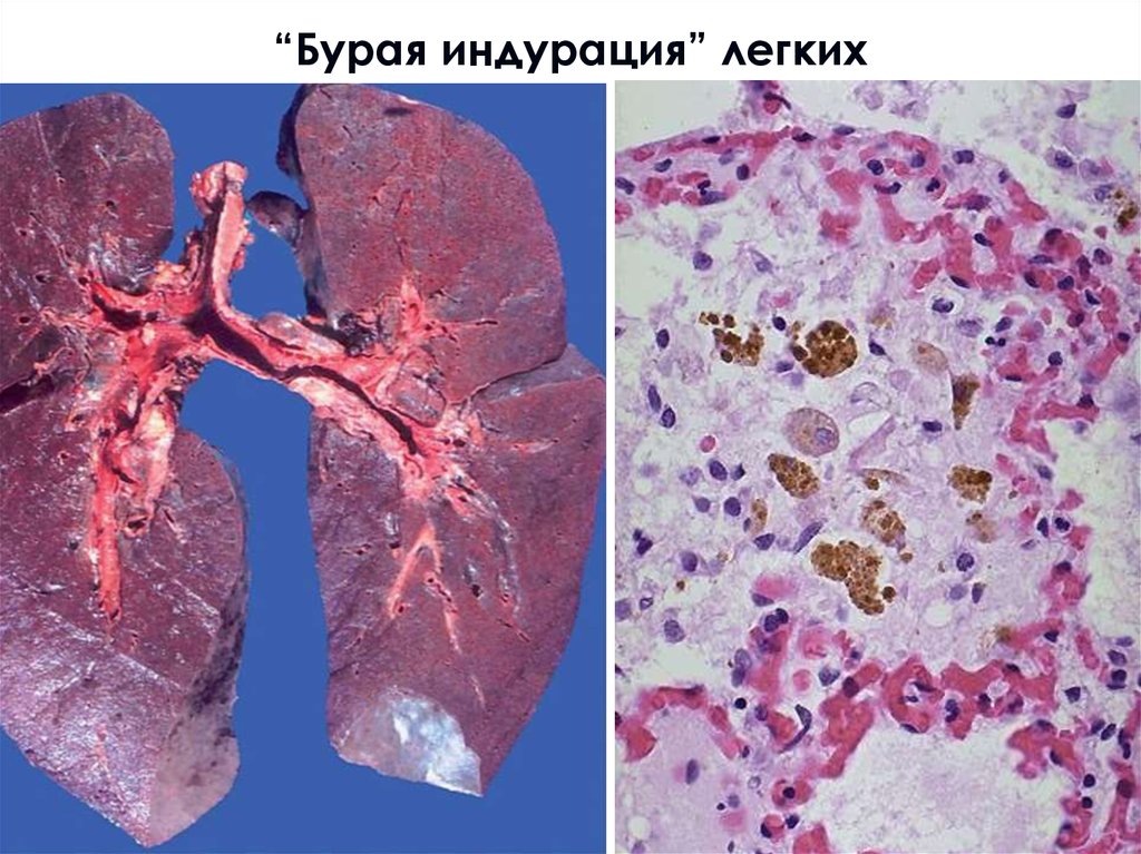
“Бурая индурация” легких26.
Цианотическая индурация, спленомегалия27.
Надоели пробки? Одевай кроссовки!28.
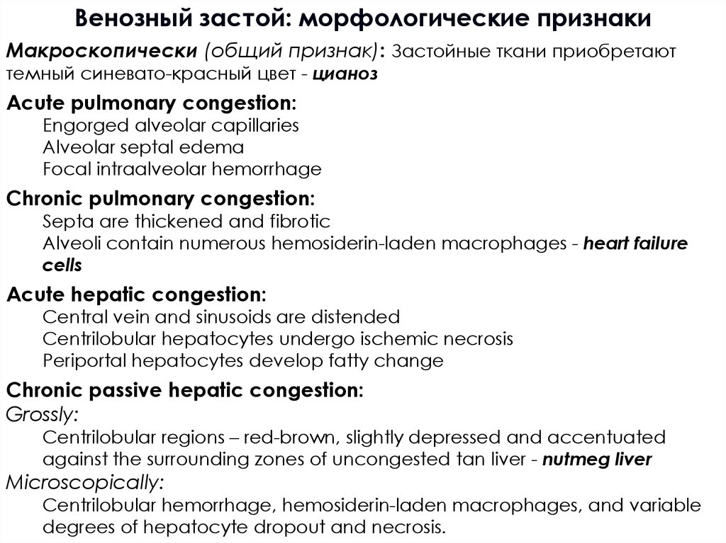
Венозный застой: морфологические признакиМакроскопически (общий признак): Застойные ткани приобретают
темный синевато-красный цвет - цианоз
Acute pulmonary congestion:
Engorged alveolar capillaries
Alveolar septal edema
Focal intraalveolar hemorrhage
Chronic pulmonary congestion:
Septa are thickened and fibrotic
Alveoli contain numerous hemosiderin-laden macrophages - heart failure
cells
Acute hepatic congestion:
Central vein and sinusoids are distended
Centrilobular hepatocytes undergo ischemic necrosis
Periportal hepatocytes develop fatty change
Chronic passive hepatic congestion:
Grossly:
Centrilobular regions – red-brown, slightly depressed and accentuated
against the surrounding zones of uncongested tan liver - nutmeg liver
Microscopically:
Centrilobular hemorrhage, hemosiderin-laden macrophages, and variable
degrees of hepatocyte dropout and necrosis.
29.
Кровоизлияния30.
Hemorrhagic Disorders■ Disorders associated with abnormal bleeding inevitably stem from
primary or secondary defects in vessel walls, platelets, or coagulation
factors, all of which must function properly to ensure hemostasis
massive bleeds associated with ruptures or erosion of large vessels
Aortal aneurism rupture
Myocardial infarction rupture (hemopericardium)
Bleeding from eroded vessels (e.g., peptic ulcer of stomach)
Trauma (incl., hemothorax, hemoperitoneum)
defects in clotting
Defects of primary hemostasis (von Willebrand disease): bleeds in skin or
mucosal membranes - petechiae, 1- to 2-mm hemorrhages, or purpura ≥3 mm
(nasal bleedings – epistaxis, gastrointestinal bleeding, menorrhagia)
Secondary hemostasis disorders (e.g., hemophilia): hemarthrosis, soft tissue
hemorrages.
Generalized defects involving small vessels: “palpable purpura” and
ecchymoses. Ecchymoses (sometimes simply called bruises) 1 to 2 cm
hemorrages. In both purpura and ecchymoses, the volume of extravasated
blood is sufficient to create a palpable mass of blood known as a hematoma.
(e.g., systemic vasculitis, amyloidosis, scurvy)
31.
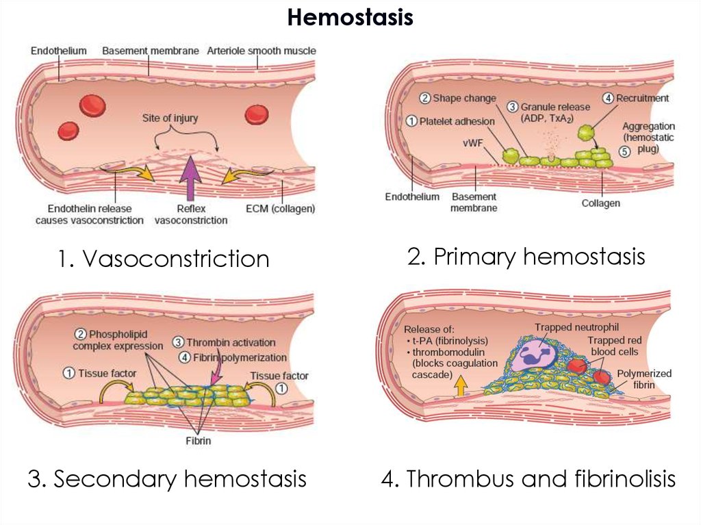
Hemostasis1. Vasoconstriction
2. Primary hemostasis
3. Secondary hemostasis
4. Thrombus and fibrinolisis
32.
33.
Thrombosis34.
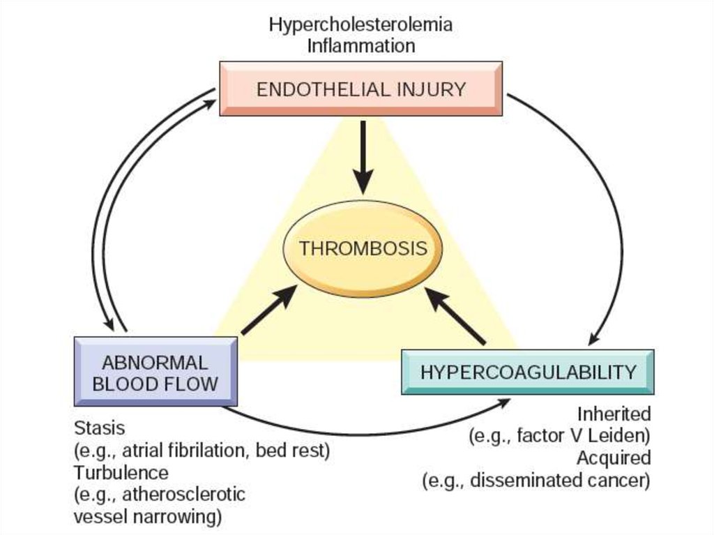
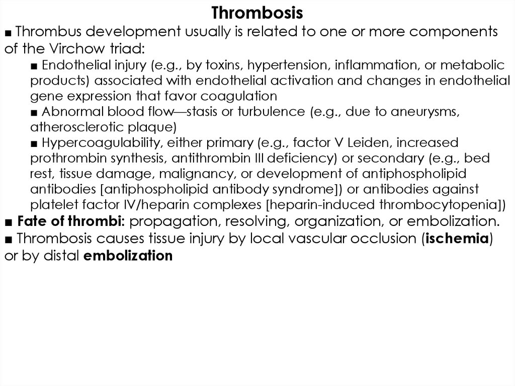
Thrombosis■ Thrombus development usually is related to one or more components
of the Virchow triad:
■ Endothelial injury (e.g., by toxins, hypertension, inflammation, or metabolic
products) associated with endothelial activation and changes in endothelial
gene expression that favor coagulation
■ Abnormal blood flow—stasis or turbulence (e.g., due to aneurysms,
atherosclerotic plaque)
■ Hypercoagulability, either primary (e.g., factor V Leiden, increased
prothrombin synthesis, antithrombin III deficiency) or secondary (e.g., bed
rest, tissue damage, malignancy, or development of antiphospholipid
antibodies [antiphospholipid antibody syndrome]) or antibodies against
platelet factor IV/heparin complexes [heparin-induced thrombocytopenia])
■ Fate of thrombi: propagation, resolving, organization, or embolization.
■ Thrombosis causes tissue injury by local vascular occlusion (ischemia)
or by distal embolization
35.
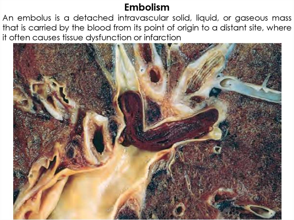
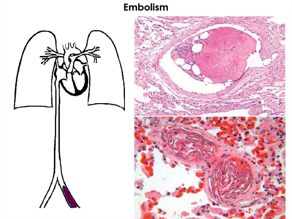
EmbolismAn embolus is a detached intravascular solid, liquid, or gaseous mass
that is carried by the blood from its point of origin to a distant site, where
it often causes tissue dysfunction or infarction
36.
Embolism37.
Infarction38.
Infarction fate39.
Infarction■ Infarcts are areas of ischemic necrosis most commonly
caused by arterial occlusion (typically due to thrombosis
or embolization); venous outflow obstruction is a less
frequent cause.
■ Infarcts caused by venous occlusion or occurring in
spongy tissues with dual blood supply and where blood
can collect typically are hemorrhagic (red); those caused
by arterial occlusion in compact tissues typically are pale
(white).
■ Whether or not vascular occlusion causes tissue
infarction is influenced by collateral blood supplies, the
rate at which an obstruction develops, intrinsic tissue
susceptibility to ischemic injury, and blood oxygenation.
40.
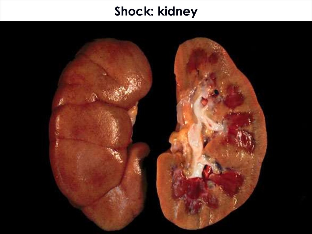
Shock• a state when diminished cardiac output or reduced
effective circulating blood volume impairs tissue perfusion
and leads to cellular hypoxia
•Cardiogenic
•Hypovolemic
•Systemic inflammatory response associated (Septic)
•Neurogenic
•Anaphylactic