
 medicine
Similar presentations:


Disseminated intravascular coagulation by Venkatesan Abinesh Group-163(2)
1.
DICDisseminated intravascular coagulation
by
Venkatesan Abinesh
Group-163(2)
2.
WH AT I S D I C ?• Disseminated intravascular coagulation (DIC) is an
acquired syndrome characterized by the intravascular
activation of coagulation with loss of localization arising
from different causes.It can originate from and cause
damage to microvasculature, which if sufficiently
severe , can produce organ dysfunction.
3.
• Normal Pregnancy – Hypercoagulable state.• After the 1st trimester there occurs a marked increase
in plasma fibrinogen( more than double the non
pregnant level ).
• Plasma fibrinolytic activity is decreased during
pregnancy and returns to normal within one hour
of delivery of placenta.
4.
TYPE S O F D I C ?• ACUTE DIC – the physical findings are those of
underlying or inciting etiology .
• Patients with acute DIC have petechiae on the soft
palate and legs from thrombocytopenia and ecchymosis
at venipuncture sites.
• Acute DIC occurs in obstetric calamities such as placental
abruption
• and amniotic fluid emboli.
5.
• Amniotic fluid has been shown to be able to activatecoagulation in vitro, and the degree of placental seperation
correlates with the extent of DIC , suggesting that leakage
of thromboplastin like material from the placental system is
responsible for the occurrence of DIC.
• Coagulation system may also be activated in patients with
pre
• eclampsia and HELLP syndrome.
6.
• CHRONIC DIC- manifestation is thrombosis fromexcess thrombin formation , the symptoms and
signs of venous thromboembolism may be present.
7.
COMMON CAUSES OFDIC
ACUTE DIC
• Abruptio Placentae
• Endotoxemia- septic abortions, chorioamnionitis,
pyelonephritis of pregnancy.
• Amniotic Fluid Embolism
• Severe pregnancy induced hypertension
• Intra-amniotic hypertonic saline
8.
• Vesicular mole• Dextran Infusion
• Hemorrhagic shock due to –PPH ,Cs
• CHRONIC DIC – IUD ( prolonged retension of dead
fetus).
9.
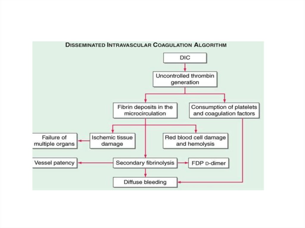
PATH O PH YSI O LO G Y• DIC is diagnosed in almost one-half of pregnant
women with abruptio placentae, or with amniotic fluid
embolism. Trauma, particularly to the brain, can also
result in DIC.
10.
PATH O PH YSI O LO G Y11.
CLINICAL MANIFESTATION• Clinical manifestations of DIC are related to the
magnitude of the imbalance of hemostasis, to the
underlying disease, or to both.
• The most common findings are bleeding ranging from
oozing from venipuncture sites, petechiae, and
ecchymoses to severe hemorrhage from the
gastrointestinal tract, lung, or into the CNS.
• In chronic DIC, the bleeding symptoms are discrete and
restricted to
• skin or mucosal surfaces.
12.
• The hypercoagulability of DIC manifests as theocclusion of vessels in the microcirculation and
resulting organ failure.
• Thrombosis of large vessels and cerebral embolism can
also
• occur.
• Hemodynamic complications and shock are common
among patients with acute DIC. The mortality ranges from
30 to >80% depending on the underlying disease, the
severity of the DIC, and the age of the patient.
13.
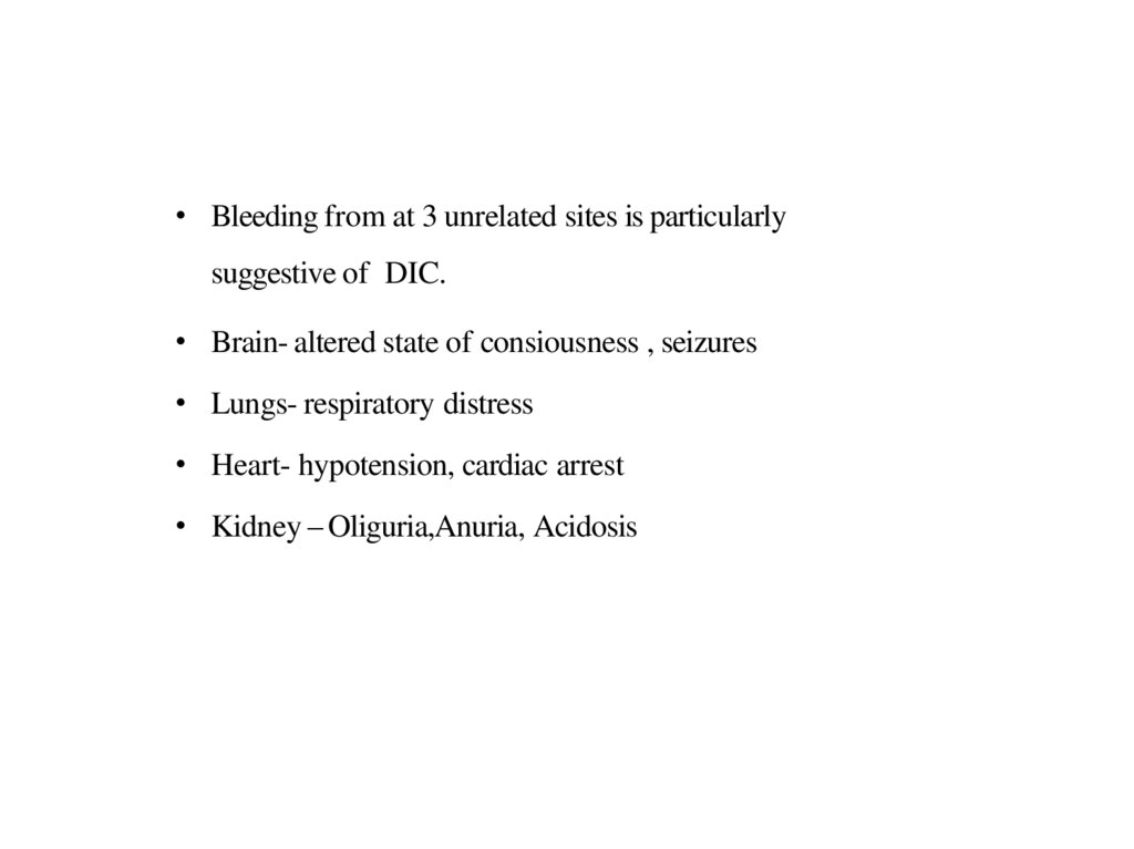
• Bleeding from at 3 unrelated sites is particularlysuggestive of DIC.
• Brain- altered state of consiousness , seizures
• Lungs- respiratory distress
• Heart- hypotension, cardiac arrest
• Kidney – Oliguria,Anuria, Acidosis
14.
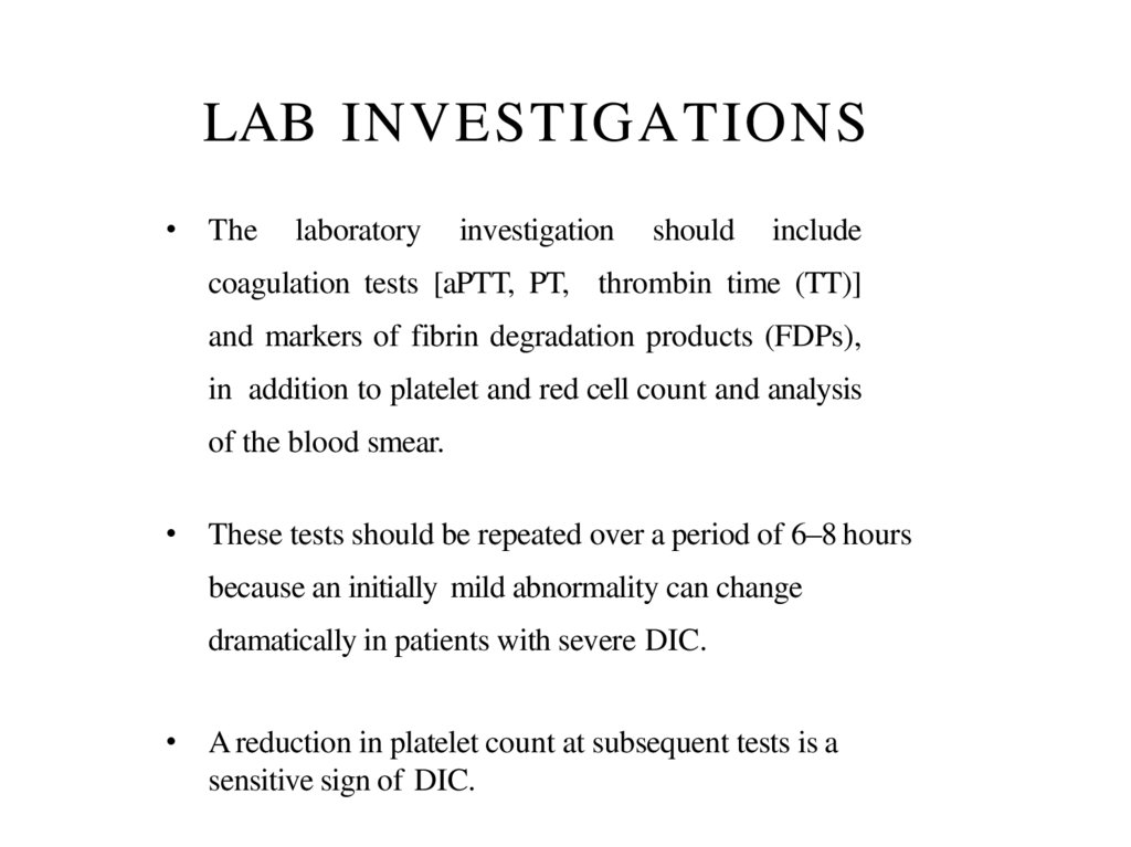
LAB INVESTIGATIONS• The
laboratory
investigation
should
include
coagulation tests [aPTT, PT, thrombin time (TT)]
and markers of fibrin degradation products (FDPs),
in addition to platelet and red cell count and analysis
of the blood smear.
• These tests should be repeated over a period of 6–8 hours
because an initially mild abnormality can change
dramatically in patients with severe DIC.
• A reduction in platelet count at subsequent tests is a
sensitive sign of DIC.
15.
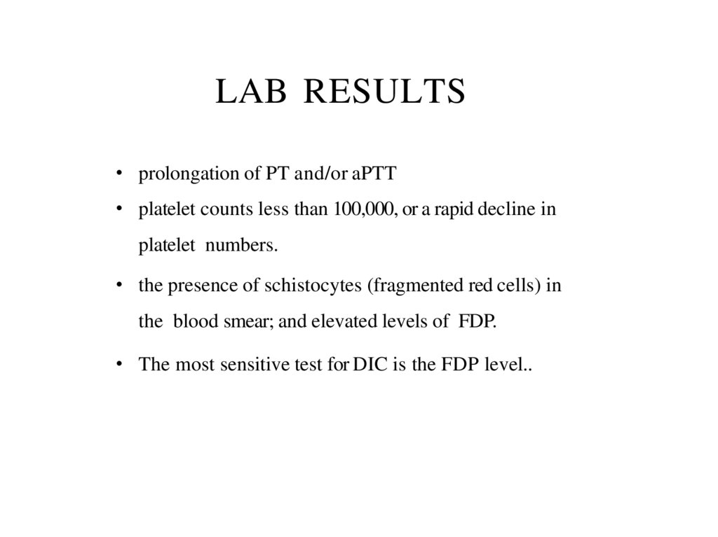
LAB RESULTS• prolongation of PT and/or aPTT
• platelet counts less than 100,000, or a rapid decline in
platelet numbers.
• the presence of schistocytes (fragmented red cells) in
the blood smear; and elevated levels of FDP.
• The most sensitive test for DIC is the FDP level..
16.
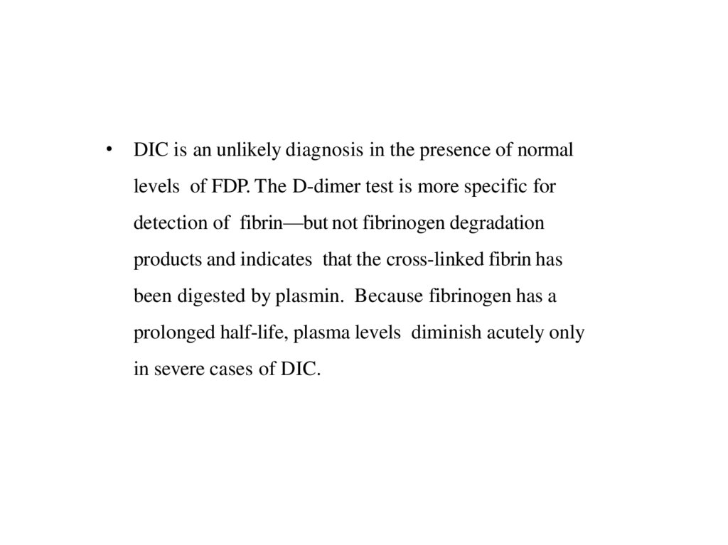
• DIC is an unlikely diagnosis in the presence of normallevels of FDP. The D-dimer test is more specific for
detection of fibrin—but not fibrinogen degradation
products and indicates that the cross-linked fibrin has
been digested by plasmin. Because fibrinogen has a
prolonged half-life, plasma levels diminish acutely only
in severe cases of DIC.
17.
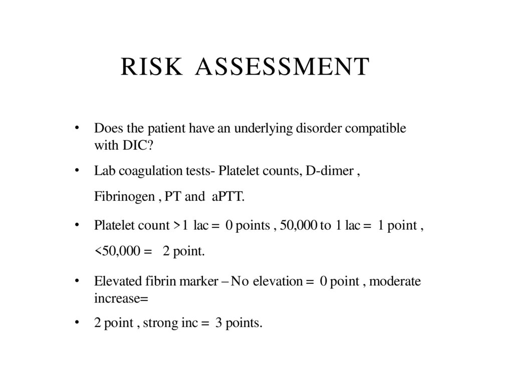
RISK ASSESSMENT• Does the patient have an underlying disorder compatible
with DIC?
• Lab coagulation tests- Platelet counts, D-dimer ,
Fibrinogen , PT and aPTT.
• Platelet count >1 lac = 0 points , 50,000 to 1 lac = 1 point ,
<50,000 = 2 point.
• Elevated fibrin marker – No elevation = 0 point , moderate
increase=
• 2 point , strong inc = 3 points.
18.
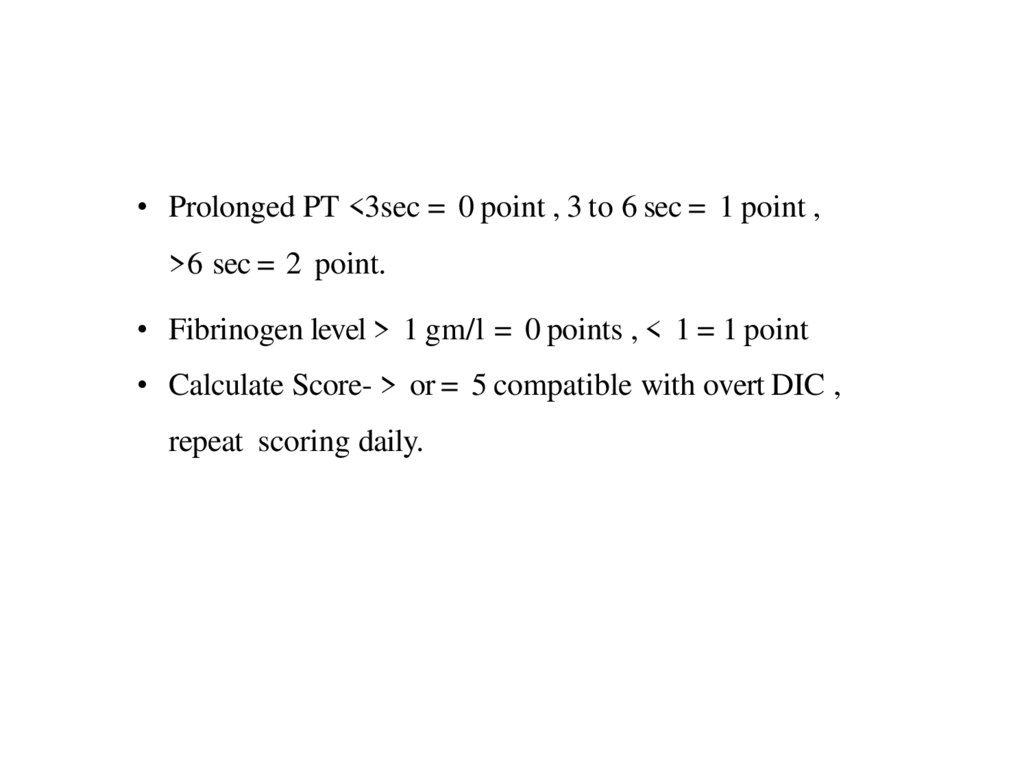
• Prolonged PT <3sec = 0 point , 3 to 6 sec = 1 point ,>6 sec = 2 point.
• Fibrinogen level > 1 gm/l = 0 points , < 1 = 1 point
• Calculate Score- > or = 5 compatible with overt DIC ,
repeat scoring daily.
19.
20.
TREATMENT• The morbidity and mortality associated with DIC are
primarily related to the underlying disease rather than
the complications of the DIC. The control or
elimination of the underlying cause should therefore be
the primary concern.
• Patients with severe DIC require control of
hemodynamic parameters, respiratory support, and
sometimes invasive surgical procedures.
• Attempts to treat DIC without accompanying treatment
of the
• causative disease are likely to fail.
21.
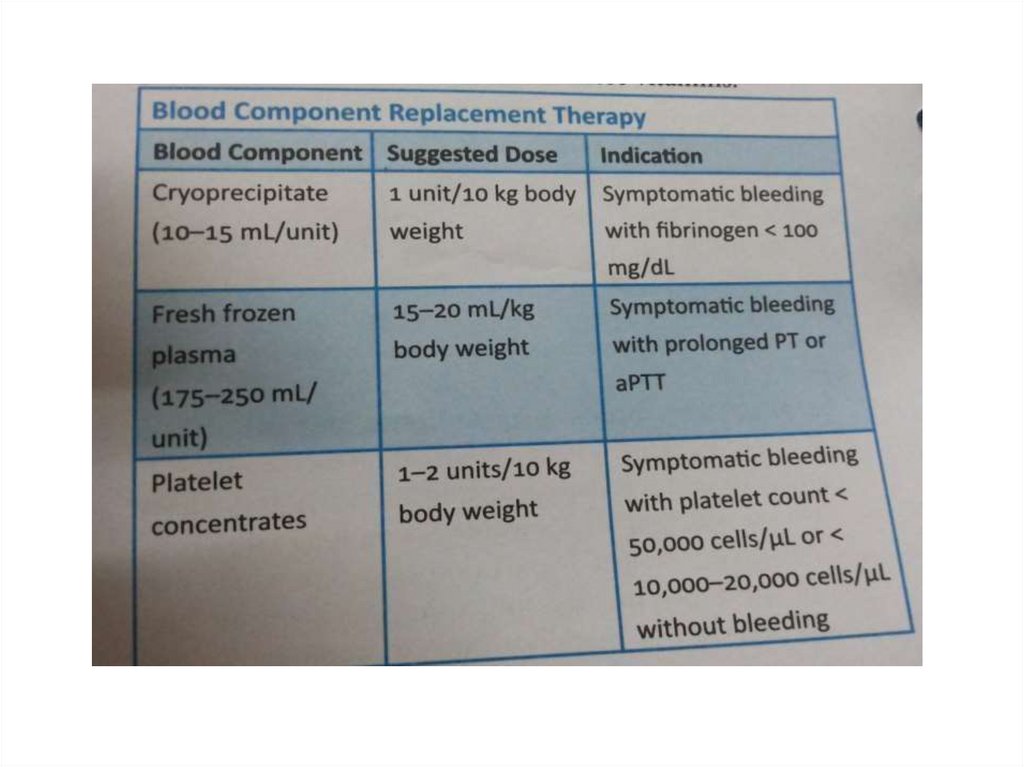
MANAGEMENT OFHEMORRHAGIC SYMPTOMS
• The control of bleeding in DIC patients with marked
thrombocytopenia (platelet counts <10,000–20,000/ L3)
and low levels of coagulation factors will require
replacement therapy.
22.
• The PT (>1.5 times the normal) provides a good indicator of theseverity of the clotting factor consumption.
• Replacement with FFP is indicated (1 unit of FFP increases
most coagulation factors by 3% in an adult without DIC).
• Low levels of fibrinogen (<100 mg/dL) or brisk
hyperfibrinolysis will require infusion of cryoprecipitate
(plasma fraction enriched for fibrinogen, FVIII, and vWF).
• The replacement of 10 U of cryoprecipitate for every 2–3 U of
FFP is sufficient to correct the hemostasis.
23.
• The transfusion must be adjusted according to thepatient's clinical and laboratory evolution.
• Platelet concentrates at a dose of 1–2 U/10 kg body
weight are
• sufficient for most DIC patients with severe
thrombocytopenia.
• Clotting factor concentrates are not recommended for
control of bleeding in DIC because of the limited
efficacy afforded by replacement of single factors
(FVIII or FIX concentrates), and the high risk of
• products containing traces of aPCCs that further
aggravate the disease.
24.
R E P L A C E M ENT OF CO AG ULATI ONOR FI BRI NO LYSI S INHI BI TO RS
• Drugs to control coagulation such as heparin, ATIII
concentrates, or antifibrinolytic drugs have all been tried
in the treatment of DIC.
• In acute DIC, the use of heparin is likely to aggravate
bleeding. To date, the use of heparin in patients with severe
DIC has no proven survival benefit.
25.
• The use of antifibrinolytic drugs, EACA, ortranexamic acid, to prevent fibrin degradation by
plasmin may reduce bleeding episodes in patients
with DIC and confirmed hyperfibrinolysis.
However, these drugs can increase the risk of
thrombosis and concomitant use of heparin is
indicated.