 biology
Similar presentations:


 and blood, muscle, nervous")


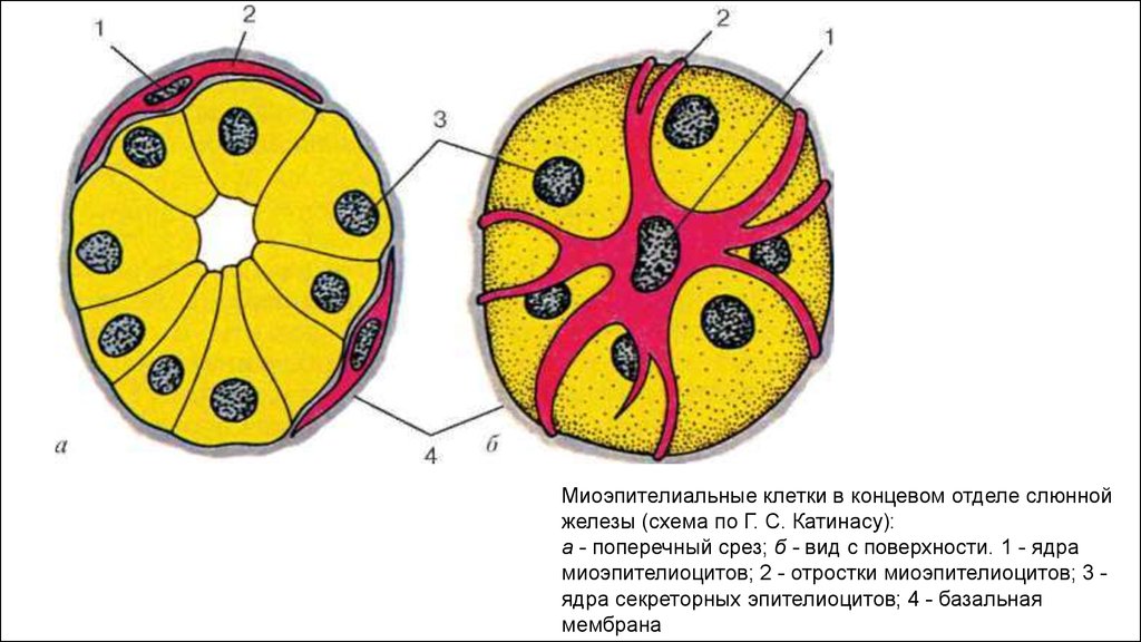
Миоэпителиальные клетки в концевом отделе слюнной железы
1.
Миоэпителиальные клетки в концевом отделе слюннойжелезы (схема по Г. С. Катинасу):
а - поперечный срез; б - вид с поверхности. 1 - ядра
миоэпителиоцитов; 2 - отростки миоэпителиоцитов; 3 ядра секреторных эпителиоцитов; 4 - базальная
мембрана
2.
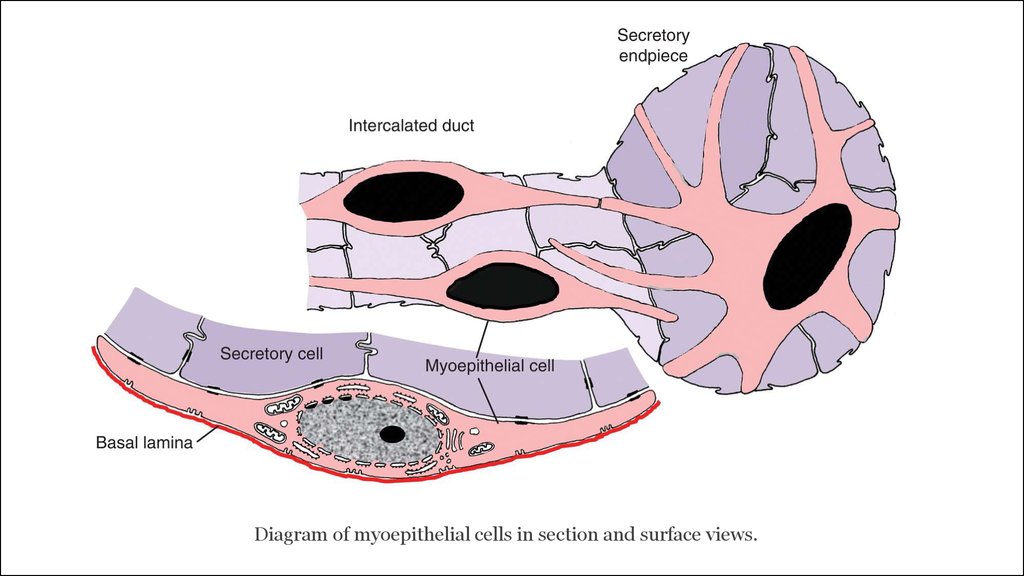
Diagram of myoepithelial cells in section and surface views.3.
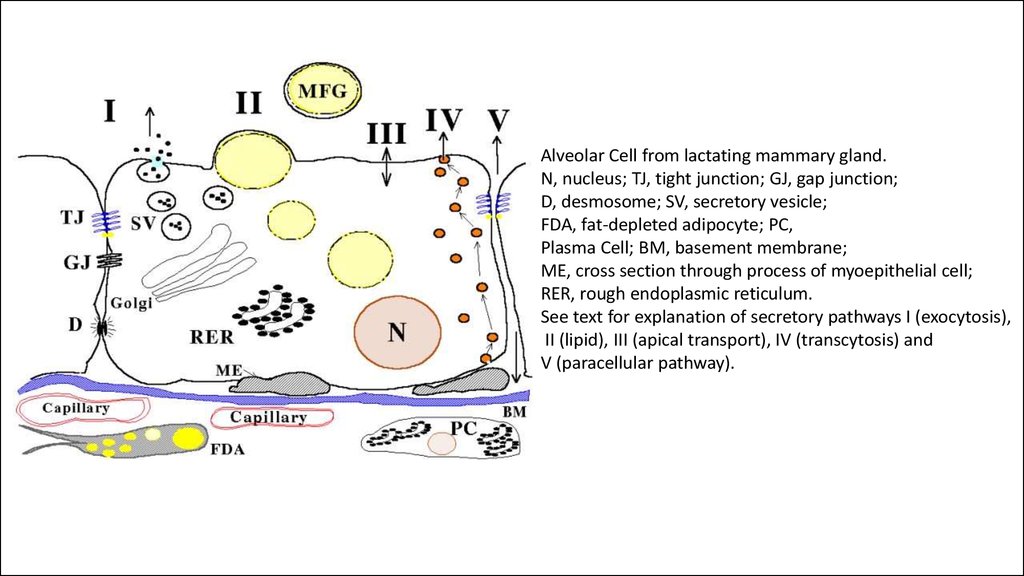
Alveolar Cell from lactating mammary gland.N, nucleus; TJ, tight junction; GJ, gap junction;
D, desmosome; SV, secretory vesicle;
FDA, fat-depleted adipocyte; PC,
Plasma Cell; BM, basement membrane;
ME, cross section through process of myoepithelial cell;
RER, rough endoplasmic reticulum.
See text for explanation of secretory pathways I (exocytosis),
II (lipid), III (apical transport), IV (transcytosis) and
V (paracellular pathway).
4.
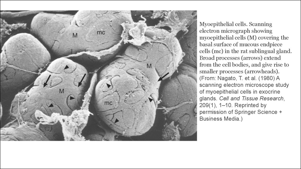
Myoepithelial cells. Scanningelectron micrograph showing
myoepithelial cells (M) covering the
basal surface of mucous endpiece
cells (mc) in the rat sublingual gland.
Broad processes (arrows) extend
from the cell bodies, and give rise to
smaller processes (arrowheads).
(From: Nagato, T. et al. (1980) A
scanning electron microscope study
of myoepithelial cells in exocrine
glands. Cell and Tissue Research,
209(1), 1–10. Reprinted by
permission of Springer Science +
Business Media.)