


 medicine
Similar presentations:


 of thebreast")


Dermatology. Basal Cell Carcinoma (BCC)
1.
DERMATOLOGY3.001-3.003
Handbook
2.
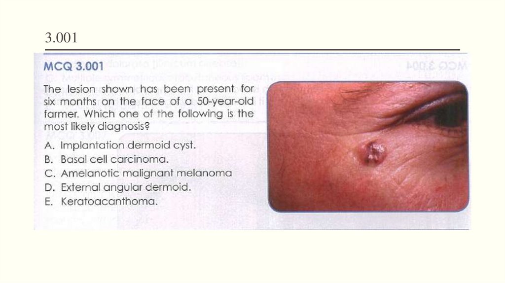
3.0013.
Basal Cell Carcinoma (BCC)Age: usually >35 years
Management:
More frequent in males
Simple elliptical excision (3–4 mm
margin) is best.
Mostly on sun-exposed areas: face (mainly),
neck, upper trunk, limbs
May ulcerate easily = ‘rodent ulcer’
Slow-growing over years
Has various forms: nodular, pigmented,
ulcerated, etc.
Does not metastasise via lymph nodes or
bloodstream
Photodynamic therapy—response rate is
>90% for nodular and superficial BCCs.
Cryotherapy is suitable for well-defined,
histologically confirmed, superficial
tumours at sites away from head and
neck.
4.
Basal Cell Carcinoma (BCC)Pearly edge
5.
Implantation dermoid cystsas the result of implantation of epidermal
fragments into the dermis by a
penetrating injury.
The epidermis continues to grow and
forms a cyst lined with stratified
squamous epithelium and filled with
keratin
6.
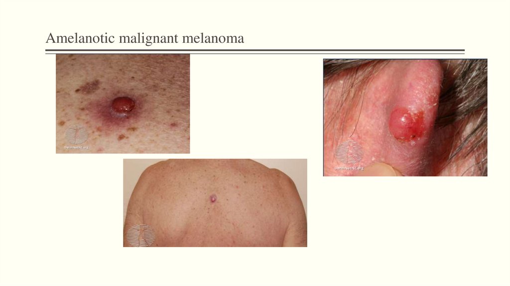
Amelanotic malignant melanoma• Amelanotic melanoma is a form of
melanoma
Treatment:
wide local excision of the wound with a
10–20 mm margin of normal tissue
• The malignant cells have little to no
pigment
• Risk factors: Increasing age, Sunexposed skin
Amelanotic melanoma can metastasis.
These cases require individualized
treatment that may include surgery,
• May present as an erythematous scaly
macule, plaque, or nodule with irregular
borders
radiotherapy, chemotherapy
7.
Amelanotic malignant melanoma8.
External angular dermoidsLooks like subcutaneous lumps at the
lateral angle of the eye
9.
KeratoacanthomaTumour of keratinocytes
Occur singly on light-exposed areas
Raised crater with central keratin plug
Grows to 2 cm or more
Can be confused with SCC
Treatment is surgical excision and
histological examination. Ensure a 2–3
mm margin for excision.
10.
3.00211.
Malignant melanoma12.
Melanoma• Typical age range 30–50 years (average
40 years)
• Can occur anywhere on the body—more
common: lower limbs in women, upper
back in men
• Often asymptomatic
• Can bleed or itch
13.
MelanomaPrognosis
thickness (Breslow classification)
level or depth (worse in level IV or V)
site (worse on head and neck, trunk)
sex (worse for men)
age (worse >50 years)
amelanotic melanoma
ulceration
14.
Management points for naevi and melanomas15.
Neuropathic ulcerA neuropathic ulcer is one that occurs as a result of
peripheral neuropathy
Neuropathic ulcers can develop with any condition with
peripheral neuropathy
Associated with diabetes, syphilis, leprosy and other
neuropathies.
16.
Diabetic foot ulcerA feature is a deep, punched-out lesion over
pressure points.
The ulcers may extend to the bone and into
joints.
They are prone to secondary infection.
Treatment is based on controlling the diabetes
and clearing infection with appropriate
antibiotics, but referral for surgical
management is usually essential.
17.
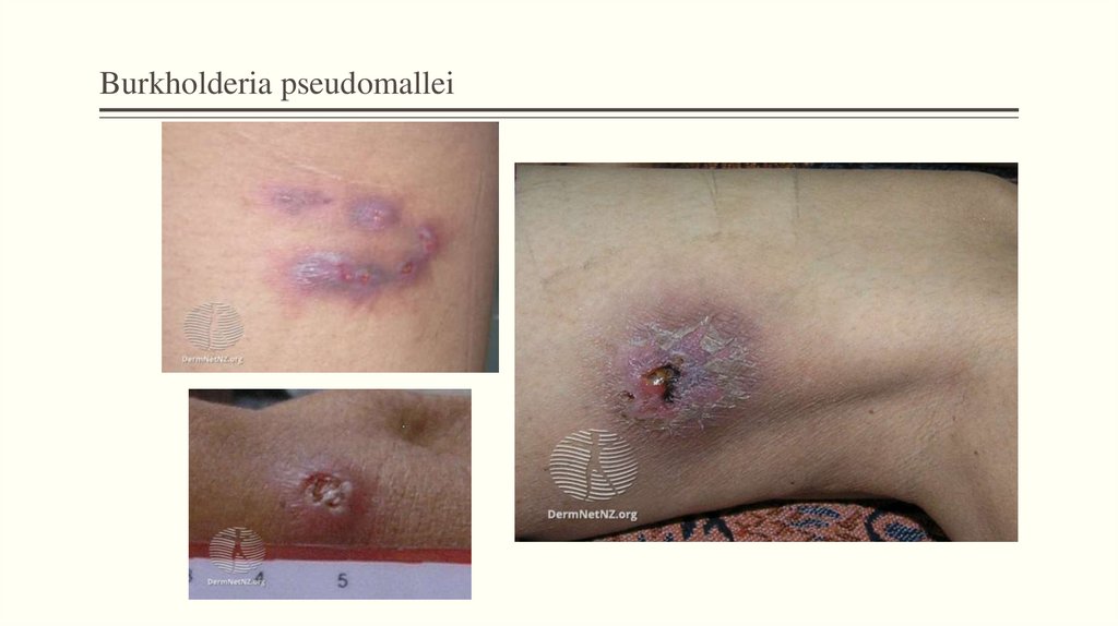
Burkholderia pseudomalleiMelioidosis is an uncommon tropical disease caused by the bacterium,
Burkholderia pseudomallei
a soil saprophyte that infects humans mainly by penetrating through
skin wounds, especially abrasions
It is mostly acquired while wading in rice paddies
fever + pneumonia + myalgia → melioidosis
Treatment: antibiotics are given intravenously initially for the first 10–14
days.
18.
Burkholderia pseudomallei19.
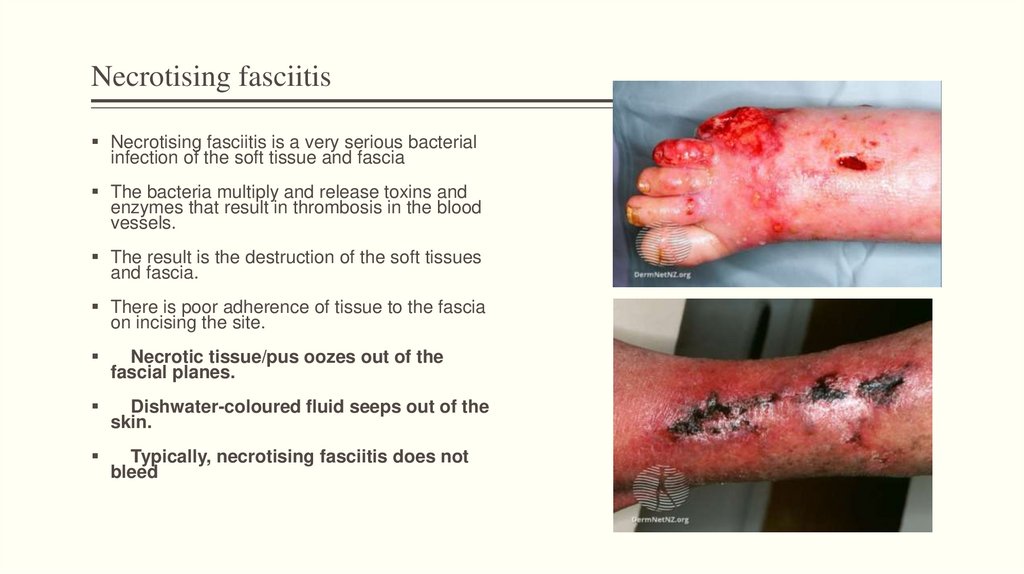
Necrotising fasciitisNecrotising fasciitis is a very serious bacterial
infection of the soft tissue and fascia
The bacteria multiply and release toxins and
enzymes that result in thrombosis in the blood
vessels.
The result is the destruction of the soft tissues
and fascia.
There is poor adherence of tissue to the fascia
on incising the site.
Necrotic tissue/pus oozes out of the
fascial planes.
Dishwater-coloured fluid seeps out of the
skin.
Typically, necrotising fasciitis does not
bleed
20.
Erythema ab igneErythema ab igne (EAI) is a skin reaction caused
by chronic exposure to infrared radiation in the
form of heat
Causes a mild and transient red rash resembling
lacework or a fishing net
the condition will resolve by itself over several
months.
If there is a persistent sore that doesn't heal or a
growing lump within the rash, a skin biopsy
should be performed to rule out the possibility of
skin cancer