


 medicine
 biology
Similar presentations:

")


. Non-steroidal anti-inflammatory drugs (NSAID)")


")
Inflammaione. (Subject 4)
1. Inflammation
January 20, 20172. Causes of inflammation
Exogenous Infectious factorsExogenous Non-infectious factors:
physical
chemical
biological
Endogenous products of tissue decay
Endogenous chemical agents
3.
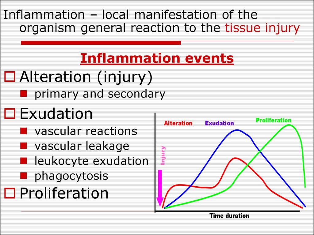
Inflammation – local manifestation of theorganism general reaction to the tissue injury
Inflammation events
Alteration (injury)
Exudation
vascular reactions
vascular leakage
leukocyte exudation
phagocytosis
Alteration
Exudation
Injury
primary and
Interrelation between the
secondary
inflammatory events
Proliferation
Time duration
Proliferation
4. Signs of inflammation
Systemic:peripheral blood
Calor - heat
leukocytosis
Rubor - redness
fever
Dolor - pain
globulins blood level
erythrocytes
Tumor - swelling
sedimentation rate
Functio laesa cateholamines and
loss of function
corticosteroids
Local:
5. Alteration
Primary alteration - direct action ofpathogenic factor (functional and structural
injury of the cells)
Secondary alteration mechanisms:
disturbances of local nervous regulation
and blood circulation;
influence of inflammatory mediators;
alteration of T 0 , pH, oncotic, osmotic
pressure;
lysosomal effect.
6. Metabolism changes
Prevalence of catabolic processes inthe early stages
High speed of metabolic reaction
(heat)
Metabolic acidosis
osmotic and oncotic pressure
Intracellular and extracellular
hyperhydration (swelling)
Prevalence of anabolism – final stages
7. Inflammatory mediators
8. Arachidonic acid metabolites
Cell membrane phospholipidsPhospholipases
- Inflammation
- Activation of
the complement
Arachidonic Acid
Lipooxygenase
Leukotrienes
Non-Steroid Anti
Inflammatory drugs
Cyclooxygenase
Thromboxane
Prostacycline
Prostoglandins
9. Arachidonic acids metabolites
Thromboxane A2 - platelet aggregator andvasoconstrictor
Prostacyclin - platelet aggregation and
vasodilator.
Prostaglandins:
dilation of vessels , vessels permeability
aggregation and adhesion of blood cells
fever, pain
Leukotrienes :
smooth muscles tone (GIT, bronchi, blood
vessels)
vessels permeability
chemotaxins for neutrophiles
10. Cellular mediators
Active oxygen radicals:endothelial cells damage ( vessels permeability)
other cells injury
Platelet activating factor (PAF):
Platelet aggregation and release
smooth muscles tone (bronchi, vessels)
leukocyte adhesion to endothelium
leukocyte chemotaxis, degranulation and
oxidative burst
11. Cellular mediators
Lysosomal enzymes:mediate tissue injury
activate bradykinine synthesis
mast cells degranulation
chemotaxis
Nitric oxide:
vasodilation
cytotoxic effect
Cytokines:
interleukins
TNF
interpherone
12. Plasma mediators
Bradykinin:Complement
vasodilation and vascular permeability
chemotaxis
mast cells degranulation
opsonisation
activate AA cascade
Clotting system
mobilization of molecules of adherence
activation of cyclooxygenase
production of NO and PAF
13. The summary of inflammatory mediators’ activity
Vasodilationof blood vessels permeability
Leukocyte adhesion
Chemotaxis
Fever
Tissue damage
Pain
14. Changes in vascular flow
1. Arterioles constriction (activation ofsympathetic nerves, mediators
influence) -localization of injuring
agent
2. Arterial hyperemia (dilatation of
arterioles due to BAS) - increase the
general rate of metabolism
15. Changes in vascular flow
3. Venous hyperemia and pre-stasis(dilation of venules and postcapillaries):
increased blood viscosity
swollen vessel walls
squeezing with inflammatory exudates
leukocytes margination along the vessels
walls
4. Stasis - complete stop of blood flow.
16.
Venous hyperemia andstasis prevent the
spreading of the
damage to surrounding
tissues.
Arterial and venous
hyperemia result in the
increase of vessels
permeability and
promote exudate
formation.
17. Mechanisms of exudation
vascular permeability (vascularleakage).
intravascular hydrostatic
pressure
osmotic and oncotic pressure of
interstitial fluid
18. Increase of vascular permeability
Endothelial cellscontraction
•histamine,
bradykinin
•occurs rapidly after
exposure to mediator
•reversible
Direct endothelial
injury
•severe non-specific
injuries (burns or
bacterial infection)
•leakage lasts until
vessels are
thrombosed or repaired
Leukocyte-dependent
endothelial injury
•toxic oxygen radicals
and proteolytic
enzymes
19. Mechanisms of exudation
hydrostatic pressure - filtration offluid from capillaries.
Ultrafiltrate of blood plasma with protein
less then 2 % - transudate.
Inflammatory - more then 2 % protein.
osmotic and oncotic pressure
Inflow of protein rich fluid from plasma
to the site of inflammation.
Destruction of molecules by the enzymes
20. The role of exudation
Negative•squeezing of
tissues and
organs
•exudate outflow
to body cavities
and big vessels
•abscess and
phlegmon
formation
Positive
•transport of antibodies,
inflammatory mediators
•elimination of toxins
and metabolites from
inflammatory site
•localization of the
inflammatory agents
21. Extravasation of leukocytes
22. Leukocytes migration
•Move pseudopods into thejunctions between the
endothelial cells
•Squeeze through
interendothelial junctions
•Release proteolytic
lysosomal enzymes making
gaps in vessels walls
•Order of migration:
neutrophiles, monocytes,
lymphocytes
23. Chemotaxis
Chemotactic agents:bacterial membrane lipopolysaccharides
components of the complement (3b,
5a,5b,6,7
leukotrienes
products of tissue decay
Mechanism
Binding to receptors
calcium mobilisation
contraction of microfilaments
movement
24. Leukocytes role in inflammation
Protective function – phagocytosis.Synthesis and secretion of inflammatory
mediators.
Processing and presentation of foreign
agents for the immune systems.
Tissue damage with :
Lysosomal enzymes
Active oxygen radicals
Products of AA metabolism
(prostaglandins and leukotrienes)
25. Stages of phagocytosis
1.Chemotaxis2. Adherence
(opsonins IgM, IgG, C3b)
3. Phagosome
formation
4. Killing or
degradation of
the ingested
material
26. Two mechanisms of bacterial killing
Oxygen-dependent mechanismreactive oxygen species – superoxide
anion, hydroxyl ion, hydroperoxide
Oxygen Independent Mechanisms
– using the content of granules
(lysozyme, proteins influencing
bacterial cell wall)
27. Proliferation in inflammation
Regeneration - replacement of deadcells with new ones; the function is
restored.
Repair - replacement with fibrous
connective tissue cells and fibers; the
functions is not restored.
28. The steps of repair
PhagocytosisProliferation of endothelial cells and
fibroblasts in the damaged area.
The growth of new vessels to
establish blood circulation in the
healing area
Fibroblasts produce collagen.
Mature scar is produced.
29. Factors influencing proliferation
Local:Persisting infection, foreign material
Inadequate blood supply
Excessive movement
Irradiation
Systemic:
Age
Nutritional deficiencies
Metabolic diseases
Catabolic state associated with malignancies
Substances:
Growth factors, TNF – activation
Chalones, glucocorticoids - inhibition
30. Classification of inflammation
Classification based on the cause ofinflammation:
Infectious: non-specific (cocci) and
specific (tuberculosis, syphilis)
Non-infectious (aseptic) – caused by
infarctions, hemorrhages, salt deposition
31. Classification of inflammation
Classification based on the prevailingmechanism:
Alterative –prevailing alteration develops in
parenchymal organs (myocardium, liver,
kidneys).
Exudative - prevailing exudate formation.
Proliferative (productive) - prevalence of
reparative process; proceeds chronically
32. Types of exudative inflammation
Serous inflammation - 3-8% of protein,single neutrophiles in exudate.
Catarrhal inflammation presence of mucus
in exudates.
Fibrinous inflammation presence of fibrin
in exudate
Croupous inflammation - fibrinous
pericarditis (hairy heart), croupous
pneumonia.
Diphtheritic – throat, pharynx, tonsils
33. Types of exudative inflammation
Purulent (suppurative) inflammationproduction of pus - pyogenic bacteria
(staphylococci).
Abscesses are localized collections of pus.
Phlegmon and empyema are diffuse pus
infiltrations.
Putrefactive inflammation - a result of
putrefactive bacteria injury.
Haemorrhagic inflammation - presence of
erythrocytes in exudates. (anthrax, plague,
influenza).
34. Neural and hormonal control of inflammation
Pro-inflammatory hormones - growthhormone, mineralocorticoids
Glucocorticoids, catecholamines anti-inflammatory effect
Violation of peripheral innervation
leads to chronic inflammation
development
35. Inflammation outcomes
Complete resolution - the injury islimited
Healing by scarring – impossibility of
regeneration or s abundant fibrin
exudation.
Abscess formation - pyogenic
microorganisms.
Progression to chronic inflammation
36. Chronic inflammation
follow acute inflammationchronic from the onset due to:
disturbances of phagocytosis
high level of glucocorticoids and
catecholamines
persistent infections or intoxications.
prolonged exposure to nondegradable
material (silica particles – silicosis)
autoimmune diseases.