
 medicine
Similar presentations:


Radiological research methods and radiological semiotics of acute cerebrovascular accident
1.
DEPARTMENT OF RADIOLOGYRadiological research methods and radiological
semiotics of acute cerebrovascular accident
Almaty 2021
2.
ACUTE CEREBROVASCULAR ACCIDENT(CVA,STROKE)
• – acute and severe brain disease. Blood may be interrupted
or stop moving through an artery, because the artery is
blocked (ischaemic stroke) or bursts (haemorrhagic
stroke).
3.
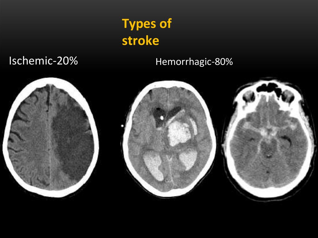
Types ofstroke
Ischemic-20%
Hemorrhagic-80%
4.
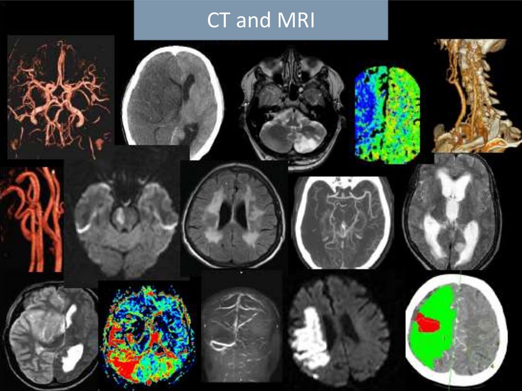
CT and MRI5.
Neuroimaging tasksDefine:
1. Intracerebral hemorrhage/infarction
2. Localization and size of the lesion
3. Time from the beginning progression infaction
4. The state of intracranial vessels and collateral blood
flow
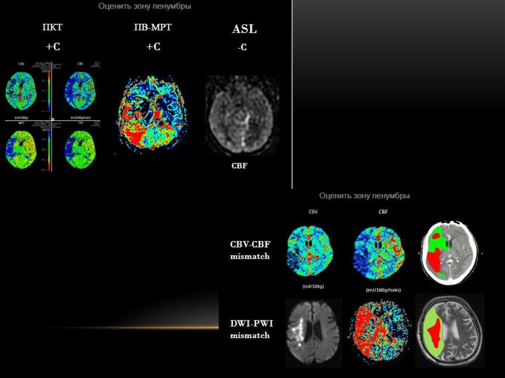
5. Penumbra zone
6. The effectiveness of the therapy
6.
CT or MRI?Exclude ICH
7.
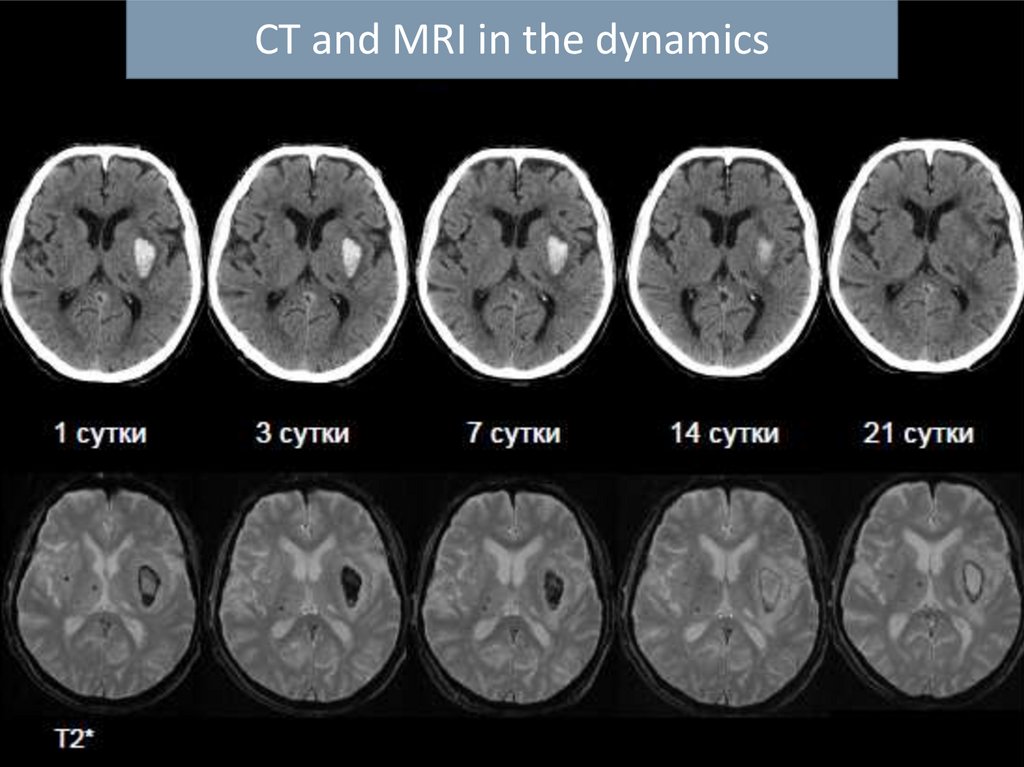
CT and MRI in the dynamics8.
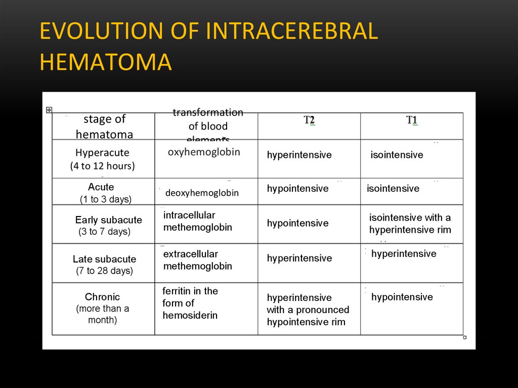
EVOLUTION OF INTRACEREBRALHEMATOMA
stage of
hematoma
Hyperacute
transformation
of blood
elements
oxyhemoglobin
hyperintensive
deoxyhemoglobin
hypointensive
isointensive
intracellular
methemoglobin
hypointensive
isointensive with a
hyperintensive rim
extracellular
methemoglobin
hyperintensive
hyperintensive
hyperintensive
with a pronounced
hypointensive rim
hypointensive
isointensive
(4 to 12 hours)
Acute
(1 to 3 days)
Early subacute
(3 to 7 days)
Late subacute
(7 to 28 days)
Chronic
(more than a
month)
ferritin in the
form of
hemosiderin
9.
Hemorrhagic strokes• CT - is a “gold standard” of diagnosis.
• Hemorrhage is a focus of increased density of matter
(hyperdense zone) of the brain of a rounded or oval shape,
sometimes there are signs of a volumetric effect on the
cerebrospinal fluid spaces and the ventricular system of varying
degrees of severity, depending on the size and localization of
the hemorrhage.
• In the dynamics of the hemorrhage focus, there is a gradual
decrease in the density of the hemorrhage focus in the brain
tissue – the phenomenon of a "melting sugar cube".
10.
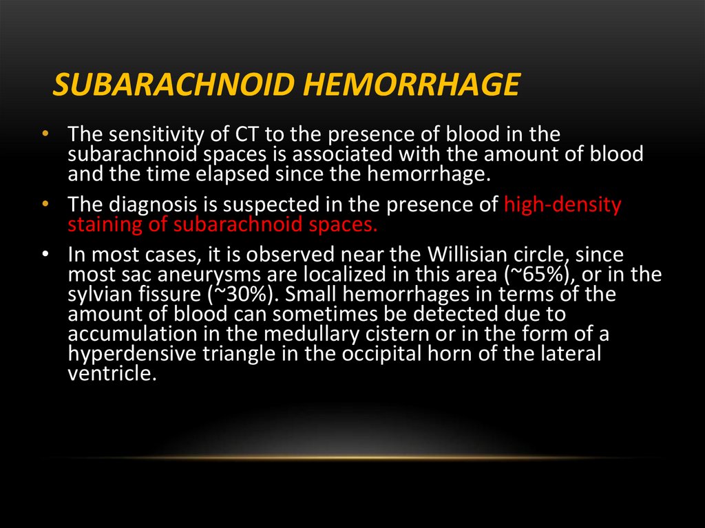
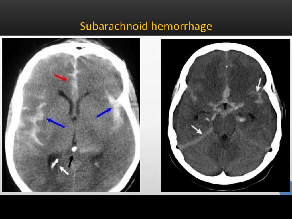
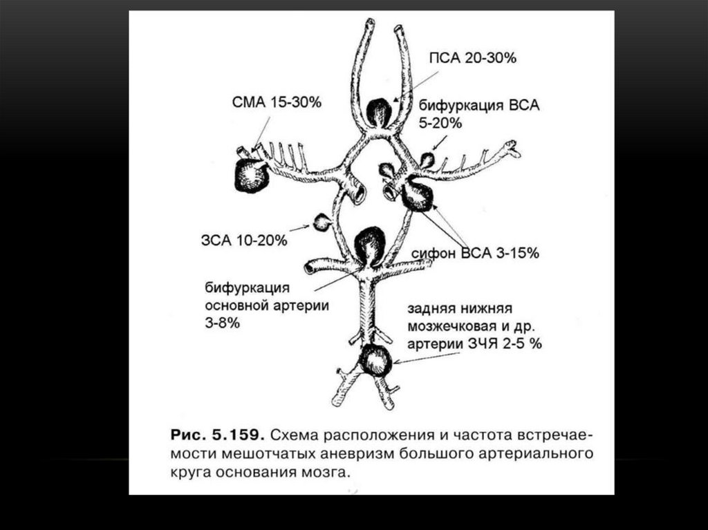
SUBARACHNOID HEMORRHAGE• The sensitivity of CT to the presence of blood in the
subarachnoid spaces is associated with the amount of blood
and the time elapsed since the hemorrhage.
• The diagnosis is suspected in the presence of high-density
staining of subarachnoid spaces.
• In most cases, it is observed near the Willisian circle, since
most sac aneurysms are localized in this area (~65%), or in the
sylvian fissure (~30%). Small hemorrhages in terms of the
amount of blood can sometimes be detected due to
accumulation in the medullary cistern or in the form of a
hyperdensive triangle in the occipital horn of the lateral
ventricle.
11.
Subarachnoid hemorrhage12.
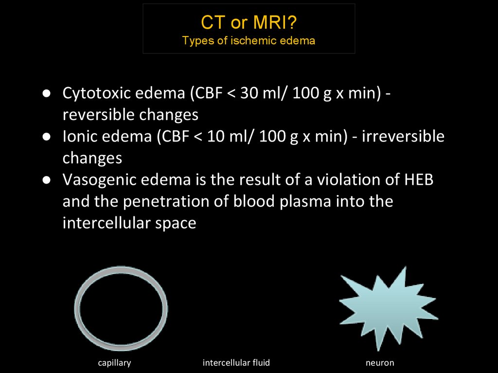
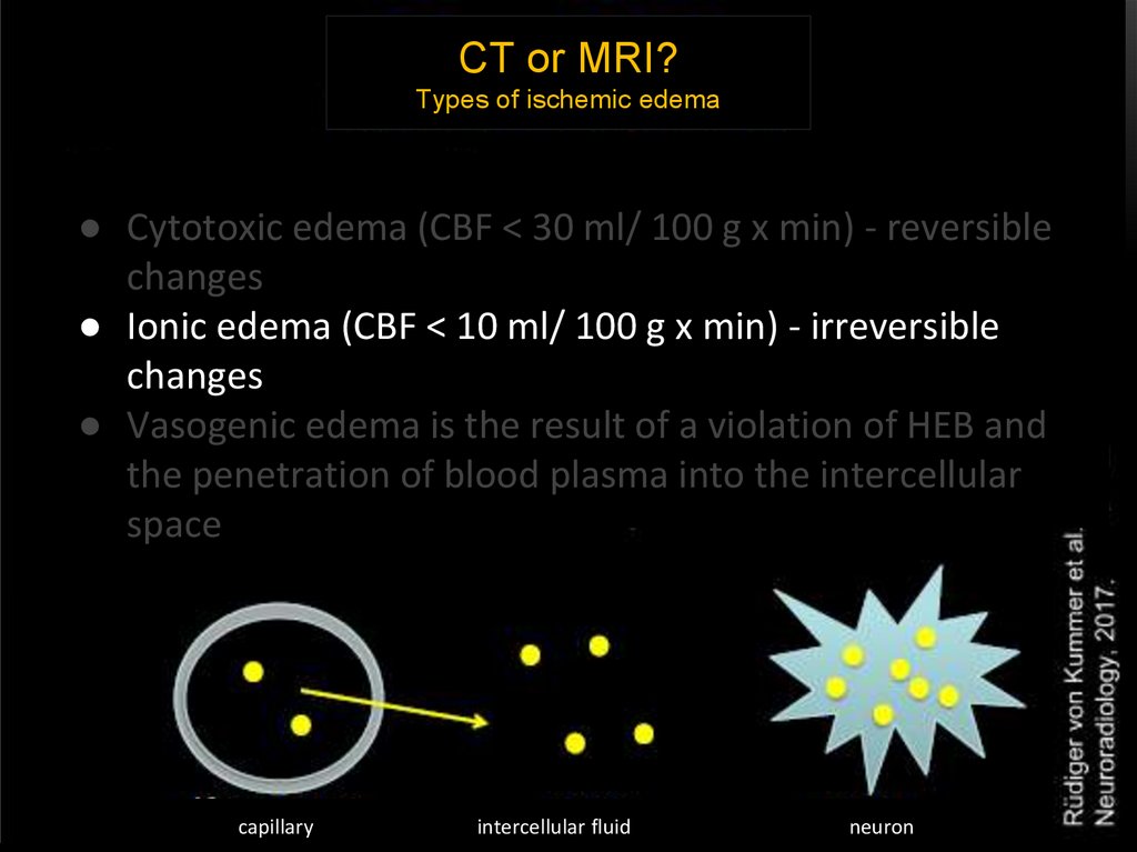
CT or MRI?Types of ischemic edema
● Cytotoxic edema (CBF < 30 ml/ 100 g x min) reversible changes
● Ionic edema (CBF < 10 ml/ 100 g x min) - irreversible
changes
● Vasogenic edema is the result of a violation of HEB
and the penetration of blood plasma into the
intercellular space
capillary
intercellular fluid
neuron
13.
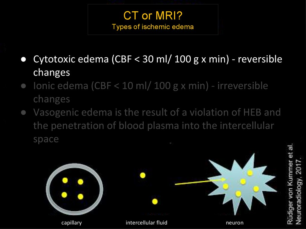
CT or MRI?Types of ischemic edema
● Cytotoxic edema (CBF < 30 ml/ 100 g x min) - reversible
changes
● Ionic edema (CBF < 10 ml/ 100 g x min) - irreversible
changes
● Vasogenic edema is the result of a violation of HEB and
the penetration of blood plasma into the intercellular
space
capillarycapillary
intercellular fluid neuronintercellular fluid
neuron
14.
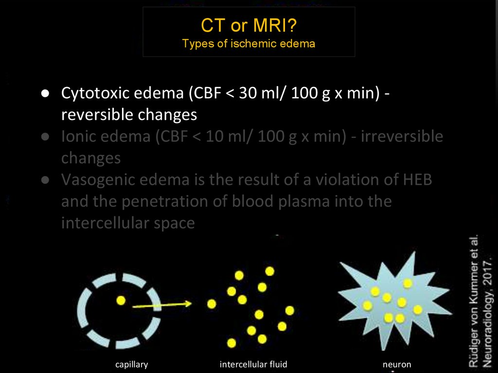
CT or MRI?Types of ischemic edema
● Cytotoxic edema (CBF < 30 ml/ 100 g x min) - reversible
changes
● Ionic edema (CBF < 10 ml/ 100 g x min) - irreversible
changes
● Vasogenic edema is the result of a violation of HEB and
the penetration of blood plasma into the intercellular
space
capillary
intercellular fluid
neuron
15.
CT or MRI?Types of ischemic edema
● Cytotoxic edema (CBF < 30 ml/ 100 g x min) reversible changes
● Ionic edema (CBF < 10 ml/ 100 g x min) - irreversible
changes
● Vasogenic edema is the result of a violation of HEB
and the penetration of blood plasma into the
intercellular space
capillary
intercellular fluid
neuron
16.
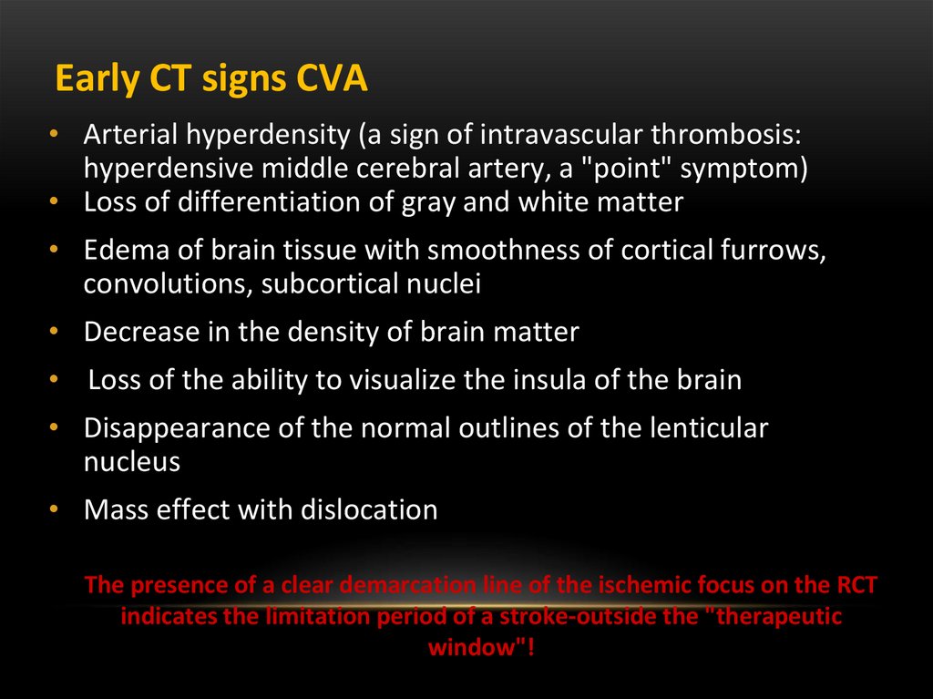
Early СТ signs CVA• Arterial hyperdensity (a sign of intravascular thrombosis:
hyperdensive middle cerebral artery, a "point" symptom)
• Loss of differentiation of gray and white matter
• Edema of brain tissue with smoothness of cortical furrows,
convolutions, subcortical nuclei
• Decrease in the density of brain matter
• Loss of the ability to visualize the insula of the brain
• Disappearance of the normal outlines of the lenticular
nucleus
• Mass effect with dislocation
The presence of a clear demarcation line of the ischemic focus on the RCT
indicates the limitation period of a stroke-outside the "therapeutic
window"!
17.
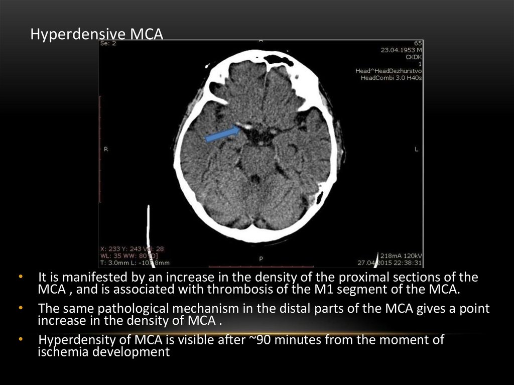
Hyperdensive MCAIt is manifested by an increase in the density of the proximal sections of the
MCA , and is associated with thrombosis of the M1 segment of the MCA.
• The same pathological mechanism in the distal parts of the MCA gives a point
increase in the density of MCA .
• Hyperdensity of MCA is visible after ~90 minutes from the moment of
ischemia development
18.
ASPECTS SCOREThe Alberta stroke programme early CT score (ASPECTS) 1 is a 10-point
quantitative topographic CT scan score used for patients with CVA
The need for the ASPECTS scale:
• the assessment of early ischemic changes is important in the assumption
of a response to thrombolysis
• thrombolysis increases the chances of a good functional outcome in
patients with small (less than 1/3 of the MCA) sizes of the hypodensive
zone on non-amplified CT scans, and quantifying the volume of one third
of the territory is inconvenient for routine practice
• ASPECTS was developed to standardize the identification of changes and
the compilation of descriptions (reports) of the degree of
hypodensiveness of ischemia
19.
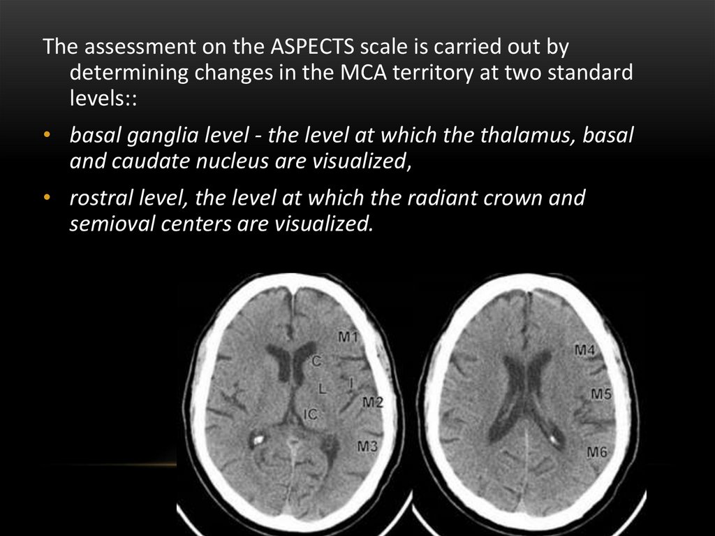
The assessment on the ASPECTS scale is carried out bydetermining changes in the MCA territory at two standard
levels::
• basal ganglia level - the level at which the thalamus, basal
and caudate nucleus are visualized,
• rostral level, the level at which the radiant crown and
semioval centers are visualized.
20.
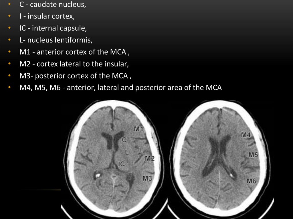
C - caudate nucleus,
I - insular cortex,
IC - internal capsule,
L- nucleus lentiformis,
M1 - anterior cortex of the MCA ,
M2 - cortex lateral to the insular,
M3- posterior cortex of the MCA ,
M4, M5, M6 - anterior, lateral and posterior area of the MCA
21.
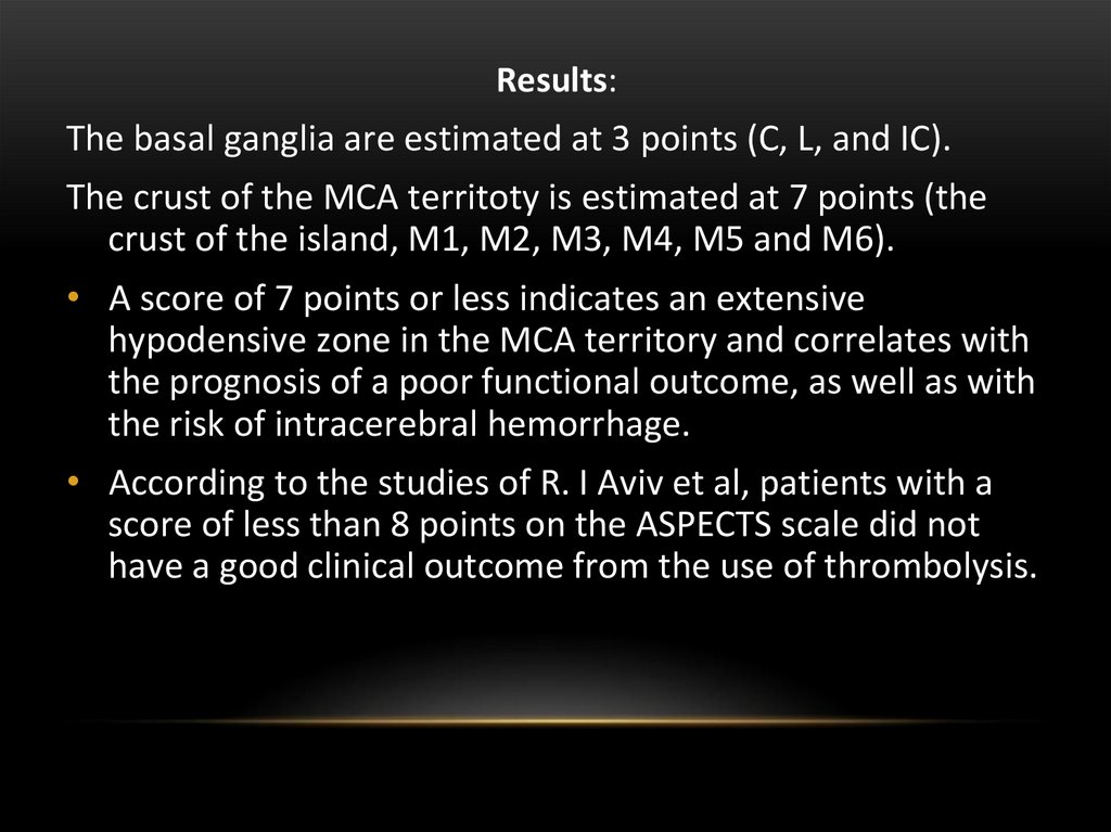
Results:The basal ganglia are estimated at 3 points (C, L, and IC).
The crust of the MCA territoty is estimated at 7 points (the
crust of the island, M1, M2, M3, M4, M5 and M6).
• A score of 7 points or less indicates an extensive
hypodensive zone in the MCA territory and correlates with
the prognosis of a poor functional outcome, as well as with
the risk of intracerebral hemorrhage.
• According to the studies of R. I Aviv et al, patients with a
score of less than 8 points on the ASPECTS scale did not
have a good clinical outcome from the use of thrombolysis.
22.
PATHOPHYSIOLOGY AND MR-PICTURE OFCEREBRAL ISCHEMIC STROKE
I
• acute stage(0-3 days).
Pathoanatomically-focal cytotoxic edema, macroscopically-thickening of the
brain gyrus and loss of clear distinctions between gray and white matter.
MRI: the focus or zone of a sufficiently uniform increase in the
signal for T2 and FLAIR, a moderate uniform decrease for T1.
Compliance with the territory (according to the form)
Violation of the differentiation of gray and white matter of
the brain + mass effect of varying degrees of severity,
depending on the volume of the lesion. .
23.
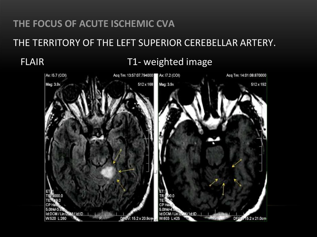
THE FOCUS OF ACUTE ISCHEMIC CVATHE TERRITORY OF THE LEFT SUPERIOR CEREBELLAR ARTERY.
FLAIR
Т1- weighted image
24.
PATHOPHYSIOLOGY AND MR-PICTURE OFCEREBRAL ISCHEMIC STROKE
• Subacute stage (3 days-10-14 days).
The combination of cytotoxic and vasogenic edema, the onset
of encephalomalacia with the formation of necrotic zones in
combination with reparative processes – the appearance of a
symptom of "giral" amplification.
MRI: focus or zone of NON-UNIFORM increase in the signal
for T2 and FLAIR, an indistinctly expressed NON-uniform
decrease for T1 or isointensive MRS (a symptom of "veiling")
. There is a violation of the differentiation of gray and white
matter of the brain, a decrease in the mass effect or its
absence.
25.
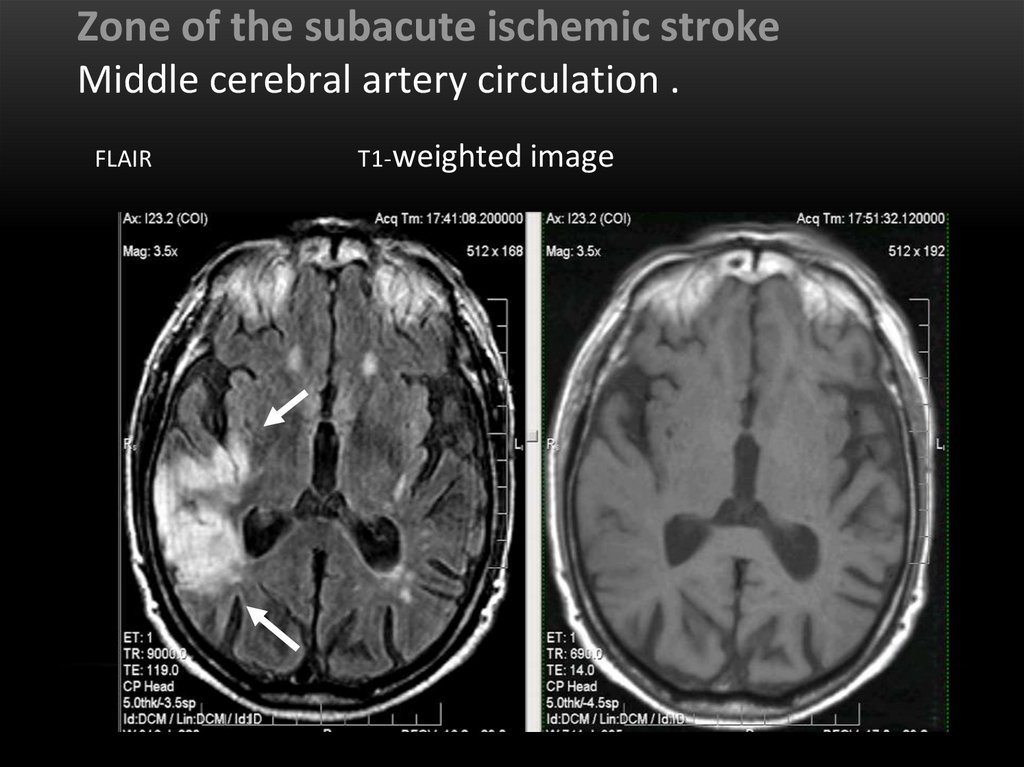
Zone of the subacute ischemic strokeMiddle cerebral artery circulation .
FLAIR
Т1-weighted image
26.
PATHOPHYSIOLOGY AND MR-PICTURE OFCEREBRAL ISCHEMIC STROKE
• Chronic stage (end of the 2nd week and beyond).
resorption of necrotic masses, the formation of a cystic cavity with
perifocal gliosis, may be accompanied by dilation of the ipsilateral
ventricle. The decrease in the intensity of the MR signal corresponds to
the prescription period of cyst formation, after 3-6 months it corresponds
to the characteristics of the cerebrospinal fluid.
MRI: foci or zones of cystic-glious or
infiltrative-glious transformation +local
atrophy
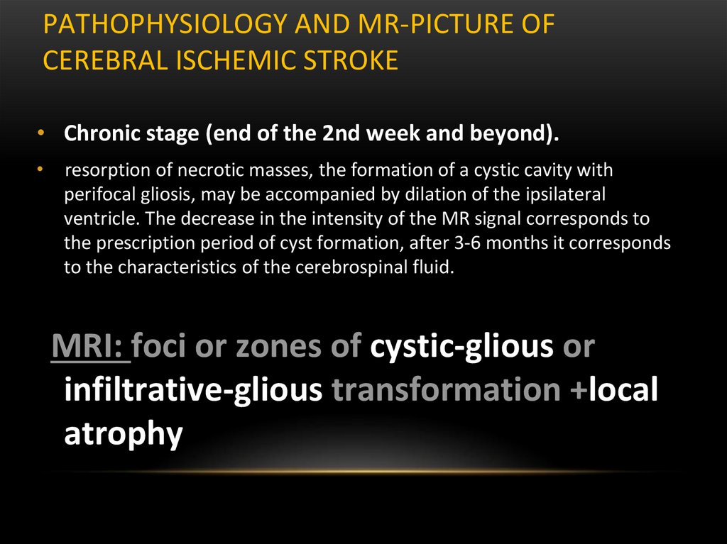
27.
Zone of the chronic ischemic strokeLeft posterior cerebral artery circulation .
FLAIR
Т1-weighted image
28.
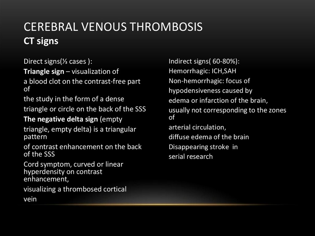
CEREBRAL VENOUS THROMBOSISCT signs
Direct signs(⅓ cases ):
Triangle sign – visualization of
a blood clot on the contrast-free part
of
the study in the form of a dense
triangle or circle on the back of the SSS
The negative delta sign (empty
triangle, empty delta) is a triangular
pattern
of contrast enhancement on the back
of the SSS
Cord symptom, curved or linear
hyperdensity on contrast
enhancement,
visualizing a thrombosed cortical
vein
Indirect signs( 60-80%):
Hemorrhagic: ICH,SAH
Non-hemorrhagic: focus of
hypodensiveness caused by
edema or infarction of the brain,
usually not corresponding to the zones
of
arterial circulation,
diffuse edema of the brain
Disappearing stroke in
serial research
29.
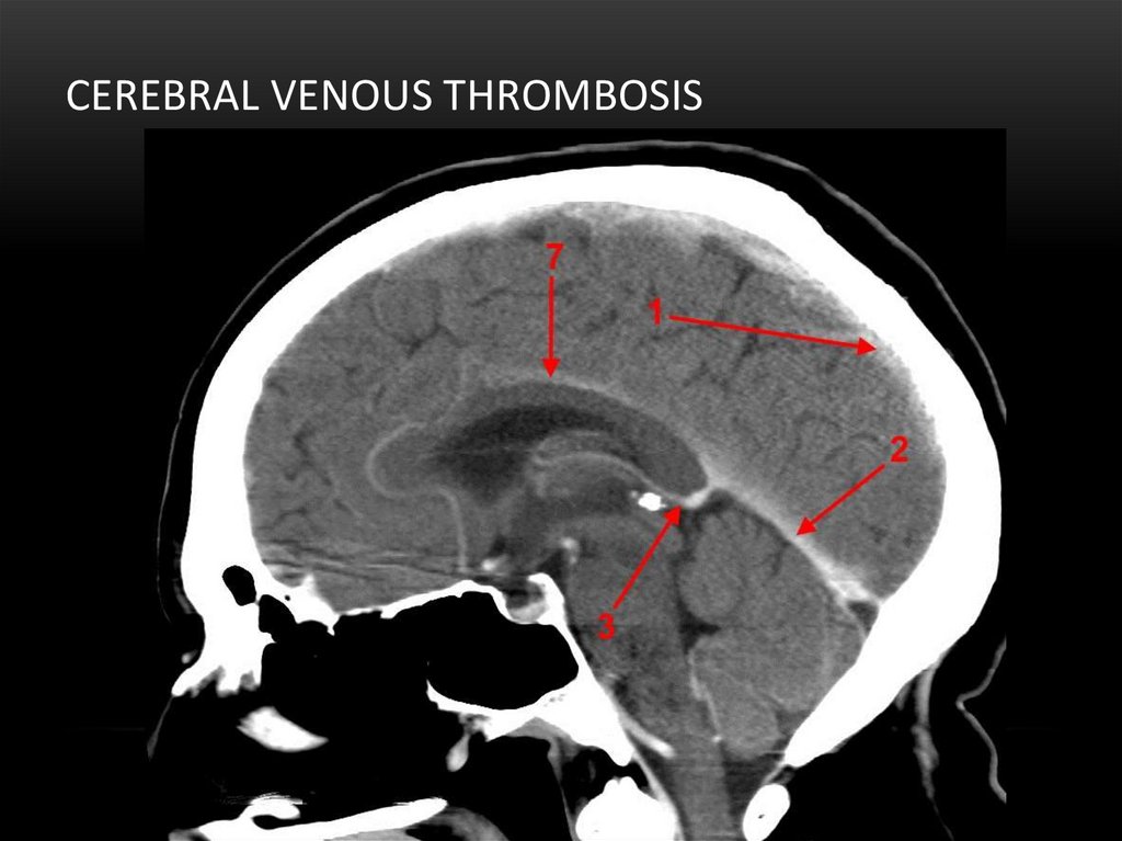
CEREBRAL VENOUS THROMBOSIS30.
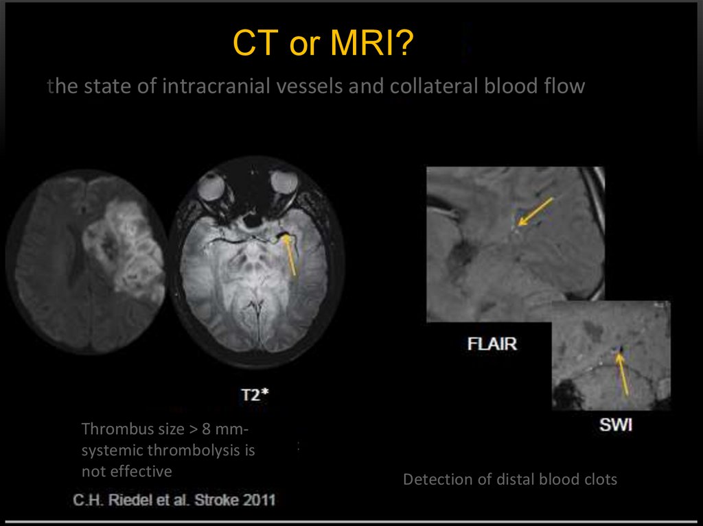
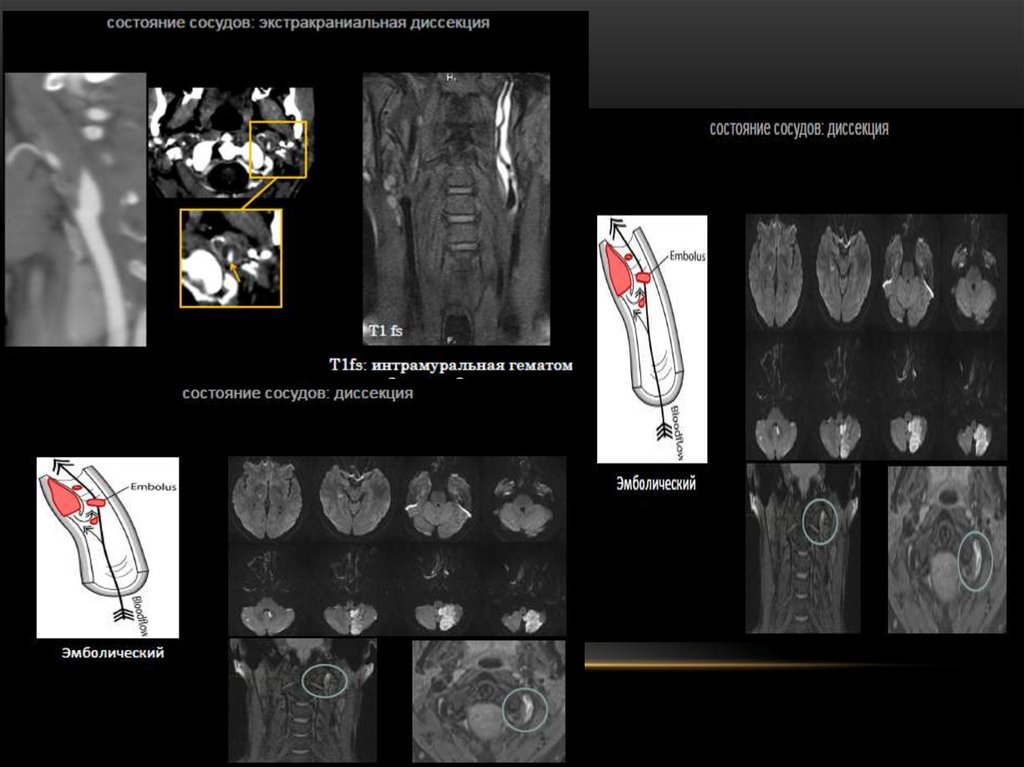
CT or MRI?the state of intracranial vessels and collateral blood flow
Thrombus size > 8 mmsystemic thrombolysis is
not effective
Detection of distal blood clots
31.
32.
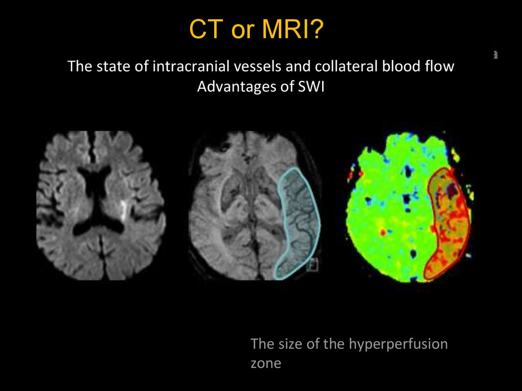
CT or MRI?The state of intracranial vessels and collateral blood flow
Advantages of SWI
The size of the hyperperfusion
zone
33.
34.
35.
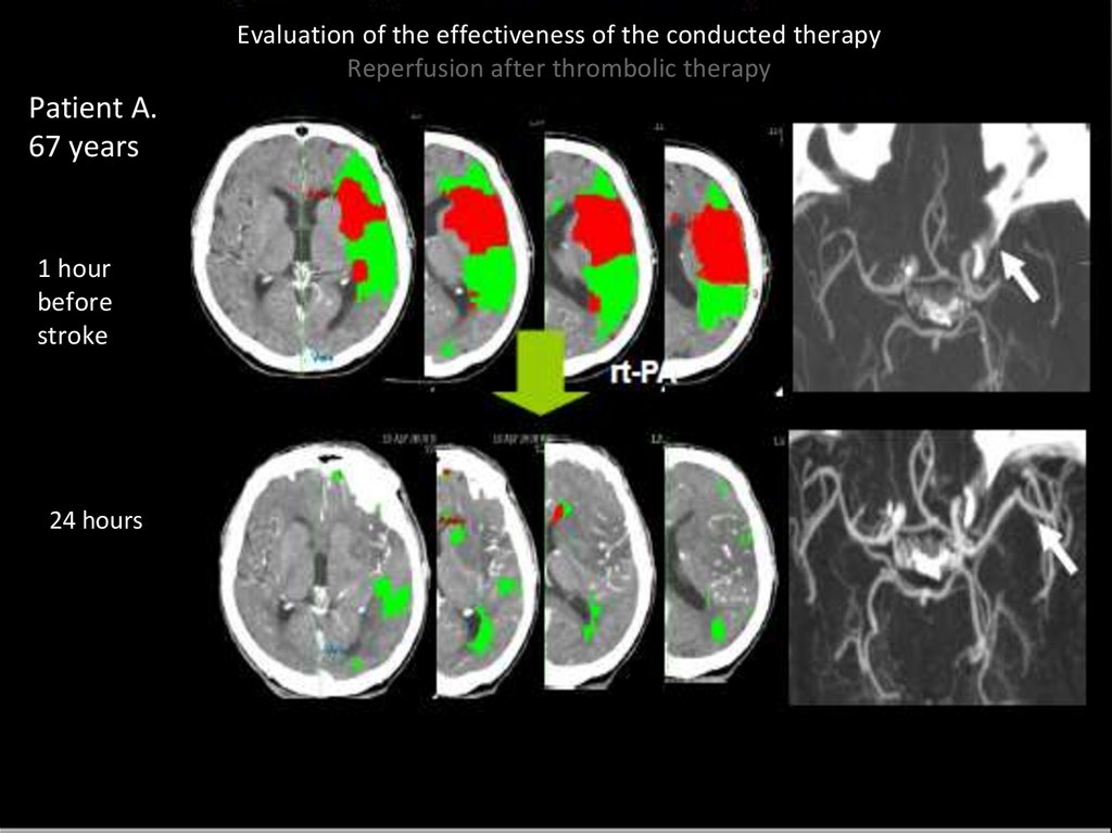
Evaluation of the effectiveness of the conducted therapyReperfusion after thrombolic therapy
Patient A.
67 years
1 hour
before
stroke
24 hours
36.
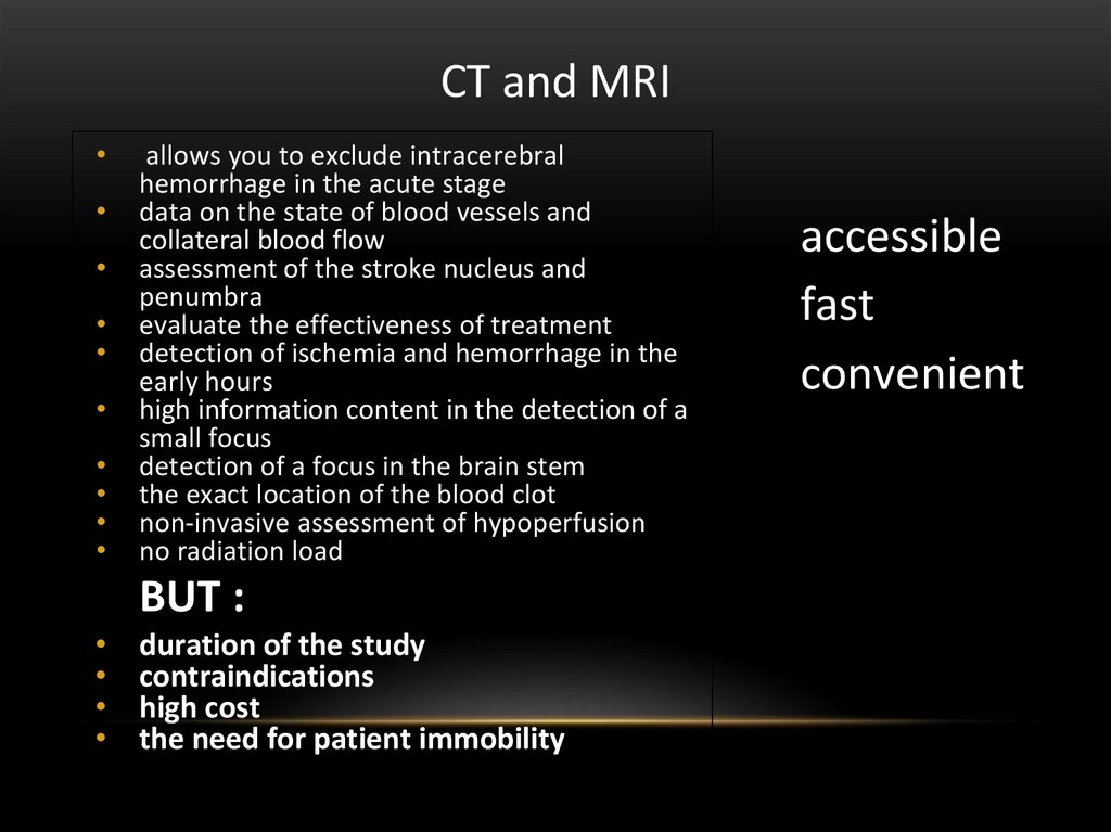
CT and MRIallows you to exclude intracerebral
hemorrhage in the acute stage
data on the state of blood vessels and
collateral blood flow
assessment of the stroke nucleus and
penumbra
evaluate the effectiveness of treatment
detection of ischemia and hemorrhage in the
early hours
high information content in the detection of a
small focus
detection of a focus in the brain stem
the exact location of the blood clot
non-invasive assessment of hypoperfusion
no radiation load
BUT :
duration of the study
contraindications
high cost
the need for patient immobility
accessible
fast
convenient