


 medicine
Similar presentations:


Peptic Ulcer Disease
1.
Peptic Ulcer DiseaseName-MOHAMMAD DANISH
ANSARI
Group-LA2-173(2)
Course-4
Teacher-IGOR YATSKOV
2.
INTRODUCTION• Peptic Ulcer is a lesion in the lining
(mucosa) of the digestive tract, typically in
the stomach or duodenum, caused by the
digestive action of pepsin and stomach
acid.
3.
Lesion may subsequently occur into the laminapropria and submucosa to cause bleeding. –
Most of peptic ulcer occur either in the
duodenum, or in the stomach – Ulcer may also
occur in the lower esophagus due to reflexing of
gastric content – Rarely in certain areas of the
small intestine
4.
PATHOPHYSIOLOGY5.
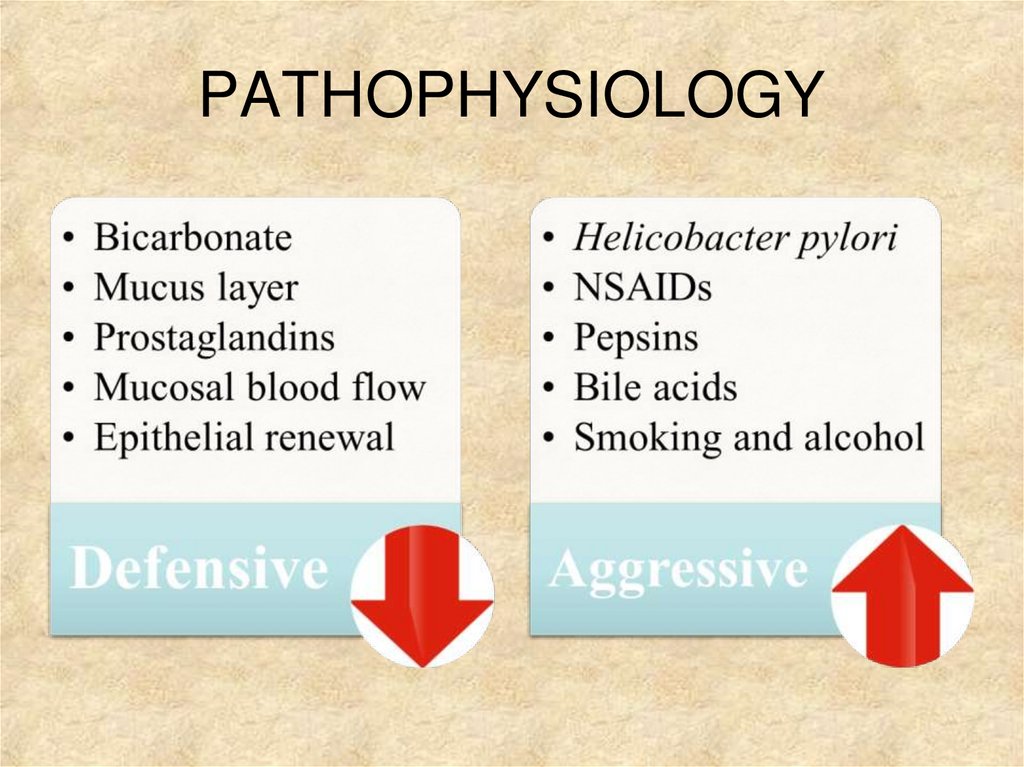
Under normal conditions, a physiologic balanceexists between gastric acid secretion and
gastroduodenal mucosal defense. Mucosal injury
and, thus, peptic ulcer occur when the balance
between the aggressive factors and the
defensive mechanisms is disrupted. Aggressive
factors, such as NSAIDs, H pylori infection,
alcohol, bile salts, acid, and pepsin, can alter the
mucosal defense by allowing back diffusion of
hydrogen ions and subsequent epithelial cell
injury.
6.
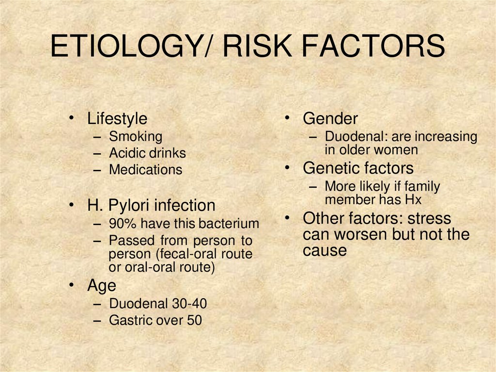
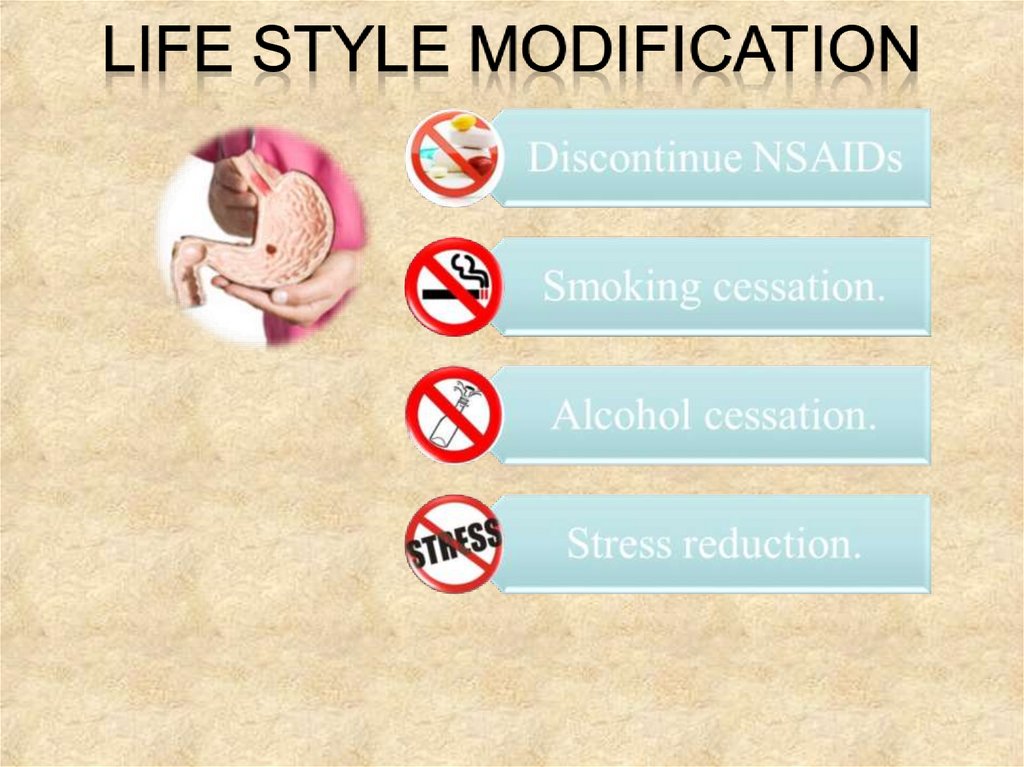
ETIOLOGY/ RISK FACTORS• Lifestyle
– Smoking
– Acidic drinks
– Medications
• H. Pylori infection
– 90% have this bacterium
– Passed from person to
person (fecal-oral route
or oral-oral route)
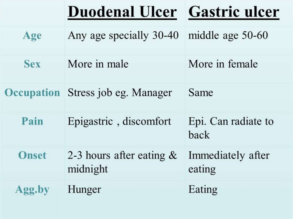
• Age
– Duodenal 30-40
– Gastric over 50
• Gender
– Duodenal: are increasing
in older women
• Genetic factors
– More likely if family
member has Hx
• Other factors: stress
can worsen but not the
cause
7.
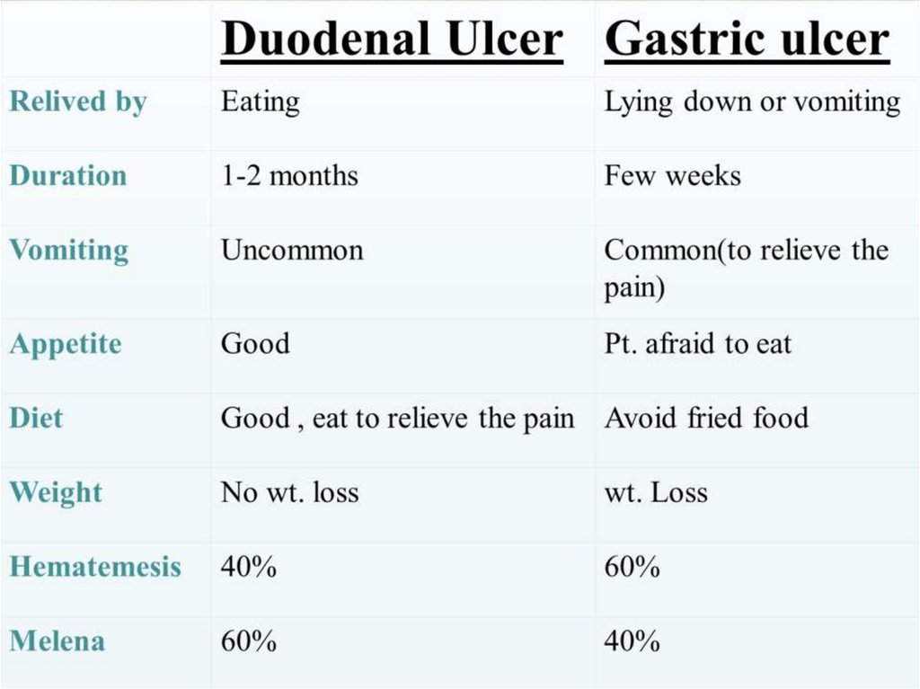
TYPES• GASTRIC PEPTIC ULCER
• DUODENAL PEPTIC ULCER
8.
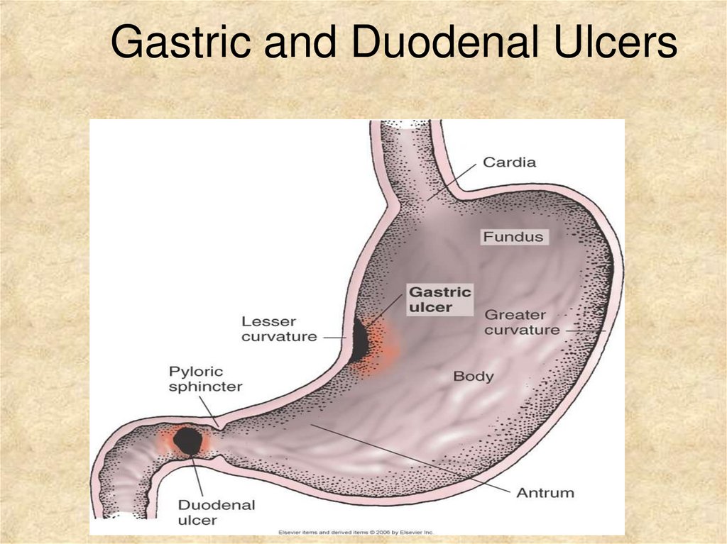
Gastric and Duodenal Ulcers9.
10.
11.
INVESTIGATION• Stool examination for fecal occult blood.
• Complete blood count (CBC) for decrease
in blood cells.
12.
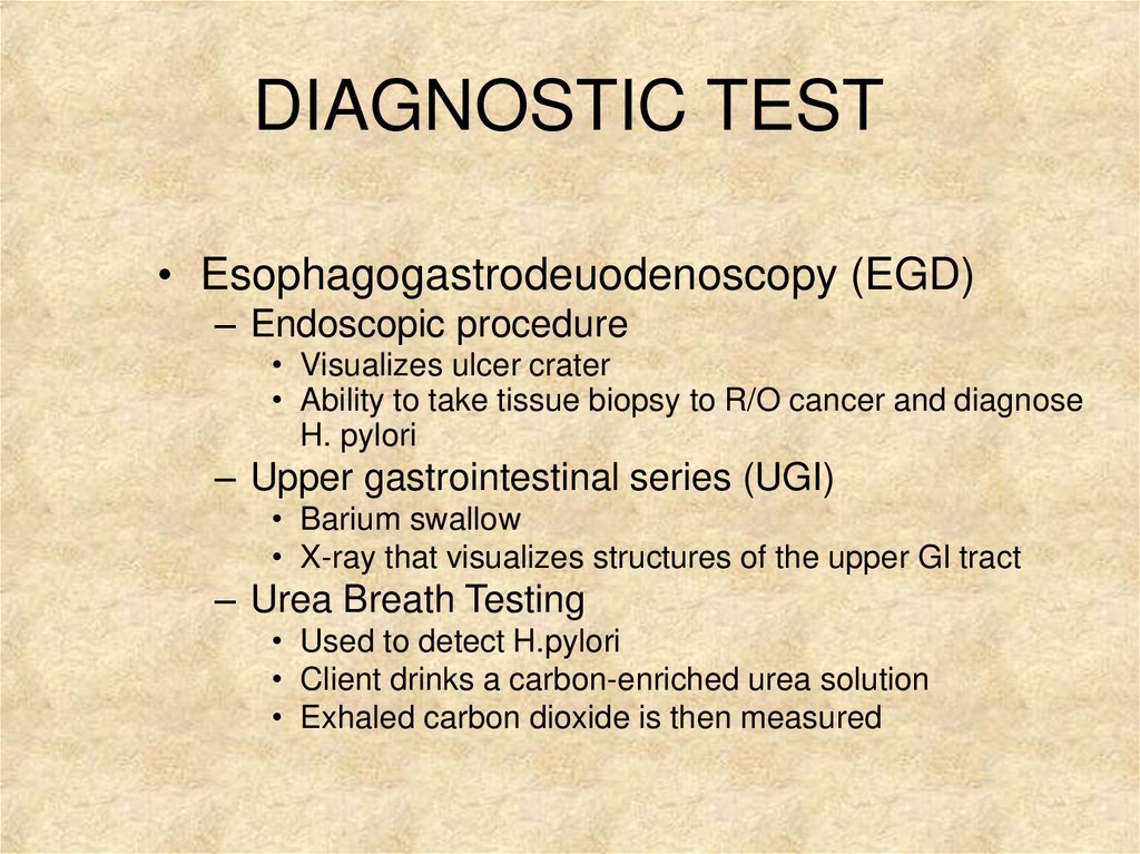
DIAGNOSTIC TEST• Esophagogastrodeuodenoscopy (EGD)
– Endoscopic procedure
• Visualizes ulcer crater
• Ability to take tissue biopsy to R/O cancer and diagnose
H. pylori
– Upper gastrointestinal series (UGI)
• Barium swallow
• X-ray that visualizes structures of the upper GI tract
– Urea Breath Testing
• Used to detect H.pylori
• Client drinks a carbon-enriched urea solution
• Exhaled carbon dioxide is then measured
13.
In all patients with “Alarming symptoms” endoscopyis required.
Dysphagia.
Weight loss.
Vomiting.
Anorexia.
Hematemesis or Melena
14.
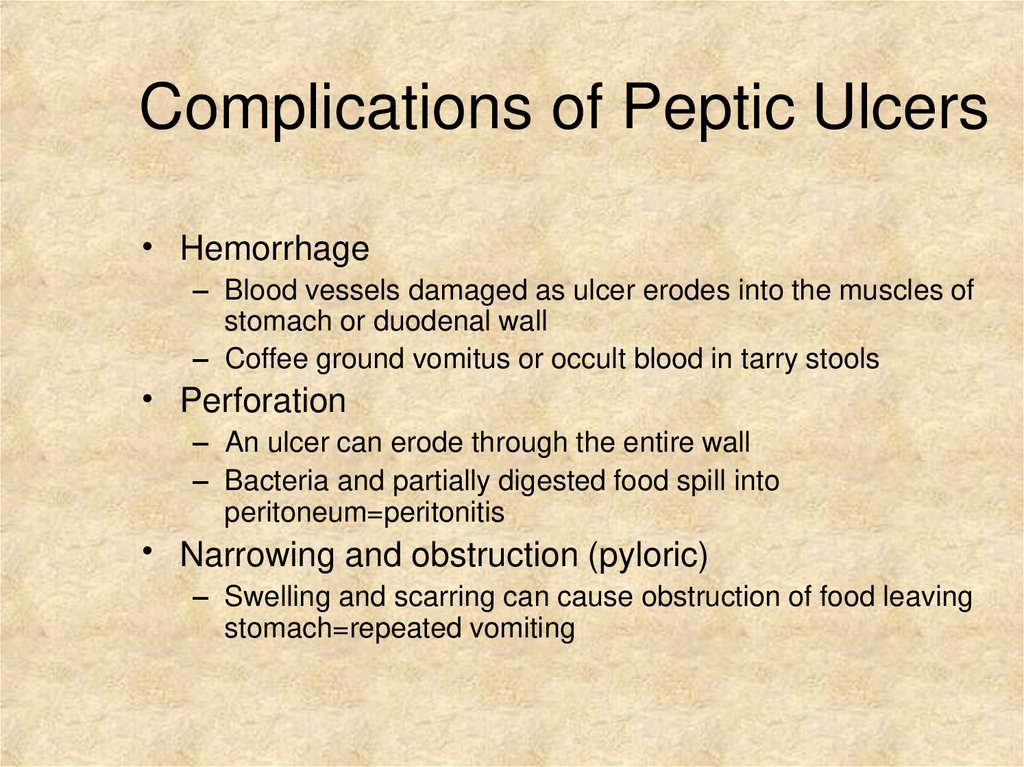
Complications of Peptic Ulcers• Hemorrhage
– Blood vessels damaged as ulcer erodes into the muscles of
stomach or duodenal wall
– Coffee ground vomitus or occult blood in tarry stools
• Perforation
– An ulcer can erode through the entire wall
– Bacteria and partially digested food spill into
peritoneum=peritonitis
• Narrowing and obstruction (pyloric)
– Swelling and scarring can cause obstruction of food leaving
stomach=repeated vomiting
15.
MANAGEMENT• LIFE STYLE MODIFICATION
• HYPOSECRETORY DRUG THERAPY
• H. pylori ERADICATION THERAPY
• SURGERY
16.
17.
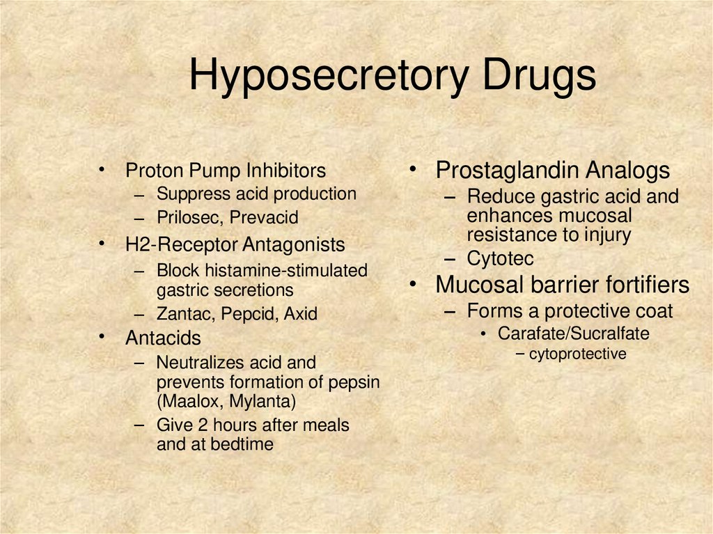
Hyposecretory Drugs• Proton Pump Inhibitors
– Suppress acid production
– Prilosec, Prevacid
• H2-Receptor Antagonists
– Block histamine-stimulated
gastric secretions
– Zantac, Pepcid, Axid
• Antacids
– Neutralizes acid and
prevents formation of pepsin
(Maalox, Mylanta)
– Give 2 hours after meals
and at bedtime
• Prostaglandin Analogs
– Reduce gastric acid and
enhances mucosal
resistance to injury
– Cytotec
• Mucosal barrier fortifiers
– Forms a protective coat
• Carafate/Sucralfate
– cytoprotective
18.
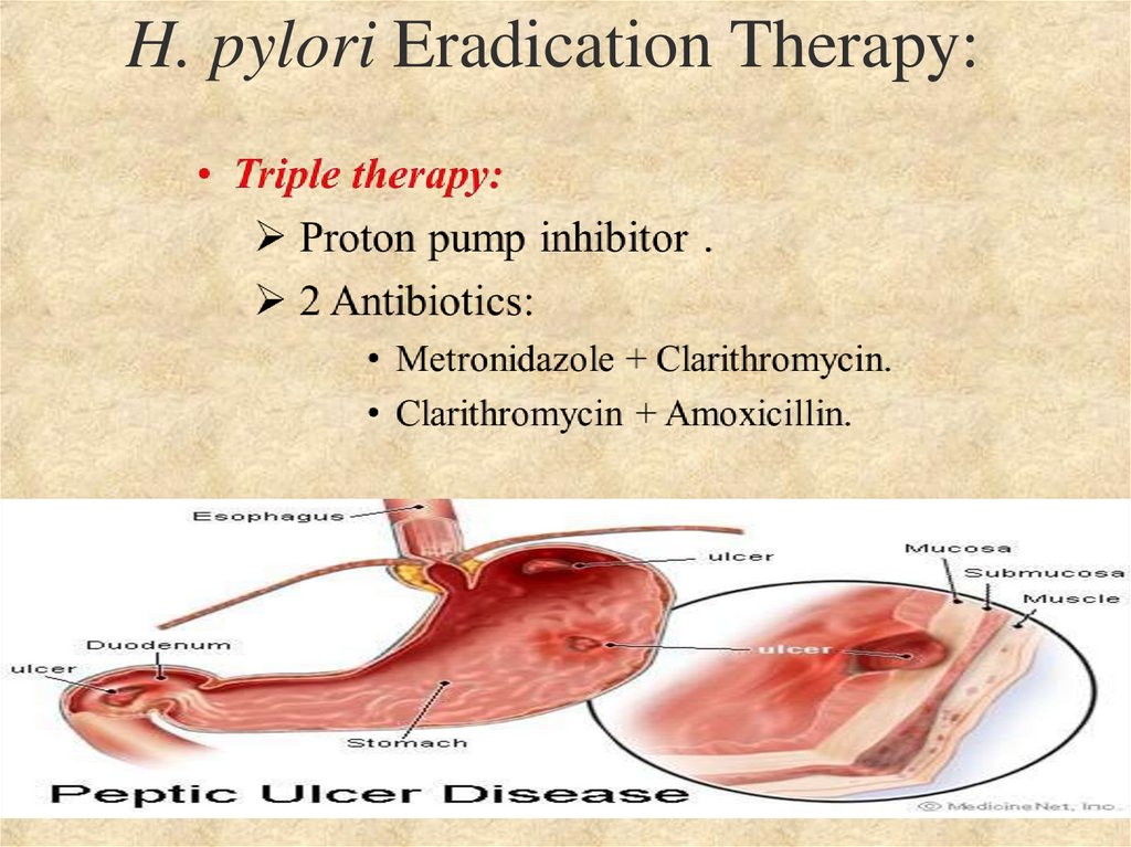
H. pylori Eradication Therapy:19.
Indications:Failure of medical treatment.
Development of complications
High level of gastric secretion and
combined duodenal and gastric ulcer.
Principle:
Reduce acid and pepsin
secretion.
20.
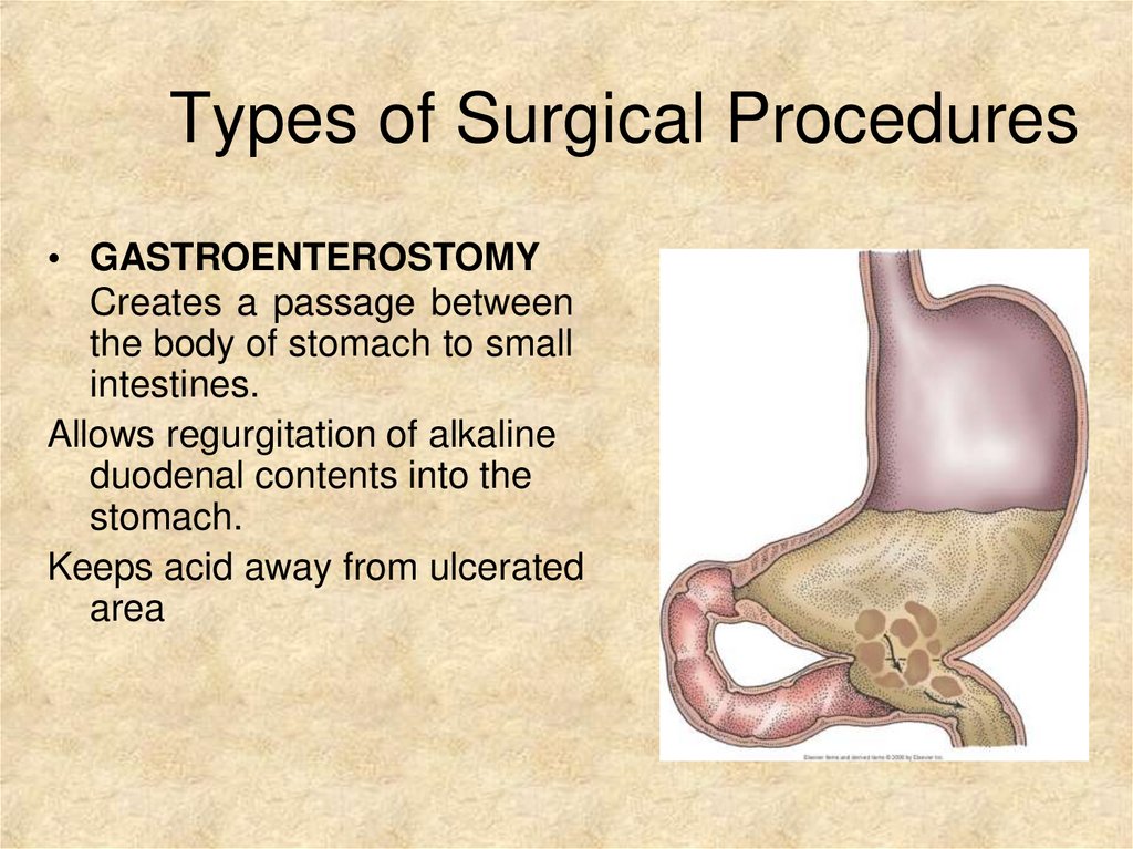
Types of Surgical Procedures• GASTROENTEROSTOMY
Creates a passage between
the body of stomach to small
intestines.
Allows regurgitation of alkaline
duodenal contents into the
stomach.
Keeps acid away from ulcerated
area
21.
Types of Surgical Procedures• VAGOTOMY
– Cuts vagus nerve
– Eliminates acidsecretion stimulus
22.
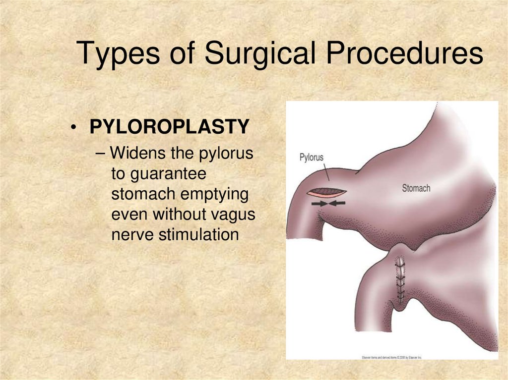
Types of Surgical Procedures• PYLOROPLASTY
– Widens the pylorus
to guarantee
stomach emptying
even without vagus
nerve stimulation
23.
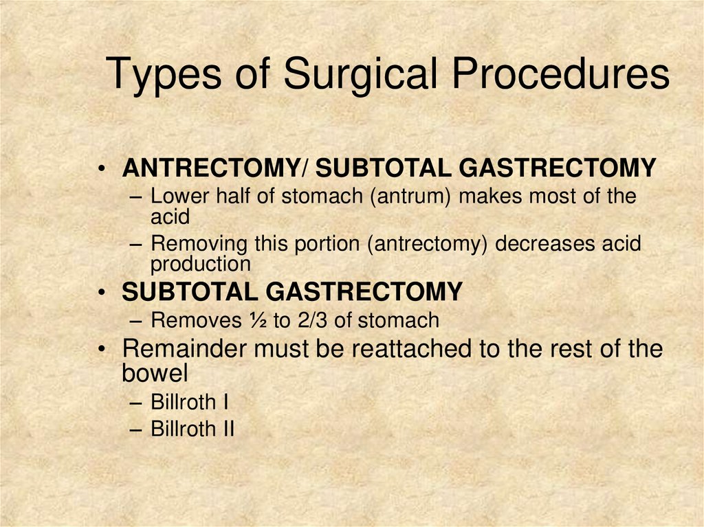
Types of Surgical Procedures• ANTRECTOMY/ SUBTOTAL GASTRECTOMY
– Lower half of stomach (antrum) makes most of the
acid
– Removing this portion (antrectomy) decreases acid
production
• SUBTOTAL GASTRECTOMY
– Removes ½ to 2/3 of stomach
• Remainder must be reattached to the rest of the
bowel
– Billroth I
– Billroth II
24.
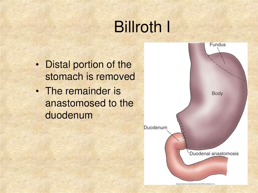
Billroth I• Distal portion of the
stomach is removed
• The remainder is
anastomosed to the
duodenum
25.
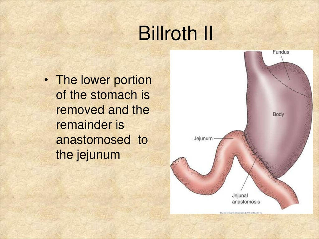
Billroth II• The lower portion
of the stomach is
removed and the
remainder is
anastomosed to
the jejunum
26.
Postoperative Care– NG tube – care and management
– Monitor for post-operative complications