


 medicine
Similar presentations:


")


Gastritis ulcer of stomach and duodenum gastric tumors
1. GASTRITIS ULCER OF STOMACH AND DUODENUM GASTRIC TUMORS
ZAPOROZHZHIAN STATE MEDICAL UNIVERSITYThe department of pathological anatomy and
forensic medicine with basis of law
GASTRITIS
ULCER OF STOMACH AND
DUODENUM
GASTRIC TUMORS
Lecture on pathological anatomy for the
3-rd year students
2. GASTRITIS
Classification of gastritis:1. Acute (erosive/hemorrhagic) gastritis
2. Chronic (non-erosive)
3. Specific forms of gastritis
4. Others (eosinophilic, allergic, granulomatous)
Diagnosis can be established only by:
at endoscopic observations
histological evaluation of biopsy specimens
ACUTE (EROSIVE OR HEMORRHAGIC) GASTRITIS
It is acute reversible disease of gastric mucous.
Reasons of development:
1. Non-steroidal anti-inflammatory drugs (NSAID’s)
2. Alcohol abuse
3. Low-flow states (shock)
4. Stress, including illness, trauma, emotional problems
5. Cigarette smoking
6. Uremia
7. Toxic substances
8. Radiation
3. Morphological forms of acute gastritis:
1. Catarrhal (simple) - edema, hyperemia, lymphoand leucocytes infiltration of mucus shell2. Erosive-hemorrhagic - edema, hyperemia,
inflammatory infiltration and formation of erosions
(superficial necrosis of mucus shell)
3. Fibrinous-necrotic - destruction of mucus shell
with formation of fibrinous exudates:
- croupous (superficial damage)
- diphteric (deep connection)
4. Purulent (phlegmonous) pan-gastritis - at deep
immune insufficiency
4. MORPHOLOGICAL FEATURES
Types of gastric epithelium damages:1. localized - involving the acid-secreting mucosa of the
fundus and body of the stomach
2. diffuse - all parts of the stomach
3. superficial inflammation not associated with significant
hemorrhage or erosions
4. deep - accompanied by focal erosions and hemorrhages.
All variants are marked by:
1. mucosal and sub-mucosal hyperemia
2. edema
3. inflammatory infiltration by lymphocytes, macrophages
and neutrophils
Acute gastritis under appropriate circumstances may
disappear within days with complete restitution of the
normal mucosa.
5.
Clinical CourseDepending on the severity of the anatomic
changes, acute gastritis may be:
asymptomatic
may cause epigastric pain, nausea, vomiting
overt hemorrhagic erosive changes
may be responsible for massive hematemesis
and potentially fatal blood loss
6.
CHRONIC (NON-EROSIVE) GASTRITISChronic gastritis is characterized by the absence of
visible mucosal erosions and by chronic inflammatory
changes leading to mucosal (gastric) atrophy and
atypical metaplasia.
The epithelial changes may become dysplastic and
possibly be transformed into carcinoma.
Asymptomatic, chronic gastritis associated with:
1. Aging
2. Helicobacter (formerly Campylobacter) pylori
3. Auto-antibodies (pernicious anemia)
4. Idiopathic
5. Peptic ulcer (gastric and duodenal ulcer)
6. Cigarette smoking
7. Alcohol abuse
8. Gastric carcinoma
7.
Main variants of chronic gastritis:I. Autoimmune chronic gastritis (Type A) it is related to
pernicious anemia, involves mainly fundus and body of
the stomach, characterized by high level of stomach
cancer development.
II. Chronic superficial gastritis (Type B) - more common
than type A, it is of non-immune origin, associated with
H.pylori, and has been further subdivided:
1. "Hypersecretory" antral gastritis, with its elevated
levels of gastric acid and pepsin, is related to duodenal
ulcer disease.
2. "Environmental" gastritis, which is multi-focal (type
AB) and often involves multiple regions of the stomach, is
associated with gastric ulcer, atypical metaplasia, and
carcinoma.
8.
Main variants of chronic gastritis:III. Chronic atrophic pan-gastritis (Type B) – it is
characterised by:
- atrophy of mucus,
- intestine metaplasia of stomach mucus – replacement
of secretory type of stomach mucus epithelium into
adsorption type of intestine epithelium
IV. Reflux gastritis (Type C) it is the inflammation of
mucus shell of gastric antrum at duodenum-stomach
reflux because of violation stomach and/or duodenum
peristalsis
9.
MORPHOLOGY1. The inflammatory changes may be limited to the
superficial zone or may extend throughout the mucosa
2. The inflammation is accompanied by variable gland loss
and mucosal atrophy. In the fundic autoimmune variant
(type A), there is particularly prominent loss of parietal
cells, owing to antibodies targeted on these cells and
intrinsic factor.
3.There are no erosions in any form of chronic gastritis, but
the surface of epithelium may undergo intestinal metaplasia
and in some instances atypical metaplasia, accounting
presumably for the increased incidence of gastric
carcinoma.
Clinical Course:
- nausea, vomiting
- upper abdominal discomfort are uncommon.
10.
STRESS ULCERSReasons of development:
Severe trauma including major surgical
procedures, serious sepsis, or grave illness of
any type
Extensive burns (referred to as Curling's
ulcers)
Traumatic or surgical injury to the central
nervous system or an intra-cerebral
hemorrhage (called Cushing's ulcers)
Long-term use of gastric irritant drugs
(aspirin, NSAIDs and corticosteroids).
11.
MORPHOLOGYMacroscopically, stress ulcers are small, round to ovoid,
sometimes irregular, mucosal defects that initially involve
only the proximal portion of the gastric mucosa then may
extend throughout the entire stomach. They rarely exceed
2.5cm in diameter, usually have a brown base of digested
blood, and only exceptionally penetrate more deeply than
the muscularis mucosa.
Histologically, the lesions appear to result from
enzymatic digestion with only a mild to moderate
inflammatory infiltration. In some cases, particularly in
association with burns and head lesions, stress ulcers may
extend into the duodenum and in this location are more
likely to be deep or penetrating.
Clinical Course. More often stress ulcers are totally silent
and come to clinical attention only when they cause
bleeding, which is sometimes massive and then carries
12.
PEPTIC ULCERA peptic ulcer can be defined as ”a hole in the
mucosa” of any portion of the gastrointestinal tract
exposed to acid-pepsin secretion.
More often peptic ulcers are located in the first
portion of the duodenum or in the stomach in a ratio
of about 4:1.
The great majority of individuals have a single
ulcer, only in certain families and in the ZollingerEllison syndrome - ulcers in the stomach and
duodenum.
Despite a remarkably uniform morphology, gastric
ulcers and duodenal ulcers may well constitute
different diseases, as will become apparent.
13.
Predisposition factors of ulcer formation1. There are hints of genetic susceptibility to duodenal
ulcers (DU) but not to gastric ulcers (GU).
2. A positive family history of DU
3. In monozygotic twins (not in di-zygotic twins).
4. The familial syndromes associated with DU, such as
the autosomal dominant hyper-pepsinogenemia-I.
5. Persons with blood group O have a greater risk for
DU than those who have other blood groups.
6. Acquired disease may also predispose to DU:
alcoholic cirrhosis
chronic obstructive pulmonary disease
chronic renal failure
hyper-parathyroidism
14.
MORPHOLOGYAll peptic ulcers are usually:
round form - 2 to 4cm in diameter
sharply punched out defects in the mucosa that
can penetrate into the submucosa, usually into
the muscularis and sometimes more deeply
Favored sites - are the anterior and posterior walls
of the first portion of the duodenum and the
lesser curvature of the stomach.
Usually it involves the entire antrum with the
ulcer crater in the margin of the affected area
close to the adjacent acid-secreting fundic
mucosa, but it may be more proximal or distal.
15.
The histological appearance varies according toactivity, chronicity and degree of healing.
During the active phase, four zones (layers) of
changes can be distinguished in epithelium:
1) the base and margins have a thin layer of
necrotic fibrinoid debris - N
2) a zone of active nonspecific inflammatory
infiltration with neutrophils - I
3) active granulation tissue - G
4) fibrous, collagenous scar that fans out widely
from the margins of the ulcer - S
16.
COMPLICATIONS OF PEPTIC ULCER DISEASEI. Ulcer-destructive:
1. Bleeding - Occurs in 25 > 33% of patients
Most frequent complication; may be massive
Accounts for about 25% of ulcer deaths
May be first indication of presence of ulcer
2. Perforation - Occurs in only 5% of patients
Accounts for 2/3 of all ulcer deaths
Rarely, is first indication of ulcer. Leads to
peritonitis
3. Penetration – ulcer bottom is in the underlying organs
(pancreas, fat tissue) with zones of destruction
17.
COMPLICATIONS OF PEPTIC ULCER DISEASEII. Inflammatory:
1. Perigastritis and/or periduodenitis
2. Phlegmon of stomach
III. Ulcer-scarring complications with stenosis
1. Stenosis or obstruction from edema or scarring of
pyloric canal or duodenum
Causes incapacitating abdominal pain
Rarely, may lead to total obstruction with intractable
vomiting, intractable pain
2. Deformations
IV. Malignisation of ulcer - transformation into
malignant tumor
18.
TUMORSI Mesenchymal neoplasms:
1. Bening - stromal cell tumors, leiomyomas,
leiomyoblastomas, neurofibromas, and lipomas
2. Malignant - sarcomas
II Epitelial neoplasms:
1. Bening – polyps, adenomas
2. Malignant - gastric carcinoma
III Carcinoids
IV Gastrointestinal lymphomas
GASTRIC POLYPS
It is nodule or mass that projects above the level of the
surrounding mucosa.
In the stomach, these lesions can be subdivided into:
1) hyperplastic polyps
2) adenomatous polyps
19.
GASTRIC CARCINOMAMorphologic types:
1. intestinal - arise from gastric mucous cells
that have undergone metaplasia into
intestinal type cells, occurs primarily after
age 50 years in 2:1 male predominance
2. diffuse - arise de novo from native gastric
mucous cells, occurs at an earlier age with
no male predominance.
20.
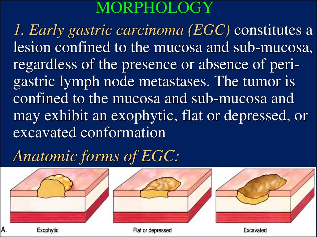
MORPHOLOGY1. Early gastric carcinoma (EGC) constitutes a
lesion confined to the mucosa and sub-mucosa,
regardless of the presence or absence of perigastric lymph node metastases. The tumor is
confined to the mucosa and sub-mucosa and
may exhibit an exophytic, flat or depressed, or
excavated conformation
Anatomic forms of EGC:
21.
MORPHOLOGY2. Advanced gastric carcinoma (AGC) is a
neoplasm that has extended below the submucosa, into the muscularis, and has spread more
widely - extends into the muscularis propria and
beyond. Linitis plastica is an extreme form of flat
or depressed advanced gastric carcinoma. Most
arise in the gastric antrum.
Anatomic forms of AGC:
22. Features of tumor propagations
All gastric carcinomas eventually penetrate thewall to involve the serosa and may spread to
regional and more distant lymph nodes and liver.
Two patterns of spread are particularly
distinctive. Gastric carcinomas frequently
metastasize to the supraclavicular sentinel
(Virchow's) node as the first clinical
manifestation of an occult neoplasm.
More uncommonly, these cancers metastasize to
one or both ovaries to cause solid tumorous
enlargements - Krukenberg tumors.